Hand Soft Tissue
Tendon transfer and peripheral nerve palsy
Tendon transfer prerequisites and reconstruction for radial and lower ulnar nerve palsy
47 questions
4 source pages
3 images
1 fact-check flags
Images appear with the first question taken from each source page — tap a question to open it.
Q1What are the prerequisites for tendon transfer?📷▸
- Patient: good motivation, willing to train
- Receiving site: tissue equilibrium (Boyes) - no infection, healthy bed, no joint contracture, joint stable
- Donor muscle FESS: strength, one transfer one function, synergistic, expendable
- Donor tendon LEP: adequate length and excursion, straight line of pull, correct tension
Q2What is the memorised summary of tendon transfer prerequisites?▸
- Transfer an expendable and good strength tendon to another synergistic action
- With straight line of pull under adequate excursion and tension
- And good soft tissue across a supple joint
- To perform single function in a compliant patient
Q3What is the aim of tendon transfer?▸
- To concentrate the available motor unit into the best working combination
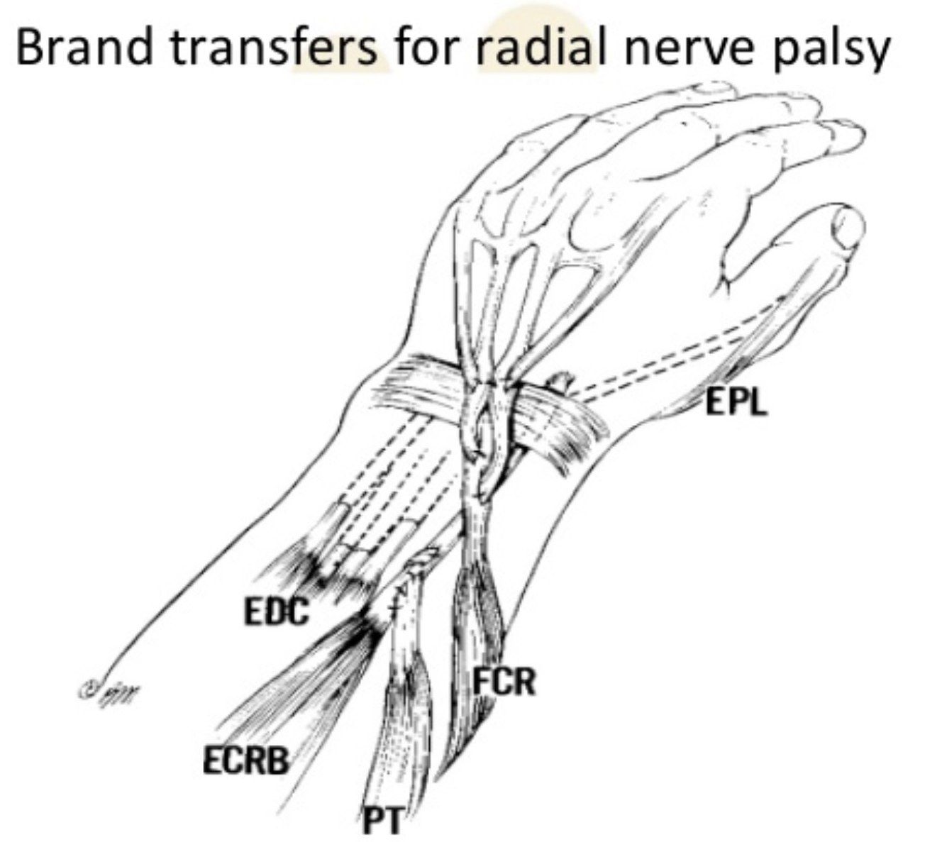
Q4What is Brand's transfer for radial nerve palsy?▸
- Wrist extension: PT to ECRB
- Thumb extension: PL to EPL
- Finger extension: FCR to EDC
Q5What tendon transfers are used for ulnar nerve palsy?▸
- Thumb adduction: ECRL/FDS4 to adductor pollicis
- 1st dorsal interosseous: EI to 1st dorsal interossei
- Little finger abduction: EDM to ADM
- Claw hand: Bouvier test - if positive, capsulodesis; if negative, Zancolli lasso (split FDS to lateral bands of ulnar digits)
Q6What tendon transfers are used for median nerve (AIN) palsy?▸
- Thumb flexion: brachioradialis to FPL
- Thumb opposition: EI to APB
- Finger flexion: FDP of R and L/F to FDP of index and middle finger (side-to-side transfer)
Q7What opponensplasties are used in carpal tunnel syndrome?▸
- Transfers to APB at MCPJ level
- Camitz (PL + palmar aponeurosis)
- Huber (ADM)
- Riordan opponensplasty (ring/little FDS, FCU slip as pulley)
- EI transfer
Q8Which muscles are supplied by the proper radial nerve?📷▸
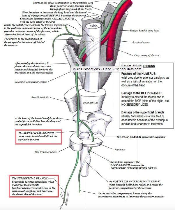
- Triceps, brachioradialis, 2/3 of brachialis, anconeus and ECRL
- Its palsy is caused by trauma or is iatrogenic
Q9What is Saturday night palsy and how is it managed?▸
- Compression of the proper radial nerve
- Involves triceps, brachioradialis and ECRL + PIN + sensory deficit
- Managed conservatively
Q10How is radial nerve palsy classified from proximal to distal?▸
- Proper radial nerve palsy
- Posterior interosseous nerve (PIN) palsy
- Cheiralgia paresthetica (Wartenberg syndrome) - superficial sensory branch
Q11What are the causes of posterior interosseous nerve palsy?▸
- Trauma: radial head fracture, Monteggia, elbow dislocation
- Space-occupying lesion: lipoma, ganglion, synovitis in RA
- Iatrogenic: after radial head replacement
- Entrapment (radial tunnel syndrome)
Q12What are the sites of compression in radial tunnel syndrome (FREAS)?▸
- Fascial band at the radial head
- Recurrent leash of Henry
- Edge of the ECRB
- Arcade of Frohse (most common site, proximal edge of the supinator)
- Supinator distal edge
Q13What are the symptoms of radial tunnel syndrome (PIN entrapment)?▸
- Lateral elbow pain
- Distal muscle weakness
- +/- dorsal wrist pain (dorsal wrist capsule)
Q14What are the signs of PIN palsy?▸
- Radial deviation with active wrist extension (ECRL is innervated by the proper radial nerve more proximally)
- Weakness of PIN-innervated thumb and finger extension
Q15Which muscles does the PIN innervate?▸
- 9 muscles: supinator + APL, EPB, ECRB, EPL, EI, EDC, EDM, ECU
Q16What is the nonoperative treatment and outcome of PIN palsy?▸
- Activity modification, splinting and NSAIDs
- Outcome at 3 months (OT@ 3m): 85% good to excellent result
Q17What is cheiralgia paresthetica (Wartenberg syndrome)?▸
- Compressive neuropathy of the superficial sensory branch of the radial nerve
- Compressed between brachioradialis and ECRL with forearm pronation (scissor-like action between the tendons)
- Symptoms: pain, numbness and paresthesias over the dorsoradial hand
Q18How is cheiralgia paresthetica examined and what is the outcome?▸
- Forceful forearm pronation for 60 seconds
- Tinel sign over the nerve
- Outcome at 6 months
Q19What does the clinical photo show and what is being assessed?📷▸
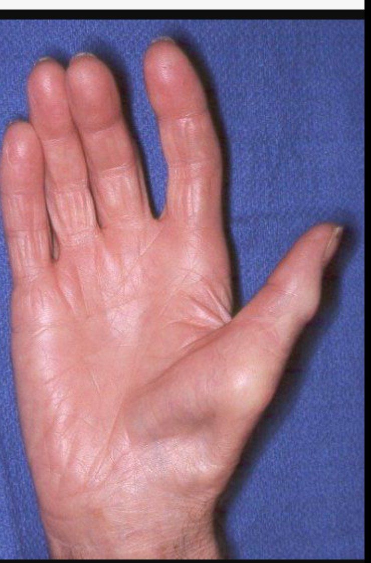
- Thenar wasting
- Carpal tunnel anatomy
- NCV result
- Carpal tunnel view to look for osteophytes
Q20What are the boundaries of the carpal tunnel?▸
- Scaphoid tubercle, trapezium ridge, pisiform, hamate hook
- Transverse carpal ligament (proximally the volar carpal ligament)
- Carpal row
Q21What structures pass through the carpal tunnel?▸
- 9 tendons: FDS, FDP and FPL
- 1 nerve: the median nerve
Q22What is the diagnostic approach to carpal tunnel syndrome (PC Ho)?▸
- Clinical diagnosis (2/3): Phalen, DCT, Tinel + NCV +/- USG
- Look for risk factors, secondary causes, and rule out other diagnoses
Q23What are the typical history features of carpal tunnel syndrome?▸
- Nocturnal numbness over the radial 3 and a half fingers
- Improves with shaking the hand
- Look for cause: mass (ganglion), inflammation (gout, RA), fluid (hypothyroidism, renal failure), post-trauma
- R/O DDx: high median nerve palsy, brachial plexus, C5/6 nerve root; double crush syndrome
Q24What are the clinical tests for carpal tunnel syndrome?▸
- Phalen, direct compression, Tinel
- High specificity: sensory loss, CT compression (Durkan's test, press over CT x 30 sec)
- High sensitivity: Phalen (flexion 90 x 60 sec), CT compression
- Thenar wasting + weak APB
Q25What are the NCV findings in carpal tunnel syndrome?▸
- Early: decreased velocity <50 m/sec, increased distal motor latency >4.2 ms
- Late: conduction block, decreased amplitude
- Sensitivity and specificity ~80%
- USG for space-occupying lesions
Q26What is the conservative management of carpal tunnel syndrome?▸
- Night splint, nerve gliding exercises
- Steroid injection: transient relief in 80%, 20% symptom free after 1 year
- INSTINCTS trial (Lancet 2018): steroid superior clinical effectiveness vs splintage at 6 weeks
Q27What predicts a poor response to nonoperative management of carpal tunnel syndrome?▸
- Age >50
- Symptoms >10 months
- Persistent paresthesia
- Trigger finger
- +ve Phalen test after 30 seconds
Q28What are the operative options for carpal tunnel syndrome?▸
- Open carpal tunnel release
- ECTR (endoscopic carpal tunnel release): 1 portal or 2 portal
Q29What are the efficacy and complications of endoscopic carpal tunnel release (ECTR)?▸
- Similar symptom relief, electrophysiology result and carpal tunnel pressure relief
- Earlier return to work (CORR 2015 Sayegh meta-analysis), no long term difference
- Higher nerve injury (2x open; open release risk 0.16%)
- Inadequate release
Q30What is required before performing a 2-portal ECTR?▸
- Patient selection, confirmation of diagnosis of carpal tunnel syndrome and r/o other DDx and secondary causes
- R/O contraindications: inflammatory cause, limited wrist extension, SOL
- Consent, Bier's block with double cuff
Q31Where are the incisions placed for a 2-portal ECTR?▸
- Distal incision: Kaplan's line (RMB) / hyperabduction thumb line (SPA), line extrapolated from the 3rd finger web
- Proximal incision: just ulnar to PL, at the proximal palmar crease
Q32Which structures must be avoided during a 2-portal ECTR?▸
- Palmar cutaneous nerve (branch of median nerve 5cm from wrist): stay ulnar to PL
- Recurrent motor branch (extra-ligamentous 50%, sub-ligamentous 30%, trans-ligamentous 20%): incise TCL on the ulnar side
- Guyon's canal (hook of hamate and pisiform): stay radial to the line from the 4th webspace
- Superficial palmar arch (average 15mm distal to Kaplan line; DPA 6mm): stay proximal to Kaplan's line
- Riche-Cannieu median-to-ulnar connection at the distal portal: blunt dissection
Q33How is a 2-portal ECTR performed?▸
- Open proximal incision, blunt dissect forearm fascia, insert McDonald to feel the washboard texture of TCL
- Hyperextend wrist, insert T&C and open the distal incision; visualise TCL and free soft tissue
- Incise TCL from distal to proximal with a retrograde knife, staying ulnar
- Success confirmed by prolapse of fat and transillumination seen on the skin
Q34What are the named nerve anastomoses in the hand?▸
- Berrettini: digital nerve anastomosis
- Martin-Gruber: median to ulnar
- Marinacci: reverse Martin-Gruber
- Riche-Cannieu anastomosis: motor ulnar and median nerve anastomosis in the thenar region
Q35How is an open carpal tunnel release performed?▸
- Confirm diagnosis, assess secondary causes; Bier's block
- Long incision: distal end proximal to Kaplan's line, just ulnar to thenar crease, curve ulnar slightly distal to the distal palmar crease, cross the crease at an angle
- Proximal end at proximal palmar crease, just ulnar to PL
- Structures to release: deep forearm fascia, TCL, volar carpal ligament
- Short incision: difficult to tackle the volar carpal ligament, but less hypertrophic scar
Q36What are the intra-operative complications of carpal tunnel release?▸
- Anaesthesia related
- Injury to median nerve, palmar cutaneous nerve, recurrent motor branch
- Injury to superficial palmar arch
- Injury to ulnar nerve and artery
- Injury to tendon
Q37What are the post-operative complications of carpal tunnel release?▸
- Pillar pain, scar pain
- Infection
- Recurrence of symptoms
- Stiffness, bowstringing of tendon
- Painful scar = injury to communicating branch of ulnar/median nerve (retract and cut)
Q38What are the causes of symptoms persisting after carpal tunnel release?▸
- Wrong diagnosis
- Double crush
- Inadequate release
- Injury to nerve
- SOL
Q39Describe the claw hand deformity and its differential diagnosis.▸
- Little finger clawing with flexed IPJs and extended MCPJs
- Unilateral DDx: ulnar nerve palsy, Volkmann's contracture (volkman’s contracture), thoracic outlet syndrome
- Bilateral DDx: CMT, cervical myelopathy
Q40What is the pathomechanism of claw hand in ulnar nerve palsy?▸
- Imbalance of intrinsic and extrinsic muscles
- Unopposed MCPJ extension due to weak intrinsic muscles
- Used up excursion of long extensors + weak intrinsics, so cannot extend IPJ
- Unopposed long flexors
Q41Why is the little finger more clawed than the others?▸
- MCPJ of L/F more hyperextendable (the MCPJ of the little finger is more hyperextendable)
- The lumbrical of the ring finger has a median nerve contribution
Q42Describe the anatomy and contents of Guyon's canal.▸
- 4 cm long tunnel
- Floor: transverse carpal and pisohamate ligaments
- Roof: volar carpal ligament
- Walls: pisiform and hook of hamate
- Contains ulnar artery and nerve; nerve bifurcates into sensory (superficial/ulnar) and motor (deep/radial)
Q43What are the three zones of Guyon's canal and their causes?▸
- Zone 1 (proximal to bifurcation): ganglion, fracture hook of hamate; contains both motor and sensory
- Zone 2: motor branch (ganglion, fracture hook of hamate)
- Zone 3: sensory branch (ulnar artery aneurysm)
- Operative treatment: decompress all three zones and address the primary cause
Q44How is lower ulnar nerve palsy investigated?▸
- Investigation: thorough examination
- NCV
- CT/MRI for the underlying cause
Q45What is the conservative management of Guyon canal syndrome?▸
- NSAIDs
- Splint
- Activity modification
Q46How is claw hand managed?▸
- Claw hand splint: prevent MCPJ extension so extensor force is transmitted to the digits
- Dynamic: Zancolli lasso (FDS around A1 and A2 then onto itself), Bunnell (FDS into 4 slips inserted into lateral bands)
- Static: volar capsulodesis
Q47What is the tendon transfer strategy for combined ulnar and median nerve palsy?▸
- Most common is combined low palsy
- Low: ECRB or FDS for key pinch; FDS or EIP opponensplasty; ECRL or BR for finger flexion
- High: ECRB, BR or EIP for key pinch; ECRL to FDP for finger flexion and grip; EIP, EPL, ECU for opponensplasty
Fact check
In carpal tunnel syndrome NCV shows increased distal motor latency >4.2s — unit error — distal motor latency is measured in milliseconds; normal median DML is <=4.2 ms and >4.2 ms is abnormal — source