Hand Soft Tissue
Nerve compression syndromes and brachial neuritis
Median and ulnar nerve compression at elbow and wrist, Guyon canal, and brachial neuritis
29 questions
4 source pages
2 images
1 fact-check flags
Images appear with the first question taken from each source page — tap a question to open it.
Q1What are the causes of compression in pronator syndrome?📷▸
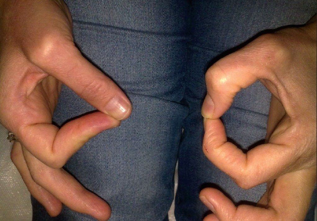
- Supracondylar process
- Ligament of Struthers
- Lacertus fibrosus
- Deep head of pronator teres
- FDS arch
Q2What is pronator syndrome and how does it present?▸
- Compression of the median nerve at the elbow level
- Numbness of the medial 3 and a half fingers
- Numbness of the thenar region
Q3What are the causes of compression in AIN (anterior interosseous nerve) syndrome?▸
- Lacertus fibrosus
- Deep head of pronator teres
- FDS arch
- Accessory head of FPL
Q4What is the presentation of AIN syndrome?▸
- Weakness of FPL
- Weakness of index and middle FDP
- Weakness of pronator quadratus
Q5How do you distinguish pronator quadratus from pronator teres weakness?▸
- PQ: resisted pronation with elbow in full flexion (least mechanical advantage to PT)
- PT: resisted pronation with elbow slightly flexed
Q6What does the photo show and what is the differential diagnosis?▸
- Failed abduction of the little finger (unilateral)
- No claw hand, no interossei muscle wasting
- DDx: Wartenberg sign, cervical myelopathy (bilateral)
Q7What is Wartenberg's sign?▸
- Weak interossei
- Ulnarly deviated pull of EDM + EDC
Q8How do you assess suspected cubital tunnel syndrome?▸
- Delineate high vs low ulnar nerve palsy
- R/O other causes with full neuro exam: C8/T1 root (I/F FDP, APB), sensory, myelopathic hand signs, LL involvement
- High: cubital valgum (malunited lateral condyle fracture), limited elbow range (OA elbow)
- Check Tinel and elbow flexion test (>60 sec)
- McGowan grading: 1 sensory, 2 weakness, 3 wasting
Q9List the sites of ulnar nerve compression around the elbow.▸
- Around the medial intermuscular septum and arcade of Struthers (medial intermuscular septum to medial head of triceps)
- Medial epicondyle in cubital valgum
- Epicondylar groove
- Cubital tunnel: SOL, Osborne ligament (medial epicondyle to olecranon), FCU aponeurosis between the two heads
- The flexor pronator aponeurosis (fascia btw FDP and FDS), anconeus epitrochlearis
Q10Outline the conservative management of cubital tunnel syndrome.▸
- Splint with elbow in 30 degrees flexion, wrist in neutral to relax FCU
- NSAIDs
Q11What are the indications and pros/cons of ulnar nerve decompression in situ?▸
- CI: subluxable nerve, SOL, scar, severe elbow deformity
- Adv: minimal dissection, can do under LA, protects the vascular mesoneurium, avoids scarring
- Disadv: if released more proximal to the epicondylar groove, risk of nerve subluxation and a new site of compression
Q12What are the indications and pros/cons of medial epicondylectomy?▸
- Releases compression against the epicondyle
- For visible and symptomatic subluxating nerve, or thin patients where subcutaneous anterior transposition may not be feasible
- Pros: smaller wound, nerve can seek its own course of least resistance, less ischaemic injury, no need to sacrifice the articular branch
- No need to immobilize elbow
- Cons: injury to MCL (prevent by not going too basally, <70%), pain, weakness of flexor origin, too little resection may not fully relieve traction
Q13How do you perform a medial epicondylectomy?▸
- GA, supine, shoulder abducted and externally rotated; incision centred on medial epicondyle; protect the medial antebrachial nerve
- Proximally release arcade of Struthers and medial intermuscular septum, then Osborne ligament, distally the FCU aponeurosis and deep flexor pronator aponeurosis
- Expose the medial epicondyle subperiosteally and remove 3-4mm
- Reattach periosteum in 'pant over vest' manner to the CFP
- Identify the ulnar nerve posteriorly in its groove
Q14Compare the types of anterior transposition of the ulnar nerve.▸
- Subcutaneous: technically easiest; nerve becomes superficial, more dissection of blood supply
- Submuscular: least recurrence, good protection and vascular bed; needs 3 weeks immobilisation, risk of recompression (improve with Z-plasty), not good in severe OA, too close to joint may cause impingement
- Intramuscular: nerve buried in a groove within flexor-pronator muscle; risk of recurrence, more bleeding, better preserves nerve blood supply
Q15What is the evidence for in-situ decompression versus transposition?▸
- Ilyas JHS 2019 meta-analysis: functionally in-situ decompression = transposition; transposition has more complications
- JAMA Network 2020 (Wade): in-situ decompression associated with lower risk than any form of transposition
- Addition of epicondylectomy associated with increased probability of symptomatic cure without increasing complication risk
Q16How is an anterior transposition of the ulnar nerve performed?▸
- GA, supine, shoulder abducted and ER; incision centred on medial epicondyle; avoid the medial antebrachial nerve (pierces brachial fascia adjacent to basilic vein)
- Anteriorly develop skin flap to expose the CFO; posteriorly identify the ulnar nerve in its groove
- Proximally release arcade of Struthers and medial intermuscular septum, then Osborne ligament; distally release FCU aponeurosis and deep flexor pronator aponeurosis
- For submuscular: sharply divide the tendinous origin of the medial epicondyle, retract the flexor mass distally, place nerve deep to the fascial sling
- Flex and extend the elbow to ensure no kinking; release tourniquet before closure for haemostasis; immobilise elbow 3 weeks
Q17What are the boundaries of Guyon's canal?▸
- Ulnar: pisiform
- Radial: hook of hamate
- Floor: transverse carpal ligament
- Roof: volar palmar ligament and palmaris brevis
Q18What does Guyon's canal contain?▸
- Ulnar nerve and ulnar vessels (ulnar NV)
Q19Describe the three zones of Guyon's canal.▸
- 1. Before bifurcation: contains both motor and sensory
- 2. Motor, radial
- 3. Sensory, ulnar
Q20What is the epidemiology of Parsonage-Turner syndrome?📷▸
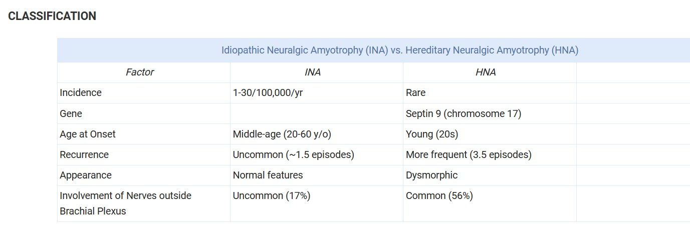
- Uncommon: 1.6-3 cases per 100,000 persons per year
- Males, middle-age (4th decade; 20-60 most common, average 41)
- Usually unilateral; bilateral in 10-30% (16% simultaneously)
Q21What are the causes and risk factors of brachial neuritis?▸
- Idiopathic (INA) or hereditary (HNA) neuralgic amyotrophy
- Multifactorial: autoimmune / genetic / infectious / environmental / biomechanical
- Viral infections 25-55% (EBV, VZV, Coxsackie B, parvovirus B19, CMV, mumps, smallpox, HIV); bacterial (Leptospira, TB, Yersinia, Salmonella, Borrelia)
- Immunizations 15% (tetanus, hepatitis B); perioperative and peripartum 14%; strenuous exercise 8%
- Drugs (abacavir, streptokinase, heroin, infliximab, interferon); iatrogenic (interscalene block, surgery, lumbar puncture, irradiation)
Q22Which nerves are most commonly affected in brachial neuritis?▸
- Any nerve or branch within the brachial plexus; extra-plexus ~17%
- Most common: long thoracic, suprascapular, axillary, musculocutaneous, radial nerves
- Other: cervical roots, AIN, PIN, lateral antebrachial cutaneous nerve (LABC)
Q23What is the proposed pathogenesis of idiopathic neuralgic amyotrophy?▸
- Autoimmune: increased blastogenic activity of lymphocytes and increased complement-fixing antibodies to peripheral nerve myelin
- Biomechanical: mobility of the upper trunk predisposes to wear-and-tear on the blood-nerve barrier
- Multifactorial: autoimmune / genetic / infectious / environmental / biomechanical
Q24Describe the three phases of brachial neuritis.▸
- Phase I: sudden severe shoulder pain (90%), radiates to proximal arm/neck; lasts days to weeks
- Phase II: painless flaccid paralysis - 24 hours (33%) to 4 weeks (80%); patchy paresis is highly characteristic
- Phase III: slow recovery of motor function over 6-18 months; duration proportional to duration of pain phase
Q25What are the physical examination features of brachial neuritis?▸
- Signs of denervation: fasciculations and atrophy
- Pain not affected by motion or palpation
- Severe weakness of shoulder, especially external rotation and abduction
- LMN signs: hypotonia and areflexia; medial scapular winging
- Sensory change in 78%; autonomic dysregulation in 15%
Q26What investigations are used in brachial neuritis?▸
- Bloods: CBC and ESR usually normal, elevated liver enzymes; positive antiganglioside antibodies and ANA
- MRI: early T2 diffuse signal hyperintensity (edema) in affected muscles; late T1 fatty infiltration and atrophy
- EMG: early PSW/fibrillation; late (3-4 months) chronic denervation and early reinnervation with polyphasic MUAPs
- NCS: reduced amplitude with preserved conduction velocity and distal latency (less useful than EMG)
- CSF: usually normal, mildly elevated protein, slight pleocytosis and oligoclonal bands
Q27What is the differential diagnosis of brachial neuritis?▸
- Cervical spine radiculopathy
- Rotator cuff pathology
- Entrapment neuropathy
- Idiopathic hypertrophic brachial neuritis (IHBN): painless upper limb weakness with brachial plexus hypertrophy
Q28What is the treatment and prognosis of brachial neuritis?▸
- Nonoperative: observation and pain control (NSAIDs/physio); oral corticosteroids may hasten pain resolution but do not affect progression or prognosis
- Operative if no regeneration or early recovery by 6-9 months on exam and EMG: neurolysis, neurorrhaphy, grafting, nerve or tendon transfers
- Recurrence rare in non-hereditary cases; poor prognosis: female, lower trunk, persistent pain and no motor recovery by 3 months, hereditary cases; age has no effect
- Recovery: 66% show motor recovery within 1 month; excellent in 36% at 1 year, 75% at 2 years, 89% at 3 years; may take up to 8 years
Q29What is the operative outcome in brachial neuritis?▸
- Exploration of patients without neurologic recovery reveals hourglass-like constrictions in peripheral nerves with no external compression
- Neurolysis alone was superior to neurorrhaphy and nerve grafting
Fact check
66% have recovery of motor function within 1 month in Parsonage-Turner syndrome — misleading/unsupported — secondary sources cite 66% early partial or any recovery at 1 month; validated functional recovery is 36% at 1 year, 75% at 2 years and 89% at 3 years — (medium confidence) — source