Hand Soft Tissue
CRPS and hand rehabilitation
Complex regional pain syndrome: sympathetic mechanism, clinical hand picture and rehab programme
32 questions
3 source pages
2 images
2 fact-check flags
Images appear with the first question taken from each source page — tap a question to open it.
Q1Describe the proposed cycle in RSD.▸
- Injury -> triggering sensed by brain -> sympathetic signal sent to limb -> inflammatory response -> triggers another response (cycle) -> RSD
Q2What is the role of the sympathetic nervous system in this cycle?▸
- After the injury is sensed by the brain, it sends a sympathetic signal to the limb, which triggers an inflammatory response that perpetuates the cycle
Q3How would you describe this clinical photo?📷▸
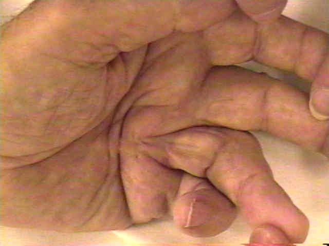
- Visible cord with flexion deformity of the ring finger MCPJ and PIPJ
- Flexion deformity of the little finger
- Thickened nodule over dorsoradial aspect of the PIPJ of the index finger (Garrod's knuckle)
- Concern: Dupuytren's contracture with diathesis
Q4What is Dupuytren's disease?▸
- Fibroproliferative disorder of the palmar fascia producing nodules/cords and causing flexion contracture of fingers
- Nodules: fibroblasts and myofibroblasts (intracellular actin filaments along long axis; extracellular fibronectin connects adjacent myofibroblasts)
- Cords: highly organised collagen, no myofibroblasts
Q5What is the pathophysiology of Dupuytren's disease?▸
- Murrell theory: local microvascular ischaemia produces free radicles -> fibroblast proliferation -> type 3 collagen (thicker than type 1) and fibrosis
- Strangulation and deficient microcirculation -> self-perpetuating cycle due to ischaemia
- Luck stages: proliferative -> involutional -> residual
Q6What is the anatomy of the palmar fascia?▸
- 3 groups of fibres based on orientation: longitudinal (pretendinous band), vertical (septum of Legueu and Juvara, separates lumbrical and flexor tendon), transverse
- Longitudinal layer has 3 layers: superficial, spiral, deep
- Spiral layer forms the pretendinous band; when pathological: pretendinous cord -> lateral digital cord -> spiral cord -> Grayson cord
Q7Which cord is responsible for contracture at each site in Dupuytren's?▸
- MCPJ: pretendinous cord
- PIPJ: spiral cord
- DIPJ: retrovascular cord
- Web: natatory cord
- 1st web: commissure cord; little finger: abductor digiti minimi cord
Q8What classifications are used in Dupuytren's disease?▸
- Woodruff: 1 no FFC, 2 MCPJ, 3 MCPJ + PIPJ, 4 more than one finger, 5 finger on palm
- Tubiana combined flexion contracture: I 0-45, II 45-90, III 90-135, IV >135
- Differentials: scars, burns, trigger finger, tendon adhesions, joint contracture, ulnar nerve palsy
Q9What are the risk factors and features of Dupuytren's diathesis?▸
- Patient factors: male, family history, Celtic/Viking, bilateral
- Disease: HIV, TB, COPD, DM; drugs: alcohol, anti-epileptics
- Diathesis BERYS: Bilateral, Ectopic (Ledderhose, Peyronie, Garrod's knuckles), Radial digit, Young <50, Sex: males
- Incidence: ring > small > middle > index
Q10What do you examine locally in Dupuytren's disease?▸
- Volar: scars, pits, confirm cord, extension of cord
- Dorsal: Garrod's knuckle pads
- Range: table top test (Hueston's test), differential ROM
- Contralateral hand; digital Allen's test + sensation
Q11When do you intervene and what are the operative options?▸
- Intervene if PIPJ FFC >20deg or MCPJ FFC >30deg
- Offer limited fasciectomy (alternatives: conservative, collagenase, percutaneous fasciotomy, segmental fasciectomy, dermofasciectomy and skin grafting)
- Counsel: recurrence ~50% at 5-10 years, possible skin graft, Long rehab (need up to 6 months of night splintage), chance of amputation
- Incision: longitudinal then multiple Z plasties for finger, transverse for palm; early mobilization; complications include recurrence, NV injury, infection, CRPS, wound dehiscence
Q12What is the aim of surgery in Dupuytren's disease?▸
- To improve the flexion contracture and to improve range of motion in the finger
Q13What is the escalating ladder of surgical options for Dupuytren's disease?▸
- Percutaneous fasciotomy -> segmental fasciectomy -> limited fasciectomy -> total fasciectomy -> dermatofasciectomy
Q14Define limited and segmental fasciectomy.▸
- Limited fasciectomy: removal of only the macroscopically diseased fascial tissue from the palm (vs abandoned total fasciectomy, where the entire palmar fascia was exenterated)
- Segmental fasciectomy: multiple small incisions targeting segments of cord most responsible for contracture, leaving intercalary fragments behind
- Surgical objective is the degree of contracture correction, not the extent of tissue removal
Q15What is the principle of Z-plasty and the gain at each angle?▸
- Local transposition flap bringing skin from an adjacent area to release contracture
- Central limb along the line of contracture and tension; peripheral limb same length at 60deg
- 30deg = 25%, 45deg = 50%, 60deg = 75% gain in length
- Gain in length = 2 x cos30 = 1.73
Q16How is collagenase injection performed?▸
- Enzymatic percutaneous fasciotomy
- 10,000 U collagenase diluted in CaCl injected percutaneously into the cord
- May inject up to 1.6mg for multiple digits
- Gentle manipulation -> extension splint for 4 months + home PT
Q17What non-operative options exist for Dupuytren's and what is the evidence?▸
- Needle aponeurectomy: perforate the cord repeatedly under tension; avoid LA to retain feedback if digital nerve encroached
- Collagenase: FDA approved (licensed for MCPJ disease), injected along the cord, manual extension 24hr-7 days later
- Hurst NEJM 2009: 60% correction to within 5deg of full extension; PIPJ disease more recurrence; Leafbald 2019: collagenase highest reintervention at 2 years
- Cochrane review 2015: insufficient evidence for relative superiority of different surgical procedures
Q18What are the recurrence rates after different Dupuytren's treatments?▸
- Post grad course figures: 80% collagenase, 60% fasciotomy, 40% selected fasciectomy, 20% open fasciectomy
- Leafbald 2019 (848 cases): collagenase had the highest reintervention rate at 2 years
- Fasciectomy has a high initial cost but the lowest reintervention rate
Q19How do you describe and assess this flexor tendon injury?📷▸
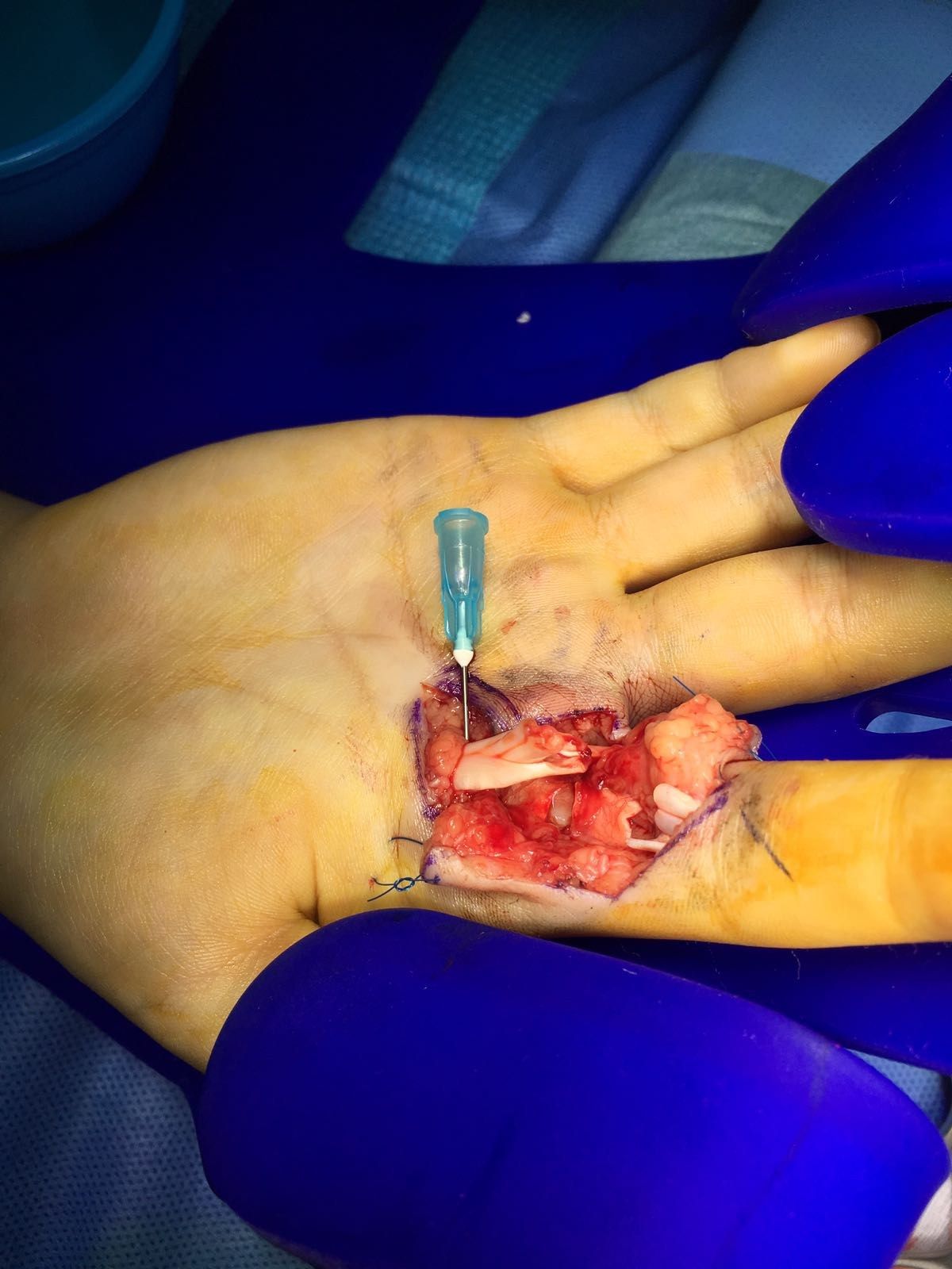
- I/F laceration with complete cut of FDS and FDP at zone 2, just at Camper's chiasma; contamination; check distal circulation
- Hx: mechanism of injury, occupation, functional demands
- PE: tenodesis, wound contamination, digital nerve 2PD, digital Allen, XR for FB/fracture
Q20What is the aim and the principles of an ideal flexor tendon repair?▸
- Aim: secure repair to allow early mobilisation for intrinsic tendon healing
- Prep: smooth tendon ends, minimal disruption of blood supply, easy passage of suture
- Repair: minimal gap, secure knot, sufficient strength for early mobilisation
Q21How do you retrieve and tunnel the flexor tendons?▸
- Retrieval: milking, reverse Esmarch, suction, tendon retriever, proximal skin incision
- Temporary locking suture to hold both ends, pass suture through pulley
- Preserve A2 (chiasma just distal to A2 at resting position); flex wrist and fingers and temporarily pin down tendon
Q22What are the key points of flexor tendon suture technique?▸
- Use non absorbable suture at least 4-0 or thicker
- Kessler (locking) better than Bunnell - superior pull-out strength when core sutures are in the dorsal half of the tendon
- At least 4 core strands (active flexion ~20N; 2 core ~34N, 4 core ~43N, 6 core ~78N)
- At least 10mm from the edge; epitendinous suture +20% strength, reduces gap and improves gliding
Q23What is the blood supply and healing of flexor tendons?▸
- Vascularised via paratenon -> travels longitudinally within endotenon; non-vascularised via synovial sheath -> mesotenon in the vincula
- Intrinsic healing from epitenon/endotenon (less adhesion) vs extrinsic healing from paratenon (promoted by immobilisation) - avoid extrinsic
- Phases: inflammatory <2 weeks (fibrin clot, strength decreases), reparative 2-6 weeks (collagen 3), remodelling >6 weeks (collagen 1, strength increases) - protect before 6 weeks
Q24Should you repair both FDS and FDP, and which pulleys are retained?▸
- Repair both for independent DIPJ and PIPJ function; FDS one slip only can prevent swan neck yet minimise adhesions
- Retain A2 and A4
- Cutting A2 causes bowstringing -> need more excursion to produce same angular movement, result in less angular movement
Q25What are the flexor tendon rehabilitation options?▸
- Static vs. dynamic program
- Dynamic: controlled passive (Kleinert/ Duran/ strictland) vs early active (Belfast) movement
Q26Describe the Kleinert rehabilitation program.▸
- Dynamic splint allowing active extension and passive flexion
- Low force, low excursion (2.5cm)
- Wrist/MCPJ flexed 30/30; rubber band and pulley system; 1 pulley at midpalm to increase DIPJ flexion for better differential gliding; release at night with finger strap to prevent PIPJ contracture
- Change to night extension splint at 4 weeks, gentle active fisting; resistive exercises at 6 weeks
Q27Describe the Duran rehabilitation program.▸
- Static wrist splint (wrist 20deg, MCPJ 50deg)
- Patient alternately passively extends DIPJ and PIPJ with other joints flexed to allow differential gliding
- Moderate force, low excursion
- Off splint at 4-6 weeks to wrist cuff with elastic traction (active extension, passive flexion); off cuff at 6 weeks, active fisting
Q28Describe the Strickland rehabilitation program.▸
- Adds a tenodesis splint (wrist 30deg, MCPJ 50deg)
- Couples active wrist extension with passive digital flexion, and wrist flexion with digital extension
- Low force, high excursion (9cm)
Q29Describe the Belfast early active rehabilitation program.▸
- Early active movement; dorsal blocking splint limiting wrist extension (wrist 40deg flexion, MCPJ 90deg flexion, IPJ 0)
- Place and hold exercises every 2 hours, started immediately
- 3 active flexion (palm + hook + flat fist), 2 active extension (PIPJ + wrist), 2 passive motion exercises (into palm + hook)
- Supported by Star et al Am J Hand Surg 2013 meta-analysis: active motion had significantly fewer flexion contractures, better satisfaction and ROM, no difference in re-rupture rates
Q30When do you use a static rehabilitation program?▸
- Children, mental deficit / abnormal muscle tone
Q31Why use a dynamic rehabilitation program?▸
- Stressed tendon heals faster, with better strength, less adhesion and better excursion
- Principle: provide motion but protect against excessive stress load which may cause rupture
Q32What are the complications of flexor tendon repair and rehabilitation?▸
- Re-rupture (15-25%)
- Contracture / adhesions
- Quadriga / lumbrical
- Swan neck
Fact check
Collagenase: may inject up to 1.6mg for multiple digits — incorrect dose — FDA label: 0.58 mg per palpable cord, up to 2 joints in the same hand per treatment visit (maximum 1.16 mg), not 1.6 mg — source
Dupuytren recurrence: 80% collagenase, 60% fasciotomy, 40% selected fasciectomy, 20% open fasciectomy — imprecise/overstated — 5-year series report lower rates: CORDLESS collagenase overall ~47% (MCP ~40%, PIP ~66%); van Rijssen RCT needle fasciotomy 85% vs limited fasciectomy 21% — (medium confidence) — source