Sports
Rotator cuff tear and cuff arthropathy
Cuff tear diagnosis, impingement types, force-couple loss, arthropathy and non-operative treatment.
78 questions
13 source pages
2 images
Images appear with the first question taken from each source page — tap a question to open it.
Q1What are the dynamic stabilizers of the shoulder?▸
- Rotator cuff
- Biceps
- Periscapular muscles
Q2What are the static stabilizers of the shoulder?▸
- Bony: osteotomy, version
- Soft tissue: ligaments, labrum
- Negative intra-articular pressure
Q3What are the roles of the glenohumeral ligaments?▸
- SGHL: inferior restraint at adduction
- MGHL: AP restraint at 0-45 degrees abduction + ER
- IGHL: AP + inferior restraint at 45-90 degrees abduction + ER
- CHL: primary ER restraint
Q4What are the functions of the rotator cuff?▸
- Dynamic stabilizer of the shoulder
- Force coupling to maintain a stable fulcrum for concentric rotation of the humeral head by neutralizing deltoid shear
- Vertical: inferior cuff (IS, TM, subscap) counteracts the superior pull of deltoid so the GHJ rotates instead of migrating superiorly
- Horizontal: IS + TM balanced by subscap
Q5What are the causes of rotator cuff tear?▸
- Impingement: subacromial -> SS, infraspinatus, teres minor (SIT); subcoracoid -> subscap
- Trauma: middle-aged after shoulder dislocation -> SIT
- Young patient after fall with hyperabduction/ER -> subscap
Q6What are the causes of impingement and what is Neer's staging?▸
- External: hooked acromion, ACJ arthritis/osteophytes
- Internal: throwers with tight posterior and lax anterior capsule; anterior subluxation on abd + ER; GT with SS impinging on posterosuperior glenoid
- Neer's staging: inflammation, fibrosis, tear with weakness
- Type 1 collagen replaced by type 2 (better compression strength, less resistant to tensile strength)
Q7What are the X-ray findings and special views in rotator cuff pathology?▸
- AP: calcific tendonitis, cuff arthropathy, acromial sclerosis, GT cystic changes, Maloney's line
- Critical shoulder angle (normal 30-35): increased in RCT, reduced in OA
- Supraspinatus outlet view (Y view with 10 degrees caudal tilt) for Bigliani acromial morphology (flat, curved, hooked)
- Also look for os acromionale (os acromiale) and ACJ osteophyte
Q8What do you look for on MRI in a rotator cuff tear?▸
- Number, size, retraction of tear; fatty infiltration/atrophy; bicep tendon pathology
- Coronal: SS tear, Patte retraction, superior migration of humeral head
- Sagittal: Goutallier fat atrophy 0-4 (0 normal, 1 fatty streak, 2 <50%, 3 50%, 4 >50%)
- Axial: biceps tendon subluxation (medial), subscapularis and IF tendon
- Ellman partial: I 3mm, II 3-6mm, III >6mm; A articular, B bursal, C intratendinous
- Size: small <1cm, medium 1-3cm, large 3-5cm, massive = 2 or more cuffs involved or >5cm
Q9What are the indications for operative management of a cuff tear?▸
- Symptomatic failure of 6-12 months conservative management
- Partial thickness >50% PASTA (Ellman 3A), or bursal side >3mm (Ellman 2B)
- Full thickness + repairable
- Young <60 with acute tear: repair within 6 weeks
- Contraindications: irreparable tear, frozen shoulder
- Irreparable: Goutallier IV, GHJ arthritis, advanced age; SS retraction beyond glenoid; cannot pull cuff beyond articular surface even at 60 degrees abduction
Q10How would you assess a patient with a suspected rotator cuff tear?▸
- Hx: history of injury, functional demand, symptoms (overhead activity, night pain) and functional limitation
- PE: identify pathology (rule out other causes of weakness e.g. nerve palsy, identify which cuff), severity of weakness
- Delineate cause (impingement) and complication (frozen shoulder)
- First-line investigation: X-ray and MRI
Q11How would you describe a rotator cuff tear on MRI and what associated findings do you comment on?▸
- Morphology: crescent, U, L, massive
- Delineate number, size and retraction of the tear; which cuff is involved
- Comment on articular cartilage, associated ACJ arthritis, superior migration and the tangent line
- Associated biceps tendon pathology
Q12What is the natural history of symptomatic cuff tears (Kim KSSTA 2017)?▸
- Symptomatic partial thickness: 26% progress at 2 years
- Symptomatic full thickness: 82% progress at 2 years
- Counsel the patient regarding the natural history
Q13What conservative management is offered for a rotator cuff tear?▸
- NSAID
- Physio: strengthen periscapular muscles + deltoid recruitment exercises
- Subacromial injection if impingement symptoms are severe
- PRP: controversial; 2022 Bhandari meta-analysis showed better long-term pain relief than steroid
Q14What is the principle of cuff repair and how is the tendon mobilised to the footprint?▸
- Principle: anatomical, secure, tension-free repair to lower retear rate
- Dissection between glenoid and cuff (not too medial 2cm or 1cm posterior - suprascapular nerve at risk)
- Anterior interval page (to coracoid, more anatomical) and posterior interval page (to glenoid, less anatomical)
- Release of the coracohumeral ligament
Q15How is a PASTA (partial articular supraspinatus tendon avulsion) tear repaired?▸
- If 25% SST remains - convert to full thickness
- Small perforation - transtendinous repair
- Young PASTA - transtendinous in situ repair
- Old PASTA - complete the tear then repair
Q16When is acromioplasty performed and how is it done safely?▸
- Not to be done routinely
- Indications: CA ligament fraying, bursal-side tear, anterolateral osteophyte, impingement on dynamic assessment
- Avoid the anteromedial corner (thoracoacromial artery bleeding)
- Together with bursectomy and biceps resection/tenodesis
Q17What does double-row repair for massive cuff tears involve?▸
- Chen 2013 meta-analysis: higher rate of intact tendon healing, especially for large tears, but no better clinical outcome
- May not be possible with severe retraction
- Conventional: tendon strangulation, worse footprint restoration, synovial fluid leakage
- Transosseous: medial tendon strangulation, failure at musculotendinous junction, highest ultimate failure load
- Knotless: lower structural strength, suture slippage
Q18What are the partial repair options for a massive cuff tear?▸
- Medialisation of footprint - no more than 10-12mm
- Patch repair (fascia lata) to share tension
- Augmentation with biceps
- Marginal convergence no longer recommended (non-anatomical)
Q19What are the surgical choices for an irreparable cuff tear?▸
- Superior capsular reconstruction (>=6mm thick graft), medial anchor to superior glenoid
- Muscle transfer: LD for posterosuperior SST/IST tear +/- simultaneous RSA to restore ER; TM for better strength; pec major for subscapular/anterosuperior defect
- Old frail patient: debridement +/- balloon spacer (poly-L-lactide); START:REACTS (Lancet 2022) does not recommend the spacer
- Reverse shoulder arthroplasty
Q20How is shoulder arthroscopy set up and performed?▸
- GA, lateral decubitus leaning 30 degrees posterior, parallel to the table; beanbag + correct head position
- Arm draped hanging out, abducted with slight flexion to relax the brachial plexus
- Landmarks: posterolateral corner of acromion and coracoid
- Posterior portal 2cm medial and inferior to the posterolateral corner, directed at the coracoid; danger - too inferior = axillary nerve, too medial = suprascapular nerve
- Rotator interval contents: coracohumeral ligament, SGHL, biceps; systematic view from anterior biceps, GHL, subscap, labrum, SS/IS
- Cuff repair needs lateral portals for anchors; Bankart repair uses anterosuperior and anteroinferior portals
Q21What is the standard rehab protocol after cuff repair (<3cm tear/single cuff)?▸
- Phase 1 (first 6 weeks): muscle protection, swelling reduction, controlled mobilisation; abduction pillow, pendulum, scapular retraction exercises
- PROM not beyond 90 degrees in the first 2 weeks, increase to 120 degrees by 4 weeks, then AAROM to 90 degrees
- Phase 2 (up to 12 weeks): full AROM/PROM as tolerated, light isometric/isotonic exercises
- Phase 3: intensive strengthening (theraband, pulleys, press-ups); Phase 4: endurance training (multi-push and lift machines)
Q22How does rehabilitation differ after massive or multiple cuff repair?▸
- Delay AROM and isotonic/isometric exercise until 10 weeks
- Can start AAROM from 4 weeks onwards
Q23Describe the footprint of the rotator cuff.▸
- Overall dimension 20 x 25mm
- SST at the anterosuperior greater tuberosity
- Medial-lateral widths (SITS): SST 12mm, IST 13mm, TM 14mm, subscapularis 18mm
- 1.5-2mm from the articular cartilage
- Anteroposterior 20mm (SST + anterior IST)
Q24What is the definition of a massive cuff tear and what are the clinical clues?▸
- >5cm (Cofield 4)
- >1 tendon involved (Europe)
- Retraction to the glenoid (Patte 3)
- >50% fatty infiltration (Goutallier 4); AHI <6mm
- Clinical clues: severe muscle atrophy, pseudoparalysis (active abduction <90 with full passive ROM and drop arm +), LHB rupture, ER lag or hornblower +
Q25Describe the cable-crescent complex in the suspension bridge theory (Burkhart).📷▸
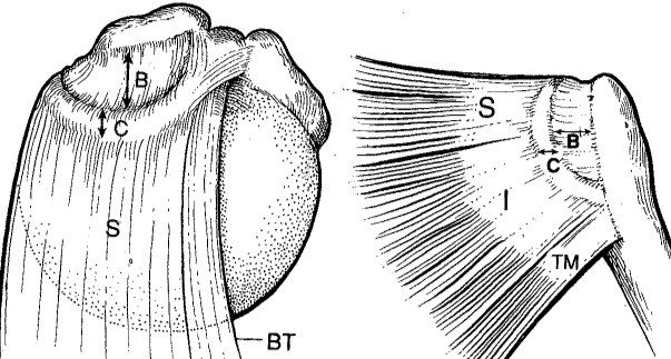
- Cable- crescent complex
- Cable: thickened part at the GT insertion of SST and IST, at the CHL avascular zone, running perpendicular to SST fibres and spanning SST and IST insertions
- Crescent: thinner avascular zone of SST and IST distal to the cable (GT attachment)
- Anterior post: LHB
- Posterior post: behind IST
Q26What is the clinical relevance of the suspension bridge theory?▸
- Forces can be transmitted as long as the anterior and posterior posts are maintained
- The cable can bypass the crescent and transfer load between the posts, shielding the crescent
- Explains why partial repair is feasible
Q27What are the effects of rotator cuff deficiency?▸
- Loss of compressive centring forces
- Superior migration of the humeral head
- GHJ cartilage destruction; superior glenoid erosion
- Synovial fluid leakage; osteoporosis
- Humeral head destruction; articulation with acromion
- CA ligament erosion; ACJ destruction
Q28What are the phases of physiotherapy for a rotator cuff tear?▸
- Phase 1: pain relief + ROM maintenance (pendular, postural exercises, active-assisted ROM, flexibility)
- Phase 2: strengthening
- Phase 3: functional training +/- work hardening
Q29What is the natural history of atraumatic rotator cuff tears (Moon group, JBJS 2014)?▸
- 75% do not require OT
- Improvement occurs in 6-12 weeks
- Beyond 3 months, likely to need OT
Q30What is the role of injections in cuff disease?▸
- Latest meta-analysis 2022 (Bhandari): steroid has short-term effects, PRP longer-term effects for the painful shoulder
- Neither alters the disease course nor facilitates tendon healing
Q31Define subcoracoid impingement.▸
- Impingement of the lesser tuberosity with attached subscapularis on the coracoid during adduction, IR and forward flexion
- Similar pathology to subacromial impingement (type 1 replaced with type 2 collagen)
- Coracohumeral distance <6mm
- Related to subscapularis tear
Q32What are the clinical features of subcoracoid impingement and how is the impingement test performed?▸
- Posture: 90 degrees FF, adduction + IR
- Impingement test: 90 degrees FF with forceful IR
- Pain = SST/SSC
- LA relief: subacromial -> SST; GHJ -> SSC
- Associated with biceps tendinitis
Q33Describe the Lafosse classification of subscapularis tears.▸
- I: upper 1/3 partial
- II: upper 1/3 complete
- III: upper 2/3
- IV: whole subscapularis, head centred, fatty infiltration 1-3
- V: whole subscapularis, anterior subluxation, fatty infiltration 4-5
Q34How is subcoracoid impingement managed?▸
- Mostly conservative: stretching +/- steroid injection
- OT indications: failure of conservative management; large subscapularis tear
- Procedure: coracoplasty (7mm clearance between coracoid and subscap) + subscap repair
- Open procedure requires reattachment of the conjoint tendon
- Type I/II: posterior portal, 1 anchor, single row; III/IV: AL portal, 2-3 anchors, double row; V: salvage
Q35What anatomy predisposes to subcoracoid impingement?▸
- long/ too lateral coracoid
- Post latarjet (lateralized coracoid)
- Cuff tear with humeral head superior escape
Q36Define internal impingement of the shoulder.▸
- Impingement of the GT with attached SST on the posterosuperior glenoid during abduction, ER and extension
Q37What is the pathogenesis of internal impingement?▸
- Tight posterior capsule causes microinstability
- GIRD (glenohumeral internal rotation deficit)
- Humeral head translates posteriorly
- Contact point shifts to the posterior and superior glenoid
- Posterior articular-sided cuff impinges on abduction + ER (GT and posterosuperior labrum in contact)
Q38What are the associated lesions, clinical features and X-ray findings?▸
- Associated lesions: SLAP, GIRD
- Clinical: same total arc but ER > IR
- Posterior pain on apprehension test, relieved with relocated test
- XR: Bennett lesion on axillary view; glenoid retroversion from posterosuperior bone loss
Q39How is internal impingement managed?▸
- Conservative: swimmer's stretch
- OT: arthroscopic debridement
- Posterior capsulotomy
- Anterior plication
Q40What is GIRD and what are the throwing phases?▸
- GIRD: IR reduced by >25 degrees compared with the contralateral side (IR >25 reduction)
- Throwing phases: wind up, early cocking, late cocking, acceleration, deceleration, follow through
- In late cocking/acceleration, GIRD changes GHJ kinematics: tight posterior capsule, posterosuperior head shift, stretched anterior capsule, GT abuts posterosuperior labrum
Q41Describe the X-ray findings in rotator cuff arthropathy.📷▸
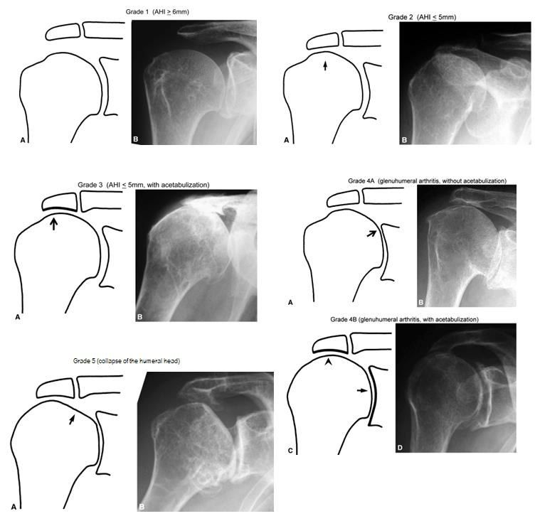
- Superior migration of the humeral head
- Acetabularization / femoralization
- Signs of loss of force couple in the shoulder
Q42Describe the Hamada classification of rotator cuff arthropathy.▸
- I: AHI >=6mm
- II: AHI <=5mm
- III: acetabulisation
- IV A: GHJ narrow, no acetabulisation
- IV B: GHJ narrow with acetabulisation
- V: head collapse
Q43What imaging is needed to work up rotator cuff arthropathy?▸
- Ask for axillary view XR to better appreciate GHJ arthritis
- MRI: massive rotator cuff tear with retraction of SS and fat atrophy
- CT for glenoid bone stock and version
Q44What history is important in rotator cuff arthropathy?▸
- Pain
- Stiffness
- Patient expectations
Q45What are the treatment options for rotator cuff arthropathy?▸
- Conservative: NSAID, periscapular muscle strengthening, deltoid training
- Arthroscopic debridement
- Reverse arthroplasty (C/I: young, poor glenoid bone stock, non-functional deltoid)
- Other: resection arthroplasty/shoulder fusion (salvage); hemiarthroplasty for pain, not function
Q46What did Gill et al (CORR 2021) conclude about aTSA vs rTSA for primary glenohumeral OA?▸
- No survivorship difference at 4 years between rTSA and aTSA
- Differences exist in revision risk between men and women
- Surgeons might select aTSA with an all-polyethylene glenoid to treat OA, despite the current popularity of rTSA
Q47What are the classical indications and prerequisites for reverse shoulder arthroplasty (RSA)?▸
- Elderly osteoporotic displaced 4-part fracture with comminuted tuberosities
- Cuff deficient (even 2 or 3 part fracture)
- Prerequisites: good glenoid bone stock, good deltoid power
Q48What are Grammont's principles of RSA?▸
- Medialised centre of rotation increases deltoid moment arm (effective lever arm from the start of movement) and recruits more deltoid fibres for FF/abduction; inferiorises to pretension deltoid
- Fixed centre of rotation decreases shearing force to the glenoid
- Reverse polarity has inherent stability: centripedal forces pass into the glenosphere; deltoid shear converted to compression
- Maintenance of external rotation (LD transfer); subscapularis + pectoralis major preserve IR
Q49What are the keys to success and the key steps of RSA?▸
- Keys: good exposure, correct sizing and version, stable fixation, satisfactory soft tissue tension
- Deltopectoral approach, biceps tenotomy, tag GT and LT fragments
- Retrieve humeral head, expose glenoid, clear labrum, guide pin, ream and screw baseplate
- Glenosphere with slight inferior overhang to prevent notching; trial
- Humeral retroversion 10 degrees; 3rd generation cementation; suture cuff to implant
Q50What are the complications of RSA and what is the Sirveaux classification?▸
- General: bleeding, NV injury, infection, haematoma
- Loosening (old lateral COR designs had high glenoid torque)
- Inferior scapular notching - Sirveaux classification: pillar, reach lower screw, cross inferior screw, central peg
- Dislocation; acromial fracture
- Cosmesis - more squaring; lengthening of upper limb; cemented - more infection
Q51Which classification is used for glenoid bone erosion in reverse shoulder arthroplasty?▸
- The Favard classification
Q52What patient and surgical factors increase notching in RSA?▸
- Patient: rotator cuff arthropathy with a narrowed AHI (76% vs 38% in post-traumatic cases)
- Glenoids with superior erosion (type E2 glenoid wear)
- MRI grade 3 or 4 fatty infiltration of the infraspinatus
- Surgical: anterosuperior approach higher than deltopectoral (86% vs 56%)
- Neutral or superiorly tilted baseplates
Q53How many histological layers make up the rotator cuff and what is each layer?▸
- Layer 1: 1mm, coracohumeral ligament (CHL) fibres
- Layer 2: 3-5mm, dense fibres parallel to the tendon
- Layer 3: 3mm, loose fibres at 45 degrees to the tendon
- Layer 4: thick collagen bands merging with CHL (rotator cable)
- Layer 5: 2mm, shoulder capsule
Q54Which layer of the cuff forms the rotator cable and what is it made of?▸
- Layer 4: thick collagen bands that merge with the CHL
- This structure is the rotator cable
Q55Which arteries supply the rotator cuff?▸
- Subscapular artery (Subscap A)
- Suprascapular artery (Suprascap A)
- AHCA and PHCA
Q56Between which layers of the cuff does the blood supply run?▸
- Between layers 2 and 3
Q57What is the critical zone of Codman?▸
- 1cm from the humeral tuberosity insertion
Q58Which side of the rotator cuff is more vascular and which side is stronger?▸
- The bursal side is more vascular
- The articular side has only half the strength of the bursal side
Q59On which side do most rotator cuff tears occur?▸
- Most tears are articular sided
Q60What did the MRI arthrogram show?▸
- Signal change/linear signal at the superior labrum, likely SLAP lesion
- Cystic lesion at the spinoglenoid notch
Q61What does SLAP stand for and what is it associated with?▸
- Superior Labrum from Anterior to Posterior tear
- Associations: cuff tear, shoulder dislocation
- Internal impingement (frequent thrower), GIRD
Q62Describe the Snyder classification of SLAP lesions.▸
- I: fraying of superior labrum
- II: biceps anchor detachment + labral fraying
- III: superior labrum bucket-handle tear
- IV: bucket-handle tear with extension into biceps
- V: SLAP + Bankart; VI: SLAP + labral flap; VII: SLAP + extension into MGHL origin
- Types 5-7 are the Maffet modification (total 7 types)
Q63What is the history and what examination tests are used for a SLAP lesion?▸
- Hx: deep shoulder pain, clicking; acute traction injury or recurrent throwing action
- PE: O'Brien's test, crank test, anterior shoulder instability
- SLAP lesion increases strain on the anterior band of IGHL
- Scapular dyskinesia
Q64Describe how the crank test is performed and what counts as positive.▸
- Sitting, arm flexed to 90 degrees; examiner adjacent to affected shoulder holds the flexed elbow/forearm
- Joint load applied along the axis of the humerus with one hand; the other hand performs humeral rotation while the shoulder is elevated in the scapular plane
- Can be repeated supine
- Positive if symptoms reproduced (usually pain), with or without a click, usually during external rotation
Q65What X-ray finding suggests internal impingement?▸
- Bennett lesion
Q66What is the function of the SLAP/biceps complex?▸
- Restraint to external rotation, anterior and inferior translation
- Depresses the humeral head in abduction and external rotation
Q67What are the normal labral variants and their frequencies?▸
- Normal 85%
- Sublabral foramen 12%: 1-3 o'clock, medial extension of contrast rather than lateral, smooth margin, <3mm, at biceps origin
- Buford complex (absent labrum + cord-like MGHL) 1.5%
- Sublabral foramen + cord-like MGHL 1%; meniscoid 1%
Q68What is the nerve supply of each rotator cuff muscle?▸
- Supraspinatus/infraspinatus: suprascapular nerve
- Teres minor: axillary nerve
- Subscapularis: upper and lower subscapular nerves
Q69Where can the suprascapular nerve be compressed and what is the effect?▸
- Suprascapular notch (suprascapular ligament): affects both SS and IS; suprascapular artery runs above the ligament; ganglion cyst from labral injury, fracture callus
- Spinoglenoid notch (spinoglenoid ligament): affects IS only; paralabral ganglion cyst from SLAP/posterior labral tear, traction injury in volleyball players
- Cyst at spinoglenoid notch: infraspinatus weakness (ER with arm at side) with no scar or winging
- Ddx: C5 palsy, brachial plexus injury
- Treatment: conservative; surgical excision of SOL/release of spinoglenoid ligament
Q70What will you look for on physical exam if there is a cyst at the spinoglenoid notch?▸
- Infraspinatus weakness (external rotation with the arm on the side) with no scar or winging
- Ddx: C5 palsy, brachial plexus injury
Q71What is the treatment and rehabilitation after a SLAP lesion?▸
- Conservative or operation: type 1/3 debridement, type 2 repair
- Type 4: debridement +/- biceps tenodesis if large biceps tendon involvement
- Rehab: 1-4 weeks active + passive assisted flexion in scapular plane; 4-12 weeks active ROM and isometrics; 12 weeks onwards strengthening
- Return to sports at 6 months
Q72What is the pathophysiology of calcific tendinitis?▸
- Local ischemia at codman zone -> fibrocartilaginous metaplasia -> CaHA deposition -> inflammation
- Resorption phase: CaHA dissolution and engulfment by macrophages
- HA - Alizarin red staining +
Q73Describe the Uhthoff classification of calcific tendinitis.▸
- Pre-calcific phase: fibrocartilaginous metaplasia leading to crystal deposition
- Calcific phase: formative, resting, resorptive (phagocytic action, most painful)
- Post-calcific phase: restoration of fibroblasts
Q74What is calcific tendinitis associated with and how is it investigated?▸
- Subacromial impingement, DM, hypothyroidism
- MRI to look for a cuff tear
Q75What is the treatment of calcific tendinitis?▸
- Conservative: NSAIDs, physiotherapy, steroid
- USG-guided aspiration and lavage (barbotage): 2 needles, one in one out
- Surgical decompression
Q76What do the photos show and what is the underlying pathology?▸
- Left: lateral winging of scapula -> trapezius weakness due to accessory nerve palsy
- Other cause of lateral winging: rhomboid weakness due to dorsal scapular nerve
- Right: medial winging -> serratus anterior weakness due to long thoracic nerve palsy
Q77What are the causes and management of medial scapular winging?▸
- Causes: overhead activity with head tilted to the other side; iatrogenic during anaesthesia
- Mx: observe for 6 months, then pect major transfer (pectoralis major transfer)
Q78What are the causes and management of lateral scapular winging?▸
- Causes: iatrogenic during lymph node dissection
- Mx: observe; if failed, Eden-Lange levator scapulae and rhomboid transfer (medial border to lateral border)