Sports
Shoulder instability and Bankart repair
Anterior dislocation mechanism, Bankart and HAGL lesions, radiographs and structure identification.
58 questions
7 source pages
4 images
Images appear with the first question taken from each source page — tap a question to open it.
Q1What history and examination findings are important in anterior shoulder dislocation?▸
- Age - redislocation rate by Rowe: <20 years >90%, <30 60%, >40 20%
- Cofield: 84% athlete vs 30% non-athlete
- Times of dislocation, mechanism, occupation and sport, comorbid NM conditions
- PE: direction of instability, generalized ligamentous laxity, associated injury (cuff tear, SLAP, axillary nerve palsy 5%), infraclavicular plexus injuries
Q2Which X-rays should you take and how are the special views performed?▸
- AP: fracture GT, Hill-Sachs lesion
- Axillary view / Velpeau axillary view
- Stryker notch (Hill-Sachs): supine, cassette under shoulder, palm on head, beam 10 degrees cephalic
- West point (Bankart): prone, shoulder on 7.5cm pad, arm 90 abducted, beam centred 25deg down and 25deg medial
- CT: glenoid inverted pear appearance, size of Hill-Sachs (70%), whether on or off track
Q3What soft tissue injuries are seen on MRI after anterior dislocation?▸
- Bankart (90%)
- Labroligamentous complex + periosteal tear: Perthes, ALPSA (medial reattachment), GLAD
- HAGL
- Cuff tear (30-80% depending on age)
- SLAP (7%)
Q4How do you reduce and immobilise an anterior shoulder dislocation?▸
- Reduction: traction-countertraction, Hippocratic, Stimson, Kocher (ER manoeuvre)
- Immobilise in IR for 2 weeks
- Itoi showed ER immobilisation decreased dislocation rate from 50% to 30%, but it could not be reproduced (compliance)
- AJSM 2015 meta-analysis: ER immobilisation no improvement, duration does not matter
- Cochrane review 2019: no difference
Q5Explain the glenoid track / on-off track concept and how it guides surgery.▸
- Glenoid track = contact point of glenoid and humeral head in functional abd + ER = 83% of glenoid width (0.83D - d)
- Hill-Sachs interval (HSI) = distance from infraspinatus attachment to medial rim of the Hill-Sachs lesion
- HSI larger than glenoid track = off-track (engaging) lesion
- Bipolar bone loss concept predicts the risk of dislocation from pathological engagement of the hillsach lesion with the glenoid bone loss
- Glenoid bone loss <25%: on track = Bankart; off track = Bankart + remplissage
- Glenoid bone loss >25%: on track = Latarjet; off track = Latarjet +/- remplissage
Q6Compare the Latarjet and Bristow procedures and describe screw fixation.▸
- Size: Bristow = coracoid tip; Latarjet = entire coracoid (anterior to CC)
- Position: Bristow <5mm from glenoid rim; Latarjet flush with rim
- Orientation: Bristow not rotated; Latarjet rotated 90 degrees (concavity along glenoid)
- Fixation: Bristow 1 screw; Latarjet 2 screws (4.0 cannulated or 4.5 cortical, below equator, <15 degrees divergent, within 5mm of rim)
- Effect: Bristow = checkrein; Latarjet = triple block (bone, conjoint tendon sling, capsule repair)
- Complications: nonunion, migration, cartilage injury, weak IR, stiff ER, lateral bone block -> OA
Q7What are the principles of definitive management in anterior shoulder instability?▸
- Definitive management depends on: patient's age and activity level, number of dislocations, and associated injury
- Aim: restore the bony and soft tissue stabilizers of the shoulder, solid post-op rehab for early return to sport
- C/I to surgery: multidirectional instability, uncontrolled epilepsy, voluntary dislocation
Q8How is an arthroscopic Bankart repair performed and when is Latarjet indicated?▸
- Arthroscopic Bankart repair restores the bumper + hammock effect of the labrum
- Latarjet if bony Bankart >25%, as soft tissue Bankart repair alone has up to 70% failure with recurrent dislocation
- Transfer coracoid with conjoint tendon (not CC ligament) through a vertical slit in subscap to the glenoid defect, fixed with screw anteroinferior
Q9What is the remplissage procedure and what are its complications?▸
- Indication: large (>25%) and engaging Hill-Sachs lesion
- Suture infraspinatus and posterior capsule to the Hill-Sachs lesion, making the lesion extra-capsular
- If >30%, may need bone graft or even osteotomy
- Complications: loss of ER (especially with Latarjet), stiff IR (posterior tightness), dehiscence (partial filling)
Q10What other soft tissue and bony procedures are used for anterior instability?▸
- Capsular shift if ligamentous laxity: tighten the anterior capsule, less ER, reduces the chance to engage (indirect)
- External osteotomy of the proximal humerus or fill with bone graft for a large Hill-Sachs lesion
- Subscapularis operations: Putti-Platt (double-breast subscap and anterior capsule), Magnuson-Stack (lateral subscap shift)
Q11What are the outcomes and indications after anterior instability surgery?▸
- Young + male more recurrence, but symptom-free in between
- If operated, recurrence 5%, may lose ER
- No difference in outcome if operated early (after 1st time) or later (after 3-4th time)
- Thus the common indications are apprehension and recurrent dislocation
Q12What is the Stanmore triangle classification of instability?▸
- 1. Structural, traumatic
- 2. Structural, atraumatic
- 3. Non-structural, muscle patterning
Q13What are the quoted association rates in anterior dislocation?▸
- Bankart: acute 94%, chronic 49%
- Hill-Sachs: 74%
- Cuff tear: 30% (40s), 80% (60s)
- Axillary nerve: 5%
- Glenoid track (Yamamoto JBJS 2007); on/off track (Di Giacomo JARS 2014)
Q14What is the instability severity index score (Boileau JBJS 2007)?▸
- Patient <20 years: 0/2 points
- Competitive sport, contact sport or forced overhead activity: 0/1 or 0/2
- Shoulder hyperlaxity: 0/1
- Hill-Sachs: 0/2, or loss of the sclerotic inferior glenoid contour: 0/2
- >6/10 = recurrence risk of 70%, so Latarjet is needed
Q15What is a Bankart lesion?▸
- Avulsion of the IGHL anterior band + torn periosteum
Q16What is a GLAD lesion?▸
- Glenoid labral articular defect
- Shearing off of a portion of articular cartilage together with the labrum
Q17What is an ALPSA lesion?▸
- Anterior labral periosteal sleeve avulsion
- Labrum-ligament complex detaches then reattaches medially and inferiorly
Q18What is a Perthes lesion?▸
- Labrum-ligament + periosteum stripped off in continuity and not reattached
Q19Describe the AP X-ray findings in posterior shoulder dislocation.📷▸
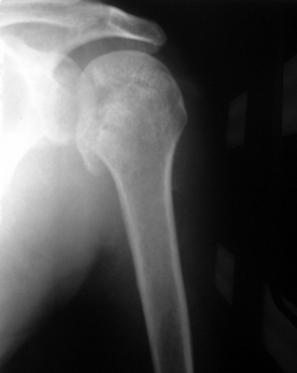
- Incongruent GHJ; disruption of Maloney's line (normal parabola)
- Light bulb sign: internal rotation with loss of humeral head profile
- Trough sign: impaction fracture over the anteromedial humeral head (reverse Hill-Sachs)
- Vacant glenoid/rim sign: medial humerus to medial glenoid clear space >6mm
- Reduced elliptical overlap; associated fractures; need scapular Y or axillary view to confirm
- Causes: trauma 50% (fall on adducted IR arm), convulsion, electrical shock
Q20What are the indications for open surgery and how is reduction performed?▸
- Initial management: ATLS/associated injury; less likely axillary nerve or cuff injury; Plain CT to look for indications for open surgery
- Major displaced LT fracture; large posterior glenoid fragment
- Reverse Hill-Sachs (anteromedial) engaged with posterior glenoid or >20% humeral head involvement
- Irreducible/impaction fracture
- CR under GA (usually muscular man) by Delpalma method (in-line traction, adduct + IR, lateral lift + ER)
- Intra-op check congruent reduction, stability and range; immobilise in ER and abduction
- Convert to open via deltopectoral approach; may address bone defect at the same time
Q21What special tests and MRI findings relate to posterior instability?▸
- MRI: cuff, reverse Bankart, reverse HAGL, POLPSA
- Kim test: seated, 90 abd + firm axial load, further 45 elevation with down/backward force; pain +/- clunk = posteroinferior labral tear
- Jerk test: 90 abd + 90 IR, axial humeral force then adduction to FF; clunk = posterior labral or posterior subluxation
- Posterior load and shift (under GA): 1 not to rim, 2 to rim, 3 beyond rim, 4 dislocation
Q22What are the surgical options for posterior instability?▸
- Indications: large bone loss, irreducible, unstable/recurrent instability
- Soft tissue (reduce intracapsular volume): reverse Bankart repair, closure of rotator interval (reverse remplissage)/inferior capsule
- Glenoid: bone graft if large reverse Bankart; posterior opening wedge osteotomy if excessive glenoid retroversion
- Humerus <30%: disimpaction + bone grafting, modified McLaughlin, McLaughlin, rotational osteotomy
- Humerus >30%: allograft, hemiarthroplasty
Q23How is a chronic (delayed more than 2 weeks) posterior dislocation managed?▸
- Pathology - bone: cartilage damage, bone loss, osteoporosis; soft tissue: capsulolabral injury, cuff tear, contracture, adherence to NV bundle
- <4 weeks: try gentle CR; >4 weeks: OR
- Humeral bone loss <40%: McLaughlin, cortical window to disimpact + bone graft, rotational osteotomy
- Humeral bone loss >40%: young = bone graft; old = hemi or RSA
- Glenoid loss >25%: reverse Bristow; redundant capsule: inferior/lateral capsular shift
- During open reduction do not do subscap tenotomy - reserve for McLaughlin
Q24Describe the McLaughlin procedure and its modification.▸
- Classically via deltopectoral approach
- A reverse remplissage: capsule + rotator interval + subscapularis filled into the defect
- Modified McLaughlin adds LT transfer: better bone healing, more secure fixation
Q25What are the primary stabilizers against posterior dislocation?▸
- Posterior band of the IGHL
- Subscapularis
- Coracohumeral ligament (CHL)
Q26How are posterior dislocations classified?▸
- By timing: acute vs chronic
- By underlying pathology: volitional, dysplastic, acquired
Q27What is the mechanism of inferior shoulder dislocation (luxatio erecta)?📷▸
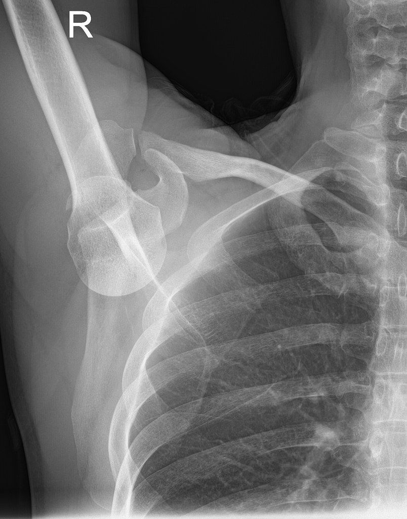
- Forceful abduction
- Levering the humeral head against the acromion out inferiorly
Q28What are the clinical features of inferior shoulder dislocation?▸
- Salute position: locked humeral head at 110-160 degrees abduction
- Palpable humeral head at lateral chest wall and axilla
- May buttonhole through the inferior capsule, necessitating open reduction
Q29What associated injuries must be assessed for in inferior shoulder dislocation?▸
- Cuff / labrum / IGHL injury
- Nerve: brachial plexus and axillary nerve injury
- Vascular: axillary artery and DCT injury
Q30How is closed reduction performed in inferior shoulder dislocation?▸
- Traction and countertraction
- Traction along the humeral shaft
- Countertraction: bedsheet across the superior shoulder/neck
- Gradual reduction with adduction
Q31What is the management of inferior shoulder dislocation?▸
- Mostly conservative
- Surgery for subcutaneous humeral head, NV injury, or recurrent dislocation
Q32What is the outcome after inferior shoulder dislocation?▸
- Groh JSES 2010 / Malon 1990: 83% excellent/good for range and pain
- NV injury NOT associated with outcome
Q33Which structures should you identify on this arthroscopic view?▸
- Long head of biceps and the biceps labral complex
- Glenoid and anterior labrum
- Subscapularis and the rotator interval
- Humeral head
Q34What is the anatomy and contents of the rotator interval?▸
- Space between the supraspinatus and subscapularis
- Contents: SGHL, coracohumeral ligament, long head of biceps, capsule
- Diseases related to the rotator interval: frozen shoulder (tightening), LHB dislocation
Q35What is frozen shoulder?▸
- Adhesive capsulitis: global loss of active and passive ROM
- Scarring and contracture of soft tissue (myofibroblast proliferation -> collagen type III), mainly involving rotator interval and coracohumeral ligament
- Presents with pain and global stiffness, especially limited ER
Q36What are the associations of frozen shoulder?▸
- Immobilisation of the shoulder
- Previous shoulder pathology (cuff tear, dislocation) or lung/breast/shoulder surgery
- DM (poor prognosis)
- Thyroid disease
- Dupuytren contracture
- Distal clavicle OT
Q37What are the Reeves stages and MRI findings in frozen shoulder?▸
- Painful phase: 3-9 months
- Frozen phase: 4-9 months
- Thawing phase: 5-26 months
- MRI: thickened IGHL
- Loss of joint volume (loss of axillary recess on arthrogram)
Q38How is frozen shoulder managed and what did the FROST trial show?▸
- Physio + injection (only in painful phase)
- MUA (FEAR: flexion-extension-abduction-rotation); cons: labral injury, fracture
- Arthroscopic capsular release after frozen phase: rotator interval release to gain ER, posterior capsule release to gain IR (270 degrees)
- FROST trial (Lancet 2020): early structured PT, MUA and arthroscopic release - none superior for pain and function at 12 months
- MUA is most cost effective; arthroscopic release carries increased risk
Q39Describe the Naviaser arthroscopic classification of frozen shoulder.▸
- Stage 1: erythematous/fibrinous synovium
- Stage 2: red, angry, thick synovium; thick contracted interval; tight joint space; adhesions in the inferior fold
- Stage 3: pink synovium
- Stage 4: no evidence of synovitis but persistent tight inferior fold and joint
Q40What is a HAGL lesion and why does it matter?📷▸
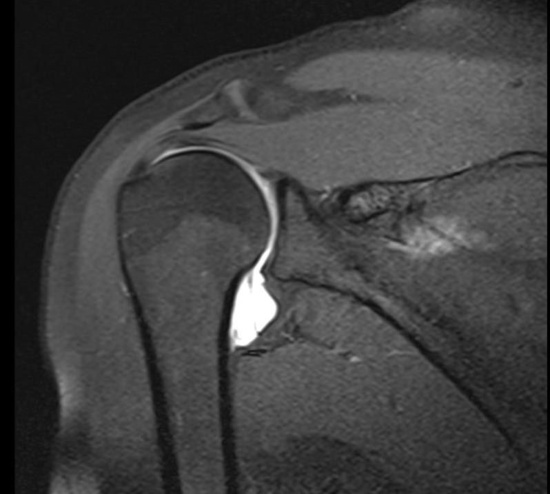
- Humeral Avulsion of the Glenohumeral Ligament
- Injury to the inferior glenohumeral ligament causing instability/pain
- Commonly a missed cause of recurrent shoulder instability
Q41Which part of the IGHL is involved and what are the patterns of failure?▸
- Anterior band most common (93%)
- Medial (glenoid) versus lateral (humerus)
- Failure at labral complex 40% > intrasubstance tear 35% > humeral insertion 25%
Q42What is the association of HAGL with instability and failed stabilization?▸
- 10% of recurrent anterior shoulder dislocators
- 20% of failed anterior stabilization
- 30% of shoulder instability patients without a Bankart lesion
Q43What MRI sign is seen in HAGL and what is the West Point classification?▸
- MRI: J sign at the inferior pouch
- West Point classification: posterior/anterior; bony/soft tissue; with or without Bankart
Q44What is the management and rehabilitation of HAGL?▸
- Conservative: 90% success
- Open or arthroscopic repair: similar outcome
- Rehab: anterior lesion limit ER; posterior lesion limit IR
- Good prognosis
Q45What does the popeye sign indicate?📷▸
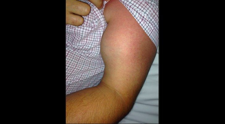
- Left: long head of biceps rupture with Popeye sign
- Right: reverse popeye sign = distal biceps tendon rupture
Q46What do you consider when managing a long head of biceps rupture?▸
- Age
- Power (supination)
- Pain due to biceps tendinitis
- Cosmesis
- Associated injury (rotator cuff tear)
Q47What are the physical examination findings in a long head of biceps rupture?▸
- Tenderness over the biceps groove
- Assess cuff power
- Elbow flexion and forearm supination power
Q48What are the physical examination findings of a distal biceps rupture?▸
- Hook test from lateral to medial (from medial blocked by lacertus fibrosis)
- Ruland biceps squeeze test
- Loss of biceps tracking with passive forearm rotation
- Supination loss 50%, flexion loss 30%, endurance loss 80-90%
Q49What factors are considered in managing a distal biceps tendon rupture?▸
- Age and function
- Power
- Cosmesis
- Chronicity of tear
- Degree of tear
Q50What are the risk factors for distal biceps tendon rupture?▸
- Steroid use and smoking
- Male, dominant hand
- Prexisting pain (radiobicipital bursitis)
Q51What investigations are performed for a distal biceps tendon rupture?▸
- XR to rule out avulsion fractures
- MRI: retraction and partial vs complete tear
Q52When is a distal biceps rupture treated conservatively versus operatively?▸
- Conservative in low functional demand (accepts 50% supination, 30% flexion loss, 80-90% endurance loss)
- Operative in young and fit patients to regain supination power
- Perform within weeks
Q53What are the treatment options for long head of biceps rupture?▸
- Tenodesis to proximal humerus (suture anchor/interference screw/keyhole procedure)
- Tenodesis: improve ~10% flexion power only, improve cosmesis
- Tenotomy for biceps tendinosis/subluxation with pain in a low-demand patient
- Approach: arthroscopic or mini-open
Q54How is distal biceps repair performed and what are the complications?▸
- Single incision: cubital fossa transverse incision (BR/PT); protect LABCN (exits between biceps and brachialis) and PIN by lateral retraction + supination; ligate recurrent radial artery lying superficial to biceps tendon; locate radial tuberosity; incise radiobicipital bursa
- Single incision: decreased risk of synostosis/HO but more LABCN injury
- Two incision: posterior approach at PL elbow (ECU/EDC, between Kocher and Kaplan); less dissection to antebrachial fossa, less radial nerve/PIN injury but more synostosis (disrupts IOM + ulnar periosteum)
- Complications: LABCN injury, SRN injury, PIN injury, synostosis, HO, proximal radius fracture
Q55What fixation options are used for distal biceps repair?▸
- Single incision: suture anchor / cortical button / bioabsorbable screw
- JBJS 2014 systematic review: lower complication rate with cortical button
- Double incision: pull-through suture
Q56What is the anatomy and blood supply of the biceps tendon relevant to rupture?▸
- LH biceps: further from the forearm rotational axis, supinator, inserts at proximal aspect of radial tuberosity
- SH biceps: flexor, inserts at distal aspect of radial tuberosity
- Blood supply: proximal brachial artery, distal posterior interosseous artery; watershed area at zone 2
Q57What are false positives for the hook test?▸
- False positive for positive hook test:
- Bicipital aponeurosis
- Partial tear
- Underlying brachialis tendon
Q58What are the biomechanical tolerances of the distal biceps tendon?▸
- Elbow at 90 degrees, no load: distal biceps sustains 50N
- Elbow at 90 degrees with 1kg load: 112N
- Force to rupture 200N
- Repair needs to withstand 50N