Sports
Radiographic interpretation of bone and joint lesions
Image-based questions on patellar, knee, talar dome and femoral radiographic abnormalities.
35 questions
5 source pages
4 images
3 fact-check flags
Images appear with the first question taken from each source page — tap a question to open it.
Q1What are the X-ray findings and additional view needed for osteochondritis dissecans of the talus?▸
- Lesion over medial talar dome with well defined radiolucent rim
- Fragmented bony lesion superficial to the rim, detached from talus but undisplaced
- No subchondral cyst, no loose body, no degenerative change, no other signs of instability including talar tilt or avulsion fractures from fibula
- Need Saltzman view (hindfoot alignment view, standing weight bearing, XR from behind 20 degrees caudally) to comment on hindfoot alignment
- AL lesions: more associated with trauma, lower spontaneous recovery, usually displaced + symptomatic; PM: more common, larger and deeper
Q2What are the differential diagnoses for a talar dome lesion?▸
- Ddx: GCT (giant cell tumour)
- Subchondral cyst
- Osteoid osteoma (most common site for OO in the foot)
Q3What are the classifications of talar OCD?▸
- Berndt and Harty (XR): I small subchondral compression, II partial fragment detachment, III complete detachment undisplaced, IV complete detachment displaced
- Frankel (CT): roof intact cyst, communication cyst, undisplaced lesion, displaced lesion
- Hepple (MRI): cartilage edema, cartilage injury +/- bone edema, undisplaced lesion, displaced lesion, subchondral cyst
Q4What history, examination and imaging are needed for talar OCD?▸
- History: trauma, premorbid and sports, pain, functional limitations
- PE: ankle ROM, tenderness, effusion, ligamentous stability (drawer test, syndesmosis), overall alignment
- CT (Ferkel) + MRI (Hepple) to delineate size, bony component and overlying cartilage integrity
- LL scannogram
Q5What is the conservative treatment and aims of surgery in talar OCD?▸
- Conservative: NSAID, rest, cast immobilisation and NWB 6 weeks (success rate 20-50%)
- Aims: relieve symptoms and improve function
- Aims of operation: restore joint congruity, increase local blood supply, Address predisposing factors such as malalignment and ligament instability
- Technique: Diagnostic scope then decide whether to repair, resurface, reconstruct
Q6What determines the choice of surgery in talar OCD?▸
- Site and size, displacement, overlying cartilage integrity
- Intact cartilage BH I or II --> retrograde drilling to stimulate growth
- Large and fixable --> fix with absorbable pin/headless screws
- Not fixable, cartilage not intact: contained --> chondroplasty/microfracture (cut off 2cm) +/- cartilage augmentation (AMIC/MACI/BMAC); uncontained --> OATS or mosaicplasty
- Wener Cartilage 2021: small stable conservative; healthy fragment fixation; necrotic excision + BMS; medium/deep autograft, BMAC, AMIC, ACI; large bone graft/allograft; failed biological - mini metal implants
- Overall quality of evidence poor: >90% of articles level III or IV
Q7What is the immediate management of a first-time lateral patellar dislocation?📷▸
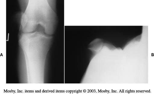
- CR under sedation (knee extended with medially directed force); X-ray shows laterally dislocated patella, no fracture or loose body
- First time: operate only if chondral lesion positive (with concomitant MPFL recon, Pedowitz AJSM 2019)
- Otherwise conservative: donut bracing, physiotherapy (VMO strengthening), ITB stretching, closed chain exercises
Q8What history and examination findings are relevant to patellar instability?▸
- History: age at first dislocation, traumatic or atraumatic, associated bone or connective tissue dysplasias, mechanical symptoms
- Owls eye patella, overall LL alignment, Q angle (M 8-10, F 15-20) vs carry angle of elbow (M 5-10, F 10-15)
- Effusion, quad wasting, ROM
- J sign (patella alta + maltracking), apprehension (push lateral at 20 degrees flexion), patella shift (>2 quadrants abnormal), patella tilt (should pass neutral), grinding test
- Rotational profile and ligamentous laxity
Q9What X-ray views and measurements are used in patellar instability?▸
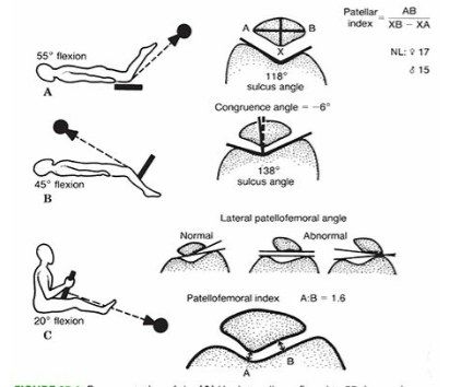
- Standing scannogram; lateral with knee flexed 30 degrees: Blumensaat line, Caton-Deschamps ratio 1.2, trochlear dysplasia signs (flat trochlea = crossing sign, convex = double contour + spur sign)
- Merchant view (knee flexed 45 degrees, caudal directed beam 60deg from horizontal): sulcus angle (N=138), congruence angle (N=-6-16)
- Laurin view (knee flexed 20 degrees, cephalad beam): lateral PFA (N = lateral opening 10)
Q10How are CT and MRI used and what predicts recurrence risk?▸
- CT: torsional malalignment, TTTG distance, trochlear dysplasia
- MRI: OC fracture in 40%
- Risk factors for recurrence: trochlear dysplasia, patellar alta, increased TTTG distance, patellar tilt
- PIS score (patellar instability severity score) >4 = high risk (age, bilateral instability, patellar tilt, patellar alta, TTTG distance, trochlear dysplasia)
Q11What are the types and anatomical causes of patellar instability?▸
- Congenital
- Acquired: acute, chronic, voluntary (party trick), habitual (dislocates every time with knee flexion within normal ROM, usually painless), recurrent (trivial sprain leads to dislocation with knee extension, painful)
- Alignment (miserable malalignment): coronal Q angle, sagittal knee recurvatum/patella alta, axial femoral anteversion/tibial torsion
- Bone: trochlear dysplasia, patella hypoplasia (Wiberg classification)
- Soft tissue: weak VMO, tight lateral retinaculum, MPFL deficiency, weak hip abductors; general ligamentous laxity
- TTTG 15mm in Asian, 20mm in Caucasian
Q12What are the surgical options for recurrent patellar dislocation and their complications?▸
- Principle: minimise destabilising forces while reconstructing bony and soft tissue restraints; recurrent --> MPFL reconstruction + correct risk factors
- Proximal: MPFL reconstruction, medial plication +/- lateral release/lengthening, VMO advancement; trochlear dysplasia --> trochleoplasty
- Distal: patella alta --> distal transfer of TT; increased TTTG --> medial transfer of TT; abnormal limb rotation --> derotational osteotomy
- Roux-Goldthwait (transfer lateral half of patellar tendon medially), Elmslie-Trillat (TT medialization), Maquet (anteriorization), Fulkerson (anteromedial + distal transfer)
- Complications: medial dislocation, medial PFJ arthritis, recurrence 5%, over-constraint leading to PFJ arthritis
Q13What is the normal function of the patella?▸
- Sesamoid bone
- Increases moment arm of quadriceps
- Aids cartilage nourishment
- Protects knee joint
Q14What is normal patella movement during knee flexion and extension?▸
- Extended: not engaged, most unstable as knee is locked in screw home mechanism with tibia ER (increases Q angle)
- Flexed 20 degrees: starts engagement, contact area moves proximal (increasing Q angle and stability)
Q15What are the details of MPFL reconstruction?▸
- The only anatomical option
- Drill through patella or suture anchor
- Interference screw to femur at the Schottle point (confluence of the posterior cortex extension line, blumensaat lines and the posterior curving line of the posterior femoral condyle)
- Graft type: gracilis (size good) or semitendinosus (longer); latest study: semiT has better outcomes
Q16What are the types of trochleoplasty and their complications?▸
- Dejour trochleoplasty (thick flap, need split): creates a gap beneath the trochlea surface, trochlea split longitudinally, two flaps collapsed into the gap and held with staples/screws/sutures
- Bereiter trochleoplasty (thin flap, no need split): elevates an osteochondral flap, thinned and moulded into a groove in the underlying bone created with osteotomes
- Resection trochleoplasty
- For Dejour B/D only; seldom done alone, technically demanding; complication: chondrolysis
Q17How do congenital/habitual patellar dislocation differ from traumatic instability?▸
- Pathology is lateral tightness and quadriceps contracture rather than a defective medial constraint
- Treatment: early relocation to allow trochlear development, TT osteotomy, MPFL imbrication/recon
- Extensive release of thick fibrous band tethering patella to lateral intermuscular septum; quadriceps VY plasty
- Congenital: look for syndromes - arthrogryposis, nail-patella syndrome
Q18How do you assess patella alta on a lateral X-ray?📷▸
- Blackburne-Peel index: ratio of perpendicular distance of patella articular surface to tibial plateau and patella articular surface (a/b); normal 0.8, alta >1.0, baja <0.5
- Caton-Deschamps: 0.6-1.3; >1.3 = alta, <0.6 = baja
- Insall-Salvati: patella tendon length / patella length (LT/LP); normal 0.8-1.2; alta >1.2, baja <0.8 (affected by patella and tibial tuberosity morphology)
- Blumensaat line: inferior pole of patella should sit on line drawn along superior aspect of intercondylar notch (rough guideline)
Q19What are the Dejour signs of trochlear dysplasia (based on CT)?▸
- A: crossing sign - sulcus too shallow and ended prematurely anterior to groove
- B: supracondylar spur - flat trochlea with lateral condyle higher than medial
- C: double contour due to lateral convex condyle
- D: spur + double contour cliff
Q20How is the Merchant's view taken and what do you measure?▸
- Knee flexed 45 degrees, beam aimed caudal
- Congruence angle: line connecting lowest pole of patella and lowest point of trochlear groove vs line bisecting the groove; normal should be negative 5 degrees (medial)
- Sulcus angle: lines along medial and lateral trochlear ridges; normal 138 degrees
Q21How is the Laurin's view taken and what do you measure?▸
- Knee flexed 20 degrees, beam aimed cephalad
- Lateral patellofemoral angle: line along top of femoral condyle and lateral facet of patella
- Normal 10 degrees (tilt)
Q22What do you look for on CT and MRI in patellar instability?▸
- CT: femoral version, trochlear dysplasia, lateral patella tilt, TTTG (lateral offset of tibial tuberosity from trochlear groove)
- MRI: MPFL injury (medial femoral epicondyle to superior medial patella)
- MRI: chondral lesion (usual medial patella and lateral femoral condyle, injured during relocation)
Q23What are the lateral knee X-ray findings in this patient with recurrent atraumatic haemarthrosis?📷▸
- Increased radiopacity over the suprapatellar region and posterior to the femoral condyles
- Well circumscribed, with no disruption of cortex, +/- nodular depression
- Cystic lesion over the femoral condyle with a sclerotic margin
- Mainly a soft tissue lesion
Q24What is the differential diagnosis for recurrent atraumatic haemarthrosis with these X-ray findings?▸
- PVNS (pigmented villonodular synovitis)
- Haemophilia
- Synovial chondromatosis
- Inflammatory synovitis
Q25What is pigmented villonodular synovitis (PVNS)?▸
- Idiopathic monoarticular neoplastic synovial disease
- Abundant proliferation of synovial villi and nodules
- CSF1 gene, genetic locus at chromosome 5q33; pexidartinib
- Can occur at any age; 50% have a history of trauma
- Presents with swelling and limited ROM; intra-articular
Q26How does the diffuse form of PVNS differ from the localized form?▸
- Both forms are most common in the knee
- Diffuse: more extra-articular involvement
- Diffuse: more likely to be painful
- Diffuse: less well circumscribed on MRI
- Diffuse: higher recurrence (30% vs 8% after synovectomy)
Q27What are the MRI and histological features of PVNS?▸
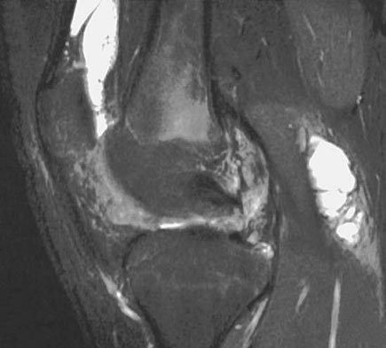
- MRI: low signal on both T1 and T2
- MRI: blooming artifact on gradient-echo sequences due to iron in hemosiderin (haemosiderin deposition)
- Histology: haemosiderin-stained multinucleated giant cells
- Mononuclear stromal cells; highly vascular villi with hyperplastic synovial cells
- Pigmented foam cells (lipid-laden histiocytes)
Q28What is the management of PVNS?▸
- Nearly no role for conservative management
- Total synovectomy (marginal excision): open, arthroscopic or combined
- Arthroscopy: brownish or reddish inflamed synovium with frond-like papillary projections
- + Irradiation reduces recurrence to 10-20% (30-35Gy in 15 fractions, or 50Gy in 25 fractions)
- + TKI (e.g. imatinib)
- Hip PVNS: joint damage occurs early, so perform synovectomy early
Q29How would you clinically assess a patient with recurrent atraumatic haemarthrosis of the knee?▸
- PE: confirm swelling, exclude a mass, document ROM
- Examine the contralateral limb for involvement
- Further imaging: MRI with gradient echo sequence
- MRI shows blooming effect over the area of concern, also involvement posterior to the knee
Q30What is the extra-articular equivalent of PVNS?▸
- Giant cell tumour of the tendon sheath
Q31Describe the arthroscopic portals used for PVNS of the knee.▸
- Posteromedial portal: 1cm above the joint line behind the MCL
- Posterolateral portal: 1cm above the joint line between the LCL and biceps tendon
Q32Describe the X-ray findings shown in this section.📷▸
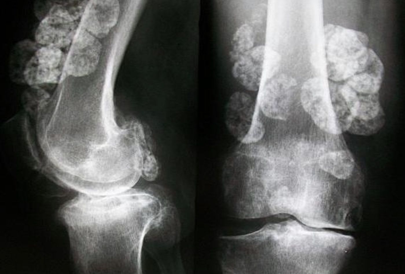
- Multiple well defined ossifications surrounding the right hip and knee joints
- Calcifications are less well defined in the hip
- No associated bone erosions
Q33What is the differential diagnosis for periarticular ossification/calcification?▸
- Dystrophic (vascular, tumour, infection, haematoma): synovial chondromatosis, synovial sarcoma, myositis ossificans
- Metastatic: renal osteodystrophy
- Calcinosis / tumoral calcinosis
Q34What is synovial chondromatosis?▸
- Proliferative disease of the synovium associated with cartilage metaplasia
- Results in multiple intra-articular loose bodies
- MRI: low T1, high T2
Q35How is synovial chondromatosis managed?▸
- Surgery as soon as possible
- Removal of loose bodies
- Synovectomy reduces recurrence rate
- Approach: posterior knee portal or open
Fact check
Merchant's view is taken with the knee flexed 45 degrees and the beam directed caudally 60 degrees from horizontal — error — Merchant's original technique inclines the beam 30 degrees from the horizontal (equivalent to 60 degrees from vertical), knee flexed 45 degrees — source
Normal congruence angle on the Merchant view is -6 to 16 — misleading — Normal mean congruence angle is -6 degrees; values greater than +16 degrees are abnormal (lateral subluxation), so -6 and 16 are not the ends of a normal range — source
PVNS is linked to the CSF1 gene on chromosome 5q33 — wrong chromosome/locus — CSF1 is on chromosome 1p13; PVNS/TGCT is driven by t(1;2)(p13;q35) fusing CSF1 to the COL6A3 promoter on 2q35. Loci at 5q22-31 are only uncommon alternative translocation partners. — source