Sports
Knee dislocation - recognition and reduction
Clinical and imaging recognition of knee dislocation and closed reduction of the subluxed knee.
31 questions
4 source pages
1 images
2 fact-check flags
Images appear with the first question taken from each source page — tap a question to open it.
Q1Describe the technique and prerequisites of the pivot shift test.▸
- IR tibia at knee extension
- Valgus force + gradual knee flexion
- Sudden glide/jump/clunk = reduction of the tibia relative to the femur
- + axial load to exaggerate
- Prerequisites: intact MCL and intact ITB
Q2What is the principle of the pivot shift?▸
- IR of the tibia exaggerates ACL deficiency (especially the PL bundle)
- Valgus stress tightens the MCL -> decreases medial tibial plateau movement -> lateral tibial plateau translates forward
- Sudden reduction by the ITB on knee flexion (axis of pull posterior to knee COR/TEA -> ITB becomes a flexor)
Q3What are the grades of pivot shift?▸
- 1: glide (may be physiological)
- 2: sublux with spontaneous reduction (jump)
- 3: subluxation +/- spontaneous reduction (clunk)
Q4What does a high-grade pivot shift indicate?▸
- Isolated ACL tear is unable to produce a high-grade pivot shift
- Meniscus: LM root tear, ramp lesion, MM posterior horn tear
- Anterolateral complex: ITB (superior and deep), ALL, AL capsule - secondary restraint to anterior translation and IR (with LM)
- Generalised ligamentous laxity (Beighton score >=5/9)
Q5What is a RAMP lesion and how is it classified (Thaunat)?▸
- 9-17% of ACL tears
- 1: capsulomeniscal junction lesions - very peripheral, low mobility at probing
- 2: partial superior lesions - stable, diagnosed only by the trans-notch approach (modified Gillquist manoeuvre)
- 3: partial inferior/hidden - not visible trans-notch, high mobility (meniscotibial ligament disruption)
- 4: complete tear in the red-red zone - very high mobility
- 5: double tear
Q6What does the clinical photo show and what is the dimple sign?📷▸
- Posterior displacement of the tibia with lateral translation
- Dimple sign: puckering of anteromedial skin from the medial femoral condyle buttonholing through the medial retinaculum/capsule
- Indicates a posterolateral dislocation
- Contraindication to closed reduction (would cause skin necrosis)
Q7How do you assess a knee dislocation acutely?▸
- ATLS protocol, AMPLE history, primary survey
- Local: rule out hip dislocation and open wound
- Distally document pulse, CR, neurology and compartment syndrome
- Systemic: secondary survey to rule out other injuries
- Recheck pulse and document nerve status after any reduction
- If stable, arrange CTA to look for an intimal tear
Q8What associated injuries occur with knee dislocation and when is an ex-fix indicated?▸
- Direct vascular injury in 10-30%
- CPN injury in 10-40%, with complete recovery in only 20%
- Ex-fix if fracture dislocation, significant soft tissue compromise/open injury, vascular compromise or compartment syndrome
Q9Scenario 2: dislocated knee with an absent pulse - what is your management?▸
- Analgesia, immediate closed reduction under sedation, splint with knee flexed 20-30 degrees, then recheck pulse
- Pulse returns: CT angiogram or serial ABI monitoring
- No return of pulse: vascular bypass, ex-fix knee, on-table angiogram, resect diseased segment and re-anastomose with reversed saphenous vein graft
- Delay >8 hours gives >80% amputation rate
- Prophylactic fasciotomy (EFFORT review 2020); Postop monitor CK for rhabdomyolysis and reperfusion syndrome
Q10What signs suggest a knee dislocation that has spontaneously reduced?▸
- 20-50% reduce spontaneously before presentation
- Hyperextension compared with the normal side
- Popliteal ecchymosis
- NV injury at presentation
- Diffuse tenderness but NO effusion
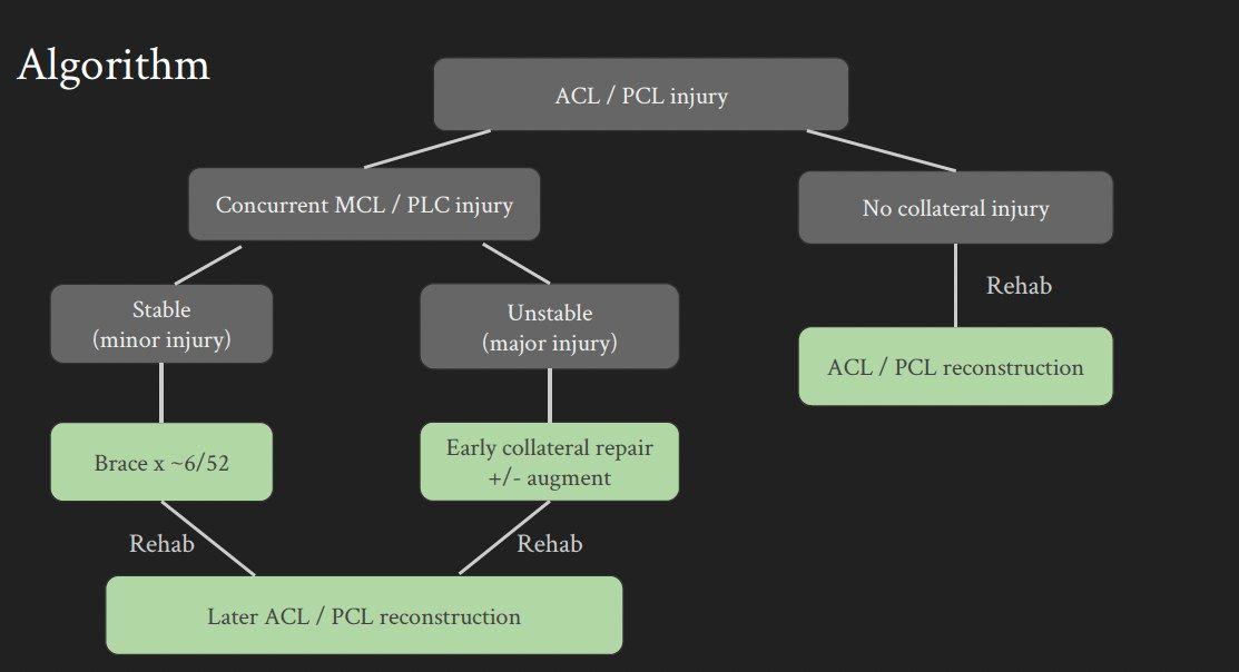
Q11Describe the definitive (staged) management of a multiligamentous knee injury.▸
- Aim: stable painless knee, full ROM, return to pre-injury activity
- Stage 1 at 2 weeks: PLC, PMC, MCL and LCL, meniscus +/- avulsion fracture fixation; hinged knee brace + NWB for 6 weeks
- Stage 2: delayed simultaneous ACL and PCL reconstruction (PCL before ACL), after good ROM and muscle bulk restored
- If vascular injury: no tourniquet, delayed surgery, inlay PCL
Q12Scenario 1: posterolateral dislocation with intact NV status - what is your management?▸
- Acute: analgesics, immobilise with a slab then proceed to open reduction
- Medial parapatellar approach; reduce then check stability + NV condition
- Then slab or external fixator
- Avoid closed reduction in posterolateral dislocation (dimple sign - risk of skin necrosis)
Q13Scenario 3: dislocated knee with a palpable pulse - what is your management?▸
- Analgesia, immediate closed reduction under sedation + splint with knee flexed 20-30 degrees, then recheck pulse
- Check symmetry of pulse and ABI
- ABI >0.9: serial monitoring; ABI <0.9 and asymmetric: CTA
- If CTA shows an intimal tear: anticoagulant, monitor, delayed OT for ligamentous reconstruction, no tourniquet, no tibial tunnel
- +/- MRI to document ligamentous laxity; classify with Schenck
Q14How do you classify a knee dislocation?▸
- Kennedy: by direction - anterior/posterior/medial/lateral/rotational
- Schenck: by number of ligaments involved + fracture
- I = single cruciate; II = bicruciate; III = bicruciate + one collateral tear; IV = all 4; V = fracture
Q15How do you reduce a knee dislocation other than posterolateral?▸
- CR under sedation, stabilise the femur and pull the tibia opposite to the direction of displacement
- Document NV status afterwards
- Apply a slab in 20-30 degrees knee flexion or an ex-fix
Q16Why is a staged approach used in multiligamentous knee injury?▸
- Shorter OT time and each stage is simplified
- Reduces the risk of arthrofibrosis
- Delay definitive OT if vascular injury is present
- Stage 1 addresses the capsular tear (risk of compartment syndrome) and soft tissue status
Q17Compare the graft choices for multiligamentous knee reconstruction.▸
- Autograft: no disease transmission, no added cost, documented healing/vascularisation; cons - donor site morbidity, longer OT
- Allograft: no donor site morbidity, shorter OT, does not further destabilise the knee; cons - availability, disease transmission, cost, biomechanics
- Synthetic: no donor site morbidity, shorter OT, readily available; cons - reactive synovitis, graft failure
Q18What are the controversies in operative treatment of knee dislocation?▸
- Timing: Hohmann Knee 2017 metanalysis - early surgery <3 weeks improved Lysholm scores compared with delayed surgery
- Sheth 2019 ISAKOS J - early surgery may provide better functional outcomes without compromising ROM when using early postop mobilisation protocols
- Mook 2009 systematic review JBJS - early <3 weeks vs staged: similar need for additional treatment for arthrofibrosis, both significantly higher than delayed; delayed >3 weeks had less residual instability and ROM deficit; staged had highest excellent/good outcomes
- Jiang 2015 KSSTA - staged operation yields the best clinical results for KDIII; Marder 2021 - neither approach superior
- Repair vs reconstruction: Stannard 2005 PLC repair failure 37% vs 9% with reconstruction; Levy 2010 40% vs 6%
- Laprade AJSM 2019 –suggest single stage anatomic based recon of all ligament (single surgeon series); bicruciate reconstruction - simultaneous preferred at PWH
Q19Describe the posterior approach to the knee.▸
- Incision: lazy S, proximal limb lateral, distal limb medial
- Open the popliteal fascia at opening of SSV and MSCN (small saphenous vein and medial sural cutaneous nerve), staying lateral to the MSCN
- Trace the MSCN proximally to protect the popliteal AV bundle
- Protect the common peroneal nerve along the posterior edge of biceps femoris
- Retract popliteal vessels laterally; ligate superior medial and middle geniculate arteries; elevate popliteus +/- soleus
Q20Define knee dislocation and explain why it is an orthopaedic emergency.▸
- Not covered in the speaker notes - the source image is the only source
Q21How is knee dislocation classified (Kennedy and Schenck)?▸
- Not covered in the speaker notes
Q22Which ligaments must be assessed in knee dislocation?▸
- Ligaments to assess: ACL, PCL, MCL, LCL, PLC
Q23What are the key complications of knee dislocation?▸
- Not covered in the speaker notes
Q24When would you suspect a knee dislocation?▸
- Multi-ligamentous injury (defined as two or more of ACL, PCL, PMC, PLC)
- High energy trauma
- Distal NV compromise
- 20-50% have spontaneously reduced by presentation
Q25What are the common mechanisms of injury causing knee dislocation?▸
- Usually high energy e.g. RTA; can occur with relatively low energy injury in athletes
- Anterior (most common): LCP, intimal tear by traction
- Posterior (2nd commonest): axial loading to flexed knee, complete tear of popliteal artery
- Lateral: varus/valgus force, bicruciate tear + peroneal nerve
- Medial: varus/valgus force, bicruciate tear
- Rotational: irreducible
Q26How do you classify knee dislocation?▸
- Kennedy: by direction (anterior/posterior/medial/lateral/rotational)
- Schenck: by number of ligaments involved + fracture
- I = single cruciate; II = bicruciate; III = bicruciate + one collateral tear; IV = all 4; V = fracture
Q27Why is the popliteal artery prone to injury in knee dislocation?▸
- Incidence quoted as 20-60%
- Tethered proximally by the adductor hiatus (trifurcation) and inferiorly by the soleus arch
- Tied down medially and laterally by the geniculate arteries
- Also tethered by the 5 geniculate arteries
Q28How do you investigate for vascular injury in knee dislocation?▸
- Document pulses; AAOS: ABI >0.9 admit + observe 24 hours, <0.9 angiogram, ischaemia explore
- Pulse -ve -> reduce -> pulse returns -> CTA
- Pulse -ve -> reduce -> pulse remains -ve -> vascular surgeon + OT
- Pulse +ve -> ABI <0.9 -> CTA +/- explore
- Pulse +ve -> ABI >0.9 -> serial monitoring for at least 72 hours
- Notes state ABI 100% sensitive, NPV 100%
Q29What is the timing of surgery for vascular injury and how is an intimal tear treated?▸
- Emergency operation
- Ischaemic time >8hr = 90% amputation rate; <6hr = 6%
- Intimal tear: anticoagulation
- Subsequent operation: pre-op no tourniquet, delayed surgery, no tibial tunnel
- Nerve injury: ~30% peroneal nerve by traction, fair prognosis; explore if open wound, otherwise conservative first
Q30Describe the approach and key steps of the posterior approach to the knee.▸
- GA, prone with bony prominences padded
- Lazy S incision with proximal limb lateral and distal limb medial to avoid the peroneal nerve
- Distally find the small saphenous vein and medial sural cutaneous nerve
- Trace proximally to reach the popliteal vein and tibial nerve in the popliteal fossa
- Incise the popliteal fossa longitudinally lateral to the vein
Q31What are the borders and contents of the popliteal fossa?▸
- Diamond-shaped space over the posterior knee
- Bounded by semitendinosus/semimembranosus, biceps femoris and the 2 heads of gastrocnemius
- Floor is the posterior capsule of the knee; roof is the popliteal fascia
- Most superficial structure is the tibial nerve
Fact check
ABI is 100% sensitive with a 100% negative predictive value for vascular injury after knee dislocation — overstated/contested — Mills et al (2004) reported 100%, but later cohort data show no single physical examination manoeuvre is 100% sensitive; a normal ABI can still miss intimal tears. Combine palpable pulses plus ABI >=0.9 and continue serial observation (48-72h). — (medium confidence) — source
Popliteal artery injury occurs in 20-60% of knee dislocations — imprecise range — Modern systematic reviews report weighted mean popliteal artery injury rates of about 8-18%; rates at the upper end (up to 60%+) are seen mainly in high-energy trauma and fracture-dislocations. — (medium confidence) — source