Sports
PCL, PLC and posteromedial corner injuries
Posterior cruciate and corner injuries with avulsion ossification signs such as Stieda-Pellegrini.
34 questions
4 source pages
3 images
Images appear with the first question taken from each source page — tap a question to open it.
Q1What are the aims of assessing a PCL injury?▸
- Assess severity
- Rule out associated injury
- Formulate management plan (also depends on patient's functional demand)
Q2What are the usual injury mechanisms of PCL injury?▸
- Hyperextension/hyperflexion
- RTA dashboard injury (look for femoral neck fracture)
- Varus force on a flexed knee
- Fall onto the ground with a plantar-flexed foot
Q3What positive physical signs should be elicited in PCL injury?▸
- Loss of medial tibial step-off with knee 90 degrees flexion
- Posterior sagging grading relative to medial femoral condyle: anterior, flush, posterior
- Posterior drawer grading: <5mm, 5-10mm, >10mm with no endpoint; rule out PLC in high grade
- Quadriceps active test: flex knee 60 degrees, stabilise foot and active extend knee
Q4What tests indicate an associated PLC injury?▸
- Dial test at 30/90 degrees (positive at 90 = combined injury, 30 only = PLC)
- Recurvatum ER test: varus = LCL injury, ER = fabellofibular/popliteus tendon, recurvatum = PCL
- Posterolateral drawer test
- Reverse pivot shift - 26% sensitivity, 95% specificity
- Gait: lateral thrust
Q5How is the reverse pivot shift performed and how does it work?▸
- Begins in flexion with ER and valgus stress applied, so tibia subluxes posteriorly
- Knee is then brought to full extension (reverse)
- ITB changes from flexor to extensor at ~30 degrees flexion, causing relocation (same principle)
- Sensitivity 26%, specificity 95%
Q6What imaging is used in PCL injury and how is stress view graded?▸
- XR: avulsion fracture
- Stress view (posterior tibial cortex to posterior end of Blumensaat line at knee flexion 70 degrees) with kneeling or Telos
- 3-8mm = partial, 8-12mm = complete, >12mm = PLC injury
Q7What associated injuries must be assessed in PCL injury?▸
- PLC injury
- Multiple ligament injury
- Knee dislocation
- Posterior hip dislocation
Q8Describe the anatomy of the PCL.▸
- 38x13mm, 30% larger than ACL; intracapsular, extrasynovial ligament
- From MFC anterolateral surface (broad crescent-shaped footprint) to posterior tibial sulcus (1cm below joint line)
- Bundles: AL (easier to rupture, tight at flexion) / PM; AL anterior to PM at femoral insertion
- Meniscofemoral ligaments: Humphrey (anterior) and Wrisberg (posterior), contribute 20-30% stability
- Type I collagen; blood supply middle geniculate artery; nerve supply posterior articular branch of tibial nerve
- Primary restraint to posterior translation; secondary restraint to ER and varus; strength 2500-3000N
Q9Why does the PCL heal better than the ACL?▸
- Mechanical: increased stability from femoromeniscal ligaments
- Biological: thick synovium sheath --> usually still extrasynovial after rupture (hematoma formation, no cytokine)
- ACL is also an intracapsular, extrasynovial ligament
Q10What is the natural history of isolated PCL injury?▸
- Shelbourne 1999: 50% same sport, 30% lower level, 20% unable to play sport
- Complication: anterior knee pain (PFJ arthritis)
- Newer evidence: Agolley (prof Fares Haddad group) BJJ 2017 91.3% return to same level or higher
Q11What is the rehab protocol for non-operative management of PCL injury?▸
- Grade 1-2: PWB + quad strengthening
- Grade 3: extension brace (Donjoy/posterior support) for 4-6 weeks, no flexion >50 degrees (PCL starts to take up tension around 50 degrees flexion)
- PWB walking + quads isometric exercises
Q12What is the indication for surgery in PCL injury?▸
- Avulsion or peel-off lesion
- Multiple ligament injury / concomitant PLC injury
- Persistent symptoms (difficulty decelerating e.g. downstairs, PFJ pain) - observe 6-12 months
Q13What factors are considered in PCL reconstruction?▸
- Type of graft (hamstring / BPTB / quadriceps + bone)
- Single/ double bundle: single bundle less demanding (reconstruct AL, tension at 90 degrees knee flexion)
- Chahla Arthroscopy 2017 systematic review - double bundle better IKDC score and posterior tibial translation/knee stability
- Inlay vs all-arthroscopic
Q14Describe the all-arthroscopic transtibial PCL reconstruction technique.▸
- Supine on tourniquet; harvest graft
- Conventional portal + PM portal (injury to saphenous nerve)
- PM portal dissection of posterior capsule (protect popliteal artery)
- Drill tibial tunnel A to P, last few cm use hand drill, knee in 90 degrees flexion
- Tibial tunnel 15mm distal to articular surface --> less killer turn
- Femoral tunnel 7mm from articular surface at 1pm/11pm (shorter than ACL tunnel; consider aperture fixation if too short); tension at 90deg flexion with anterior drawer test
Q15What are the pros and cons of all-arthroscopic versus inlay PCL reconstruction?▸
- All-arthroscopic pros: single position, can use hamstring graft; cons: difficult to pass graft, killer turn
- Inlay pros: avoid killer turn (less attrition), direct visualisation of NV bundle, better healing with bone union, biomechanically better
- Inlay cons: floppy lateral position, less graft choice (BPTB or quadriceps tendon + patella bone with bone peg)
Q16What is the post-op rehabilitation after PCL reconstruction?▸
- Hinged knee brace locked in extension for ambulation and sleep for 6 weeks
- Protect against gravity
- Early motion in prone position
- Quad strengthening; no open chain hamstring or isolated hamstring exercise for 3 months
Q17What are the static and dynamic structures of the PLC and their roles?📷▸
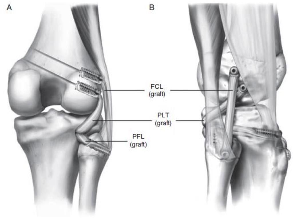
- Static (6): LCL, popliteus, popliteofibular ligament, fabellofibular ligament, arcuate ligament, capsule
- Dynamic: ITB, biceps femoris, lateral gastrocnemius
- Primary restraint to ER and varus; Secondary restrain to posterior translation
Q18What is the layer arrangement of the PLC?▸
- Layer 1: ITB/biceps femoris (CPN in between)
- Layer 2: retinaculum
- Layer 3 superficial: LCL/FFL/ALL (LGA in between)
- Deep: arcuate/popliteal/PFL/capsule
Q19What is the anatomy of the fibular head insertion and structures to the fabella?▸
- Fibular head, anterior to posterior: LCL, biceps femoris (long then short), arcuate ligament, popliteofibular, FFL
- Structures to fabella: capsular arm of short head of biceps, FFL, OPL, lateral gastrocnemius head, posterior lateral capsule
Q20What are the common mechanisms, symptoms and physical signs of PLC injury?▸
- Mechanism: blow to anteromedial knee, varus blow to flexed knee, hyperextension injury
- Symptoms: instability in knee extension and upon pivoting
- PE: dial test positive if >15 degrees difference, ER recurvatum, posterolateral drawer with foot 15 degrees ER, reverse pivot shift
- (Peroneal nerve injury in 25% of patient with PLC injury)
Q21What are the X-ray findings and varus stress grading in PLC injury?▸
- Avulsion fracture (arcuate fracture) of fibula head
- Varus stress (Laprade): <4mm = isolated PCL, 4-7mm = LCL + PCL, >7mm = LCL + PLC + XL
- Long leg standing XR: malalignment, determine mechanical axis +/- need for HTO
Q22What is the Fanelli classification and conservative management of PLC injury?▸
- Fanelli A: increase ER only (PFL + popliteus)
- Fanelli B: increase ER + varus laxity in knee flexion (above + LCL)
- Fanelli C: increase ER + varus laxity in flexion and extension (above + ACL/PCL)
- Conservative: isolated grade 1-2 injury - extension knee brace + protected weight bearing x2 weeks, then quad strengthening
Q23What are the indications for surgery, reconstruction options and evidence for repair vs reconstruction?▸
- Indications: bony avulsion, multiple ligament laxity, persistent symptoms, grade 3 injury (assess in MUA)
- Unstable (varus stress >7mm) + within 2 weeks of injury --> early repair then later staged recon; stable --> brace 6 weeks then one-stage recon (Laprade favours single stage early recon of PCL and PLC)
- Reconstruct LCL and popliteofibular ligament; graft: semitendinosus/achilles
- Larson: non-anatomical fibular-based figure of 8; LaPrade anatomical recon with 2 soft tissue grafts (LCL+PFL and popliteus); transtibial double bundle fixation
- CUHK arthroscopy paper: LaPrade technique gives better rotational stability
- Recon better than repair: Stannard failure 37% vs 9%; Levy 40% vs 6%; Geeslin AJSM 2016 metanalaysis – recon better outcomes than repairs in acute PLC injuries (meta-analysis)
Q24What is the Stieda Pellegrini sign?📷▸
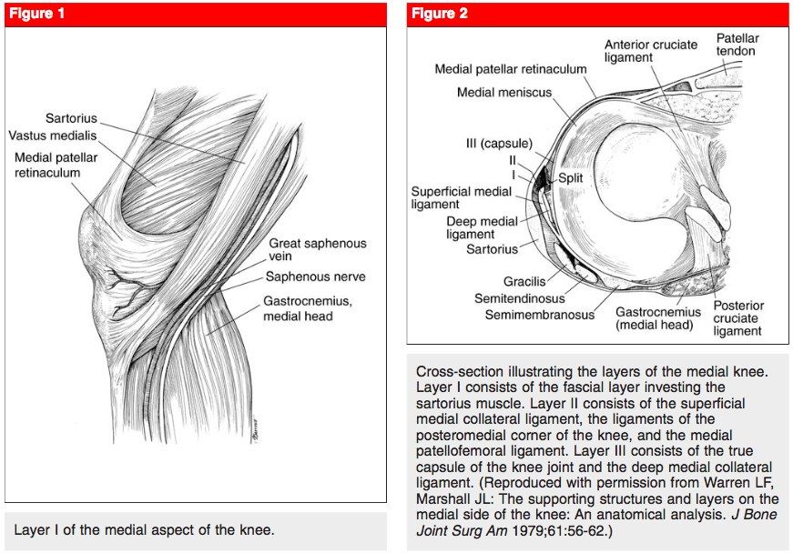
- Ossified lesion seen a few weeks after injury
- Signifies avulsion injury of MCL at the medial femoral condyle
Q25What is the function of the MCL and the usual mechanism of injury?▸
- Primary restraint to valgus stress
- Secondary restraint to ER, ant/ post translation (anterior/posterior translation)
- Usual mechanism: valgus + ER
Q26What are the layers of the medial side of the knee?▸
- Layer 1: sartorius and fascia, patella retinaculum (gracilis, semitendinosus, saphenous nerve)
- Layer 2: superficial MCL, POL, OPL, MPFL, semimembranosus
- Layer 3: deep MCL, capsule
Q27How is MCL instability graded and where do tears most commonly occur?▸
- Medial gapping compared with opposite knee: <5mm, <10mm, >=10mm
- Grade 1: stretch injury (no loss of ligamentous integrity)
- Grade 2: incomplete tear (fibres opposed, endpoint at 30 degrees flexion valgus test)
- Grade 3: complete tear (no endpoint)
- Most common: femur side tear
Q28When is operative management indicated in MCL injury?▸
- All try conservative management (hinged brace) first
- Grade III with: acute multiligamentous injury; acute displaced distal avulsion with Stener-like lesion (trapped torn end); subacute/chronic with persistent instability
- Femoral origin tears have better healing potential
Q29How is a grade 3 MCL tear with ACL tear managed?▸
- Early MCL repair (within 2 weeks) + delay ACLR
- Or conservative Mx for MCL + delay ACL reconstruction (MCL needs protection, which may cause stiffness)
Q30What are the classifications and reconstruction options for MCL tear?▸
- Taketomi (tibial side tear): under pes, Stener-like lesion, trapped inside joint
- Nakamura: type 1 femoral, type 2 tibial (none in his study), type 3 femoral with extension below the joint line
- Reconstruction: Bosworth (semitendinosus, distal intact, attached to femoral tunnel), Lind (gracilis looped over MCL), LaPrade
- Acute reconstruction is controversial
Q31What is the definition and what are the components of the posteromedial corner?📷▸
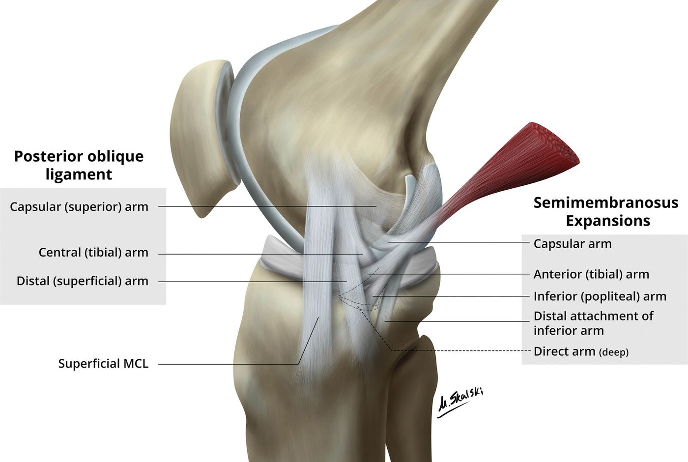
- From posterior margin of superficial MCL to PCL (does not include MCL)
- Components (2nd layer): semimembranosus expansion (5 arms: capsular, anterior, inferior, distal, direct)
- POL (ligament of Winslow), OPL, deep capsule, MM PH
Q32What is the role of the posteromedial corner?▸
- Dynamic
- Secondary restraint to ER
- Restraint to valgus stress in extension
Q33What is anteromedial rotatory instability (AMRI)?▸
- Medial tibial plateau anterior subluxation upon ER
- Signs: valgus stress + at 30 degrees ER
- Anterior drawer positive at 30 degrees ER
Q34When is early repair of the posteromedial corner indicated?▸
- In multiligament injury