Sports
Paediatric ACL injury and skeletal maturity
Paediatric ACL tears, physeal-sparing options and treatment in the skeletally mature patient.
20 questions
2 source pages
1 images
1 fact-check flags
Images appear with the first question taken from each source page — tap a question to open it.
Q1When is surgery indicated in a paediatric ACL tear and what is the evidence on delay?📷▸
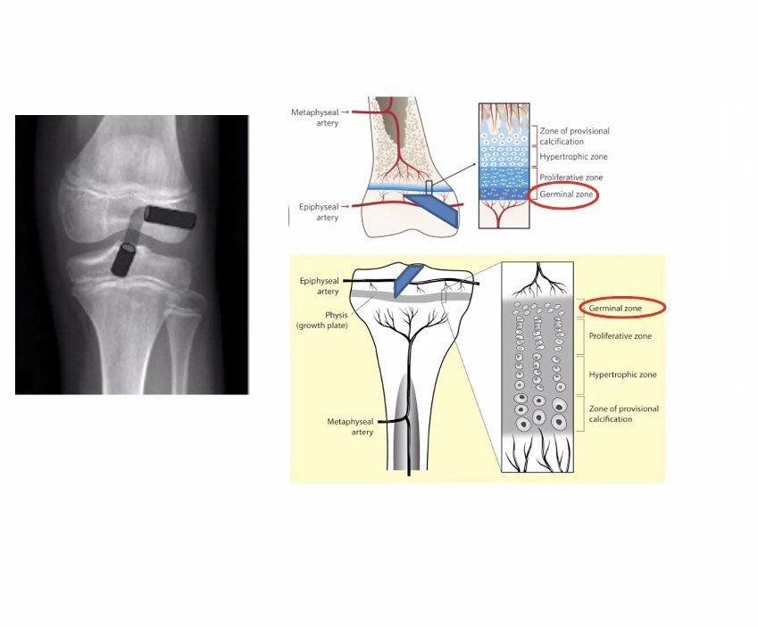
- Quoted evidence (Clin sport med 2021 metanalysis, PLUTO study group): delayed ACL reconstruction for more than 12 weeks significantly increases risk of meniscal injuries and irreparable meniscal tears
- Take into account skeletal age, growth remaining, instability symptoms, aspiration for sport and associated injury
Q2What are the surgical options for a paediatric ACL tear?▸
- Primary repair (ongoing BEAR trial)
- Extra-articular tenodesis
- Intra-articular reconstruction: transphyseal, hybrid or physeal sparing
Q3What is primary repair of a paediatric ACL tear and what is the evidence?▸
- Primary repair is one option (ongoing BEAR trial)
- Preliminary 2-year follow-up results promising
Q4What type of intra-articular reconstruction is used according to growth remaining?▸
- Transphyseal: <2 years of growth remaining
- Hybrid: 2-5 years of growth remaining (femur spared)
- Physeal sparing (over-the-top fixation): >5 years of growth remaining
Q5Why is a hybrid reconstruction used and how is growth considered?▸
- Femur is spared in hybrid as it contributes 40% of lower limb growth
- Hybrid used when 2-5 years of growth remaining
Q6What technical principles minimise physeal damage in paediatric ACL reconstruction?▸
- Drilling damages the geminal layer ( 7-9% physis cross section); 7-9% of physis cross-section is sufficient to cause growth disturbance (germinal layer)
- Aim tunnel size 3-4% of physis cross-sectional area
- Soft tissue graft; centrally located vertical tunnels
- Avoid transphyseal screws; avoid overtensioning the graft
Q7What are the differentials and how do you assess a suspected osteochondral lesion of the medial femoral condyle?▸
- Differentials: osteonecrosis (SONK, post scope, secondary), osteoid osteoma, subchondral cyst, transient osteoporosis
- History to rule out differentials: mechanical symptoms suggest displacement; trauma history
- PE: Wilson's test (knee 30-90 degrees, pain with IR, relieved by ER); overall alignment; assess for instability. Gait: ER LL
Q8What imaging is used and what are the MRI criteria for instability?▸
- X-ray: weight bearing AP, lateral and notch view (flexed 30deg) to look for detached loose bodies; scannogram for overall alignment
- MRI: position, size, attachment, displacement, loose body
- Instability criteria: increased T2 signal >=5mm deep to lesion; focal defect >=5mm2 in articular surface
Q9What is the Clanton and Delee classification of OCD?▸
- 1. Depressed
- 2. Connected by osseous bridge
- 3. Detached, undisplaced
- 4. Displaced
Q10What is the Guhl arthroscopic classification and corresponding management?▸
- 1. Intact, 1-3cm (activity modification/retrograde drilling)
- 2. Stable flap (headless screw)
- 3. Partially displaceable flap (remove fibrous tissue, chondroplasty, fix)
- 4. Complete detachment (remove, chondroplasty +/- OC allograft, ACI, MACI)
- Small (<2cm x 2cm) --> microfracture; large (>2cm x 2cm) --> chondroplasty
Q11What is the definition, epidemiology and prognosis of OCD?▸
- Definition: subchondral bone lesion with delamination + sequestration +/- articular involvement
- Most common in knee (80% medial, 40% F, 20% bilateral, 10% patella); also talus and capitellum
- 70% resolve with conservative management
- PAPPAS: I <12y, II 12-20y, III >20y (III usually OA)
Q12What are the causes of OCD and the aim of management?▸
- Constitutional: epiphyseal dysplasia with accessory nuclei separating from the epiphysis
- Vascular: watershed region from attachment of the PCL
- Traumatic: repetitive impingement of the tibial spine
- Aim: relieve symptoms and improve function; always start conservative with protected weight bearing and NSAIDs
- Surgery aims: restore joint congruity, increase local blood supply, address predisposing instability or malalignment
Q13What is the Outerbridge classification of chondral lesions?▸
- 0 intact
- 1 softening
- 2 fissure not to subchondral bone
- 3 fissure to subchondral bone + diameter >1.5cm
- 4 subchondral bone exposed
Q14How is a sizable OCD flap repaired when the physis is open?▸
- Repair/fixation for sizable flap with open physis
- Method of fixation: screw, bone peg, sutures
- Rehab: PWB 6-8 weeks
Q15What are the three broad categories of resurfacing/reconstruction options for OCD?▸
- Resurfacing/ reconstruction options fall into three broad categories: MSC stimulation, substitution, cell-based biological replacement
- MSC stimulation: microfracture, drilling, abrasion chondroplasty
- Substitution options: OATS, osteochondral allograft
- Cell based, biological replacement options: ACI, MACI, AMIC
Q16What is the microfracture technique and what tissue does it produce?▸
- OT x microfracture for fibrocartilage, others for hyaline
- Microfracture 3-4mm apart, break surface till just across the tidemark with a chondral pick with low energy
Q17What are the OAT (osteochondral autograft transfer) plug requirements?▸
- Plugs should be cylindrical
- At least 8mm long
- 4-12mm diameter
Q18How do ACI, MACI and AMIC differ?▸
- ACI: 2 procedures, expensive, can produce hyaline cartilage, not limited by size
- MACI: similar to ACI but with a matrix
- AMIC: one stage procedure
Q19What prognostic factors suggest a good outcome in OCD?▸
- Open distal femoral physis
- Size <2cm
- No sclerosis on XR
- Location at medial femoral condyle posterolateral aspect
- No synovial fluid behind lesion on MRI
- High uptake on bone scan = good healing potential (grade II-IV, uptake at lesion, femoral, tibia)
Q20How is a high tibial osteotomy (HTO) performed?▸
- Approach: direct anteromedial
- Identify the pes and release hamstrings
- Release the distal MCL fibres subperiosteally in one layer until the posterior border of the tibia
- Periosteal elevator to clear posterior structures off the tibia and place a radiolucent Homman retractor to protect the NV bundle
- Mark out biplanar osteotomy either ascending or descending; trajectory of the saw cut guided by K wires
- Apply Tomofix plate for FWB walking postop
Fact check
Clin Sport Med 2021 meta-analysis by the PLUTO study group shows delayed ACL reconstruction >12 weeks increases risk of meniscal injuries and irreparable meniscal tears — misattributed — PLUTO is a prospective multicentre descriptive epidemiology cohort of skeletally immature ACL tears, not a 2021 meta-analysis; evidence for irreparable medial meniscal tears shows risk rising by about 8 weeks (Everhart, AJSM 2019), and timing thresholds vary by study — (medium confidence) — source