Sports
ACL reconstruction - graft choice and tunnels
Graft options, tunnel size and position, fixation devices, avulsion and post-operative stiffness.
56 questions
10 source pages
2 images
Images appear with the first question taken from each source page — tap a question to open it.
Q1What are the dimensions and fixation of a BPTB graft?▸
- 10mm width, 90-100mm length
- Rigidfix/interference screw (proximal) + interference screw (distal)
Q2What is the classical placement of a BPTB graft and why?▸
- Tibial bone --> femoral tunnel; patellar bone --> tibial tunnel
- Because the tibial metaphysis is weaker, and there is increased bone contact if the intraop tunnel is too wide
Q3What is the problem if a BPTB graft is too long and how is the graft remedied?▸
- Too long: unable to use an interference screw
- Graft remedy: trim and flip 180 degrees back
- Graft remedy: self-twist 360 degrees to take up tension
Q4What are the tunnel remedies if a BPTB graft is too long?▸
- Femur: recess graft and secure at aperture with bioscrew
- Tibia: secure bone plug in trough distal to tunnel
Q5What is the problem if a BPTB graft is too short and how is it remedied?▸
- Too short: fixation not cortical
- Remedy: double fixation
Q6What happens if a BPTB graft is too wide or too narrow?▸
- Too wide: bone plug not through tunnel; remedy graft trimming +/- notchplasty
- Too narrow: loosening; remedy alternative fixation
Q7How are patellar complications prevented during BPTB harvest?▸
- Patella fracture: trapezoidal/bevel cut to increase the cross-sectional area of bone at the donor site
- Avoid transverse saw cut overruns
- Burr to round off corners
- Leave at least 1cm from the superior pole
Q8What types of graft motion occur after ACL reconstruction?📷▸
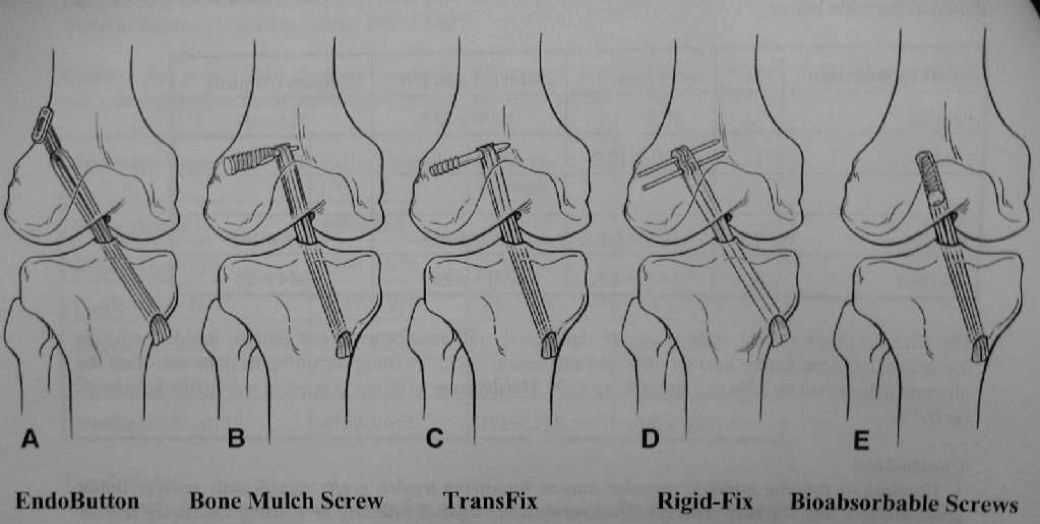
- Longitudinal motion: bungee effect
- Horizontal motion: windshield wiper effect
- Creep of graft: elongation
Q9What are the consequences of graft motion greater than 3mm?▸
- Delayed incorporation
- Tunnel widening
Q10Compare the healing time of BPTB and hamstring grafts.▸
- BPTB: healing <8-10 weeks
- Hamstring: tendon to bone >12 weeks
Q11What are the types of aperture fixation and their pros and cons?▸
- Aperture fixation: anatomical, at joint level (femoral subchondral, tibial cancellous), direct linkage
- Pros: decrease graft motion, decrease widening of canal, decrease creep
- Cons: lower margin of error, graft attrition during insertion
Q12What are the types of suspensory fixation and their pros and cons?▸
- Suspensory fixation: non-anatomical, distal level; mainly cortical (Endobutton, staple, tie on post) or cancellous (transfixation pin)
- Pros: more versatile
- Cons: increase graft motion
Q13How is the EndoButton loop length (EB-CL) calculated?▸
- Socket length is subtracted from total channel length and 6 or 7mm added for turning radius
- If the number is a multiple of 5, that is the EB-CL; if not, select the next-largest multiple of 5
- Example: channel 34mm, socket 27mm --> 7 + 6 = 13 --> 15mm EB-CL, so 34 - 15 = 19mm of graft in the femoral tunnel
Q14How does an interference screw achieve fixation and how is interference defined?▸
- Relies on friction between graft and bone tunnel
- Interference = amount by which the diameter of the screw exceeds the graft tunnel gap
Q15What are the properties of bioabsorbable interference screws?▸
- Biphasic CaPO4 (mixture of HA and tricalcium phosphate) + polylactide (more hydrophobic than polyglycolic)
- Bioabsorbable over years; no need for removal; MRI compatible
- Graft strength and stiffness decrease at 6 weeks
- Titanium screws are also used; old material was stainless steel
Q16What screw size and position give the best fixation?▸
- 1mm wider than tunnel
- Longer screw gives better fixation but only engage bone plug in BPTB (20-30mm best)
- Within 5mm proximal to joint; if too long may cause windswept effect
Q17What is the purpose of reverse thread on an interference screw?▸
- Reverse thread for less insertion torque
Q18What are the complications of interference screw use?▸
- Screw laceration of suture or graft
- Screw divergence = difference between tunnel angle and screw direction; >20 degrees = less stability
- BPTB only: bone plug advancement and plug cut off from graft
- If plug cut off: intact side to interference screw, cut side to Krakow and fix with post/button/screw in tunnel
Q19What do you describe on an X-ray after ACL reconstruction?▸
- Femoral and tibial tunnel position
- Single or double bundle
- Soft tissue or BPTB graft
- Bone quality and mode of fixation
Q20What are the three reasons for ACL reconstruction failure?▸
- Recurrent instability
- Recurrent pain
- Loss of motion
Q21What are the factors of recurrent or persistent instability (Pittsburgh classification)?▸
- Mechanical/technical (majority): wrong patient choice (knee abuser, malalignment), graft choice, graft size, tunnel position, tension (3tension in the notes), graft fixation, graft impingement, secondary stabilisers (meniscus, peripheral ligament)
- Failed graft incorporation: infection, immunological, avascularity, stress shielding
- Traumatic: reinjury, aggressive rehab
Q22How does the lecturer classify ACLR failure according to time frame?▸
- Early (<3 months): failure of mechanical fixation or early biological problem (infection)
- Midterm (3-12 months): technical problems - tunnel malposition, impingement, graft elongation, unrecognised ligamentous injury
- Later (>1 year): trauma, underlying patient factor
Q23What do you look for on X-ray in a failed ACL reconstruction?▸
- Details of surgery (single/double bundle, graft and fixation type, graft size)
- Tunnel position; tunnel widening >15mm may need 2-stage surgery; tibial slope; notch view for notch architecture
- Complications: patella fracture, posterior blowout, endobutton position, interference screw divergence
Q24What history and risk factors are relevant in a failed ACL reconstruction?▸
- Age and premorbid status
- Activity level (knee abuser)
- Risk factors: ligamentous laxity, female, pivoting sports
- Current symptoms: pain, instability, mechanical symptoms, reinjury
Q25What examination findings are relevant in a failed ACL reconstruction?▸
- Gait: varus thrusting
- Generalised ligamentous laxity
- Genu valgum
- ROM and evidence of loss of secondary restraints
- Position of previous scar
Q26What is the preop workup before revision ACL reconstruction?▸
- XR (AP/lat/tunnel/skyline/notch)
- Scannogram for varus
- CT and MRI: tunnel, graft, incorporation, bone stock; look for other concomitant injuries
- Prerequisite: knee flexion >90 degrees, fixed flexion contracture <5
Q27What are the intraoperative considerations in revision ACL reconstruction?▸
- Choice of graft (BPTB, HS for widened tunnel, graft size suboptimal, quad, allograft)
- Prepare screw/staple removal (revision ACL set)
- Tunnel planning: use old tunnel if position okay, new tunnel if severe malposition, two stage if old and new are close with not enough bone stock (dilatation >15mm)
- Revision notchplasty
- Fixation: endobutton, interference screw, tie over post, over the top, double fixation
- Send intraop culture if doubtful
Q28What are the expectations and postop plan for revision ACL reconstruction?▸
- Expectation mx: results inferior to primary ACLR (increase rerupture rate and rehab time)
- One- vs two-stage operation
- Postop: individualised, less aggressive rehab plan
Q29What should be assessed on an X-ray after ACL reconstruction?▸
- Details of surgery: number of tunnels, graft type (bone plug/harvesting) and fixation method
- Complications: patella fracture, patella alta, posterior femoral blow-out, tunnel widening
- Quality of reconstruction and tunnel position
Q30What is the optimal sagittal tunnel position after ACL reconstruction?▸
- Femur more posterior, tibia more anterior (to gain maximal AP length)
- Femoral: Blumensaat line divided into 4 parts - tunnel at the posterior 4th quadrant, must not breach the posterior cortex
- Tibia: plateau divided into 4 parts - tunnel at the anterior 2nd; too anterior causes notch impingement (double bundle can be more anterior)
Q31What is the coronal clock-face position of the femoral tunnel?▸
- 10:30 and 1:30
Q32How can femoral tunnel position be predicted in transtibial versus transportal techniques?▸
- Transtibial: can use the tibial tunnel to predict the femoral tunnel
- Transportal: by anteromedial portal
Q33What should be checked for fixation quality on a post-ACL reconstruction X-ray?▸
- Interference screw: divergence <15 degrees
- Button: on cortex (may trap ITB / within bone)
- Tunnel widening
Q34How do you recognise that a BPTB graft was used on a post-ACL reconstruction X-ray?📷▸
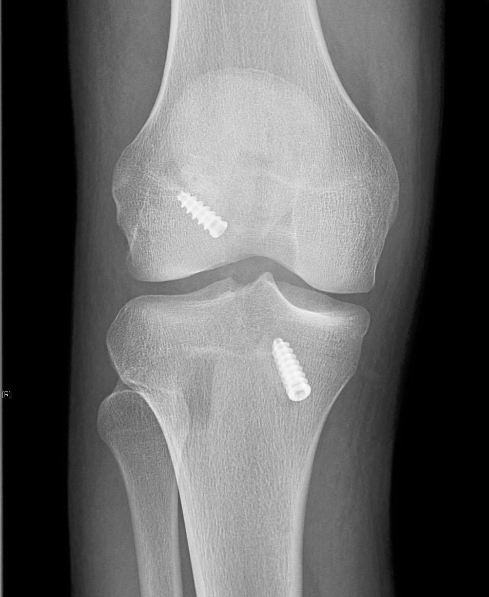
- Defect in the patella and tibial tuberosity
- Bone inside the tunnel
- Fixation by bioabsorbable screws on both sides
Q35How do you describe tunnel position on the coronal view?▸
- Graft position satisfactory; both tunnels obliquely aligned
- Femoral tunnel w.r.t. notch is at 10 o'clock position
- Tibial tunnel at 60 degrees w.r.t. knee joint (cf sagittally aligned)
Q36How do you describe tunnel position on the sagittal view?▸
- Tibial tunnel 40% from anterior (2nd quarter) with entry point posterior to Blumensaat line (ideally comment with knee in full flexion)
- Femoral tunnel adequately posteriorly placed without posterior blow-out
Q37What other findings should be commented on in this post-ACLR X-ray?▸
- There is patella Alta with Insall salvati ratio > 1.2, but I will compare it with preop XR
- No patellar fracture
- Screw position is not divergent
Q38What does the position of graft fixation represent, and when is it usually tensioned?▸
- Position of fixing the graft represents the affected motion
- Usually tensioned in 20-30 degrees flexion and posterior drawer (maximum tension) --> most stable
- If fixed in extension, ensure full extension postop
Q39What happens if the graft is fixed in extension with the femoral tunnel too anterior or too posterior?▸
- Too anterior: limit flexion
- Too posterior: lax in extension
Q40What are the consequences of a femoral tunnel placed too anterior when fixed in flexion?▸
- Tight in flexion, lax in extension
- Fails to clear the resident ridge
- Physiologically not adequate flexion; graft gradually stretches out and becomes lax (biological problem)
Q41What are the consequences of a femoral tunnel placed too posterior when fixed in flexion?▸
- Lax in flexion, tight in extension
- Rare
Q42What are the effects of tibial tunnel malposition?▸
- Anterior: notch impingement, limits extension
- Posterior: impinges PCL, tight in extension
- Medial: impingement on medial femoral condyle
- Lateral: impingement on lateral femoral condyle
Q43What is the resident ridge and what does it mark?▸
- Also called the lateral intercondylar ridge
- Marks the anterior and superior border of the ACL insertion
Q44What is the lateral bifurcate ridge?▸
- Ridge located between the AM and PL bundles
Q45How does the AM bundle position over the femur change with knee movement?▸
- More posterior on flexion
- More superior on extension
Q46What is the optimal femoral tunnel position?▸
- Coronal: right 10:30 / left 1:30
- Sagittal: B line posterior 1/4, posterior to the resident ridge with 2 mm posterior cortex remained
Q47What are the pitfalls of femoral tunnel malposition?▸
- Too anterior: tight flexion if tightened in extension
- Too posterior: blow out
- Too vertical: rotational instability
Q48What is the optimal tibial tunnel position and what are its pitfalls?▸
- Sagittal: 2nd anterior 1/4 of the tibial plateau, 7 mm anterior to PCL
- Coronal: between the tibial spines
- Too anterior: notch impingement
- Too posterior: PCL impingement + laxity?
Q49What are the global causes of stiffness after ACL reconstruction?▸
- Arthrofibrosis
- Infrapatellar contracture syndrome (fibrous hyperplasia of anterior knee - loss of flexion + extension + patellar entrapment)
- Infection
Q50What causes loss of knee flexion after ACL reconstruction?▸
- Femoral tunnel too anterior, tightened and fixed in knee extension
- Excessively tensioned graft
- Suprapatellar pouch scarring
Q51What causes loss of knee extension after ACL reconstruction?▸
- Tibial tunnel too anterior
- Notch scarring
- Cyclops lesion
Q52Why does an ACL avulsion fracture occur rather than a midsubstance tear?▸
- Ligament stronger than physeal cartilage or cancellous bone
- ACL fibres in continuity with perichondrium of tibial epiphysis/cartilage
- Mechanism: hyperflexion or direct blow to flexed knee (e.g. fall from bicycle)
Q53What is the Meyers and McKeever classification of ACL avulsion fracture?▸
- Non-displaced
- Minimally displaced with intact posterior hinge
- Completely displaced
Q54How are ACL avulsion fractures treated?▸
- Long leg cast for types I and II
- Type II: CR before casting by hyperextension to reduce
- Treat early; arthroscopic or mini-open option
- Fixation: screw (better x2), suture anchor (usually not enough strength) or pull-out suture through bone tunnel
- Rehab similar to ACL reconstruction
Q55What are the difficulties and complications of ACL avulsion treatment?▸
- Reduction difficulty: interposition of intermeniscal ligament and meniscus
- Recurvatum in children (fixation tethers anterior growth plate)
- Remove metallic screw at 3-4 mths (3-4 months) to avoid articular cartilage damage (difficult)
Q56How is the knee immobilised and what fracture is associated with ACL avulsion?▸
- Cast in 20 degrees flexion to minimise distraction force; hyperextension increases tensile force and distracts the fragment
- Position is controversial; some put in 0 degrees but usually better reduced
- 80% are type 1 or 2
- Associated Segond fracture: disruption of meniscotibial portion of lateral capsule (direct evidence of lateral capsule, indirect for ACL)