Spine Paed
Spine embryology and development
Vertebral development, spinal cord growth and vertebral level landmarks in the child.
11 questions
2 source pages
1 images
Images appear with the first question taken from each source page — tap a question to open it.
Q1Using the functional-level table, summarise the hip, knee, ankle and functional findings at each level.▸
- L1: hip ER + F (budda), knee flex, ankle equinus, flaccid
- L2: hip flex + add, knee flex, ankle equinus, +ve hip flexion
- L3: hip flex + add, knee can extend, ankle equinus, can walk (indoor), hip trdelenberg
- L4: hip flex + add, knee can extend, ankle can dorsiflex, hip tredelenberg, no equinus
- L5: hip flex (can abduct), knee can extend, ankle can dorsiflex with valgus (peronei +, PT weak), hip and knee normal, hindfoot valgus
- S1: hip, knee and ankle OK, but claw toes
Q2At which levels is the ankle in equinus and at which level can it dorsiflex?▸
- Equinus at L1-L3
- Can dorsiflex at L4-L5
- L5: dorsiflexion with valgus (peronei +, PT weak)
- L4: no equinus - the source's 'L4 calcaneus'
Q3Which levels can extend the knee and which level can walk?▸
- Knee flex at L1-L2; can extend from L3 onward
- L3: can walk (indoor), hip trdelenberg, equinus
Q4What does the source title 'L4 calcaneus' refer to?▸
- At L4 the ankle can dorsiflex with no equinus (calcaneus deformity)
- Hip is flex + add with tredelenberg; knee can extend
Q5Which functional levels have hip flexion with adduction and which can abduct?▸
- L2, L3 and L4: hip flex + add
- L5: hip flex (can abduct)
- L1: hip ER + F (budda)
Q6Which levels are Trendelenburg positive and which level has claw toes?▸
- L3 and L4: hip Trendelenburg
- S1: claw toes
Q7What are the three embryological layers and their derivatives in spine development?📷▸
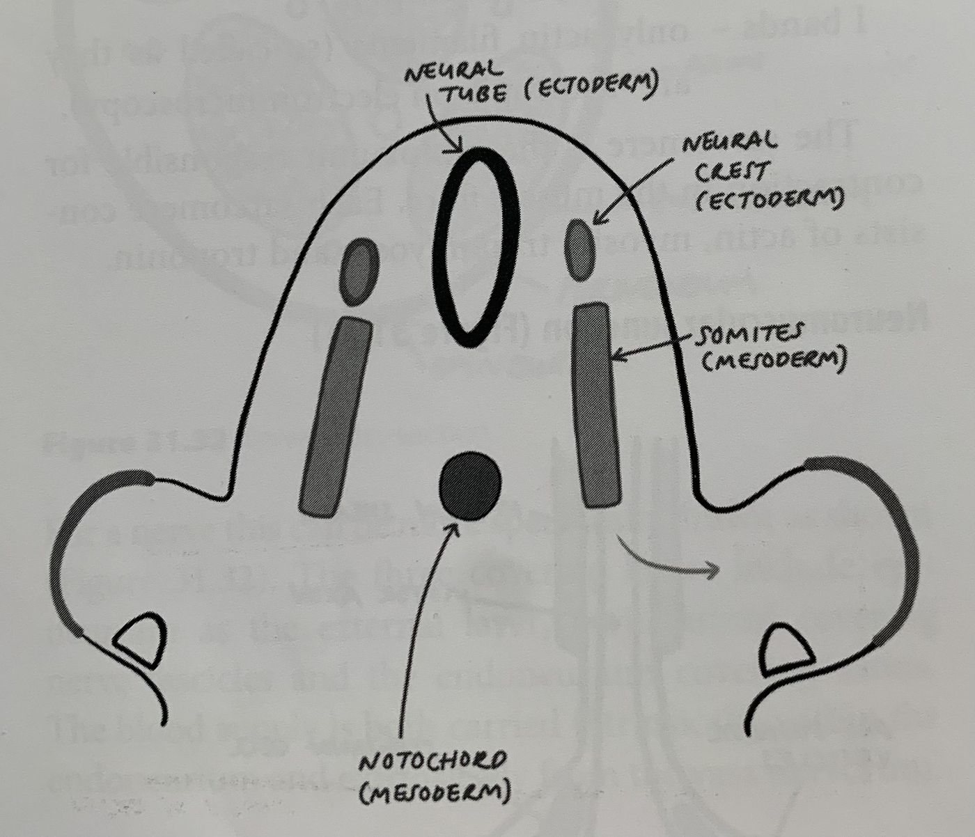
- Ectoderm infolds at the midline to form the neural tube (CNS precursor)
- Neural crest cells dorsal to the neural tube become the PNS
- Mesoderm forms the notochord, which induces neurulation and becomes the nucleus pulposus
Q8When does the neural tube form and what is the metameric shift phenomenon?▸
- At 3 weeks the ectoderm aggregates at the midline and infolds to form the neural tube
- PNS migrates between each level of somite = metameric shift phenomenon
Q9How do somites form the vertebral column?▸
- At 4 weeks paraxial mesoderm aggregates at the midline to form somites: dermatome, myotome, sclerotome
- Somites unite around the notochord to form the vertebral body and around the neural tube to form the neural arch
- Each somite forms one disc plus the adjoining cranial and caudal halves of the vertebra
- The central part undergoes apoptosis to form the disc
- von Ebner fissure = separation fissure of a single somite
Q10What is the timeline of spinal ossification?▸
- 6 weeks: chondrification of the spine
- 9 weeks: primary ossification centres - 1 at the body, 2 at the neural arch
- 2-6 years: arches fuse with the body
- 12 years: secondary ossification centres appear at spinous process, transverse process and apophyseal rings
- 25 years: secondary centres fuse with the primary centres
Q11What are the three embryological layers involved in spine development?▸
- Ectoderm, mesoderm and endoderm
- Ectoderm infolds at the midline to form the neural tube (CNS precursor); neural crest becomes the PNS
- Mesoderm forms the notochord, which induces neurulation and becomes the nucleus pulposus