Dysplasia Syndromes
Neurofibromatosis and Klippel-Trenaunay syndromes
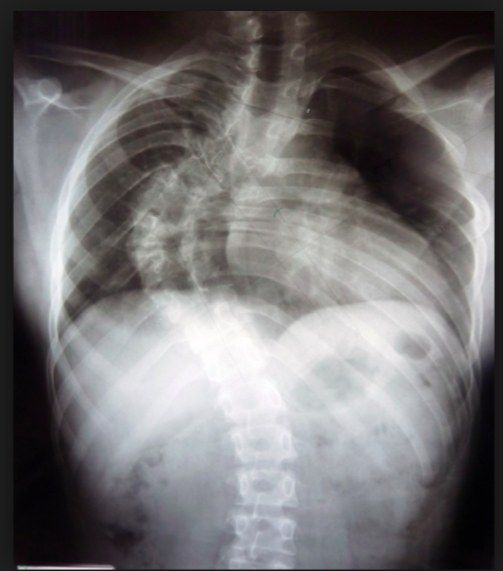
Skeletal deformity, dystrophic scoliosis and limb hypertrophy in NF and Klippel-Trenaunay.
25 questions
3 source pages
1 images
1 fact-check flags
Images appear with the first question taken from each source page — tap a question to open it.
Q1What are the three types of neurofibromatosis?📷▸
- NF1 (NIH consensus)
- NF2 with bilateral vestibular schwannomas
- Segmental NF (single body segment)
Q2What are the NIH consensus diagnostic criteria for NF1?▸
- 2 of 7 required
- Family history in a 1st degree relative
- Skin: cafe au lait spots >6 (15mm in adults, 5mm in children); axillary and groin freckling (Crowe sign)
- Nerve: neurofibroma x2 or plexiform neurofibroma
- Bone: distinctive osseous lesion (sphenoid dysplasia, long bone cortex thinning +/- pseudoarthrosis)
- Eye: Lisch nodules x2 (iris hamartoma), optic glioma; updated 2021 adds gene analysis and more specific bone/eye criteria
Q3What are the orthopaedic manifestations of NF1?▸
- Scoliosis (dystrophic / idiopathic-like form)
- Hemihypertrophy
- Atlantoaxial instability
- Plexiform neurofibroma is pathognomonic for NF1, in 4%, can progress to neurofibrosarcoma
Q4Describe the features of dystrophic scoliosis in NF1.▸
- Thoracic kyphoscoliosis with a short segmented, sharp curve and distorted ribs and vertebrae
- AP: acute short segment curve, atypical level, penciling of ribs (>=3 poor prognosis because of rapid progression), TP spindling, hypoplastic pedicles
- Lateral: posterior scalloping and wedging of VB, foramina ectasia
- Transverse: widened canal (increased interpedicular distance)
- MRI: dumbell lesion (dumbbell lesion), dural ectasia (3mm thoracic, 4mm lumbar), meningocele
Q5How is NF1 scoliosis managed?▸
- Brace is no use
- Early OT if Cobb >20
- Decompression
- ASF & PSF with instrumentation
Q6What are the genetics, prognosis and surgical difficulties in NF1?▸
- AD (50%) / sporadic (50%), 100% penetrance; NF1 gene on chromosome 17q21; abnormal neurofibromin, Affecting RAS which affect osteoclast
- Prognosis: normal life expectancy; high chance of malignancy and hypertension
- Surgery difficulties: poor bone stock, significant kyphosis, small pedicles, incidental durotomy, Bleeding due to extradural venous plexus, rib dislocation during reduction causing nerve injury, may need A+P (nonunion: PSF alone up to 40%, A+P 10%)
Q7What are the features of malignant transformation in NF1?▸
- Elephantiasis (large mass with villous skin)
- Plexiform NF lifetime risk 10% ('bag of worms', filariasis)
- Clinical: age 36-50yo, sudden pain and increase in size
- Biopsy to rule out neurofibrosarcoma
Q8What is Sprengel shoulder and what are its associations?▸
- Congenital undescended scapula (4-6th C spine pharyngeal arch; failure of caudal migration to T2-7 during 9th to 12th weeks of gestation (9th-12th weeks); interruption of embryonic subclavian blood supply)
- Associations: Klippel-Feil, congenital scoliosis, cervical spina bifida, VACTERL +/- situ invertus (situs inversus)
Q9What are the XR features of Sprengel shoulder?▸
- Omovertebral bar (best seen on lateral/oblique C spine XR)
- Clavicle uptilt
- High riding scapula
- Small scapula (transverse > vertical)
- Inferomedial corner medial tilt + glenoid downward tilt
Q10Describe the Cavendish classification.▸
- 1: Very mild, not detectable in clothing
- 2: Mild, 1-2cm higher
- 3: Moderate, 2-5cm higher
- 4: Severe, >5cm higher (superior angle near occiput)
Q11What are the principles and options for surgery in Sprengel shoulder?▸
- Excise omovertebral bar + superior trapezius release at C4
- Relocation of the scapula
- Clavicle morselized osteotomy preserving periosteum (decreases brachial plexus injury)
- Aim: improve shoulder abduction (~30 degrees, Caverndish by 1 grade)
- OT if cosmetic problem or abduction < 120 at 3-8y (too late -> more nerve injury)
- Woodward and Green procedures both improve 50 degrees
Q12What is Klippel-Feil syndrome?▸
- Fusion of more than two cervical vertebrae; (SGM 1 gene at chromosome 8
- Triad: Low posterior hair line (hairline), short webbed neck, decreased cervical movement (normal F/E, decreased side bending)
- XR triad: unsegmented C spine (commonest C2/3), ADI, degeneration (may have basilar invagination)
Q13What are the associations and surgical indications in Klippel-Feil syndrome?▸
- Renal aplasia 33%, deafness 30%, congenital heart disease 15-30%
- Atlantoaxial instability ~50%; adjacent level disease 100%
- OT if pain, instability, neurology; check CV and UG systems before OT
- If C2 involved: no contact sports
Q14What are the clinical findings of Sprengel shoulder?▸
- Hypoplastic and undescended scapula (right in the photo)
- Low hairline, short webbed neck
- No scapular winging; check for upward tilting of the clavicle from the front
Q15What should be documented on examination of Sprengel shoulder?▸
- Neck: short, stiff; document bar, shoulder movement, abduction power
- Medial wing + supero-medial angle move superiorly -> glenoid pointed inferiorly
- Look for UL abnormalities (VACTERL), periscapular muscle fibrosis/weakness, distal NV status (syringomyelia)
- Look for scoliosis/kyphosis/back pitting (spina bifida)
Q16What further imaging and workup is needed for Sprengel shoulder?▸
- CT and MRI spine for the bar and spinal problems such as dural ectasia or diastematomyelia
- Cardiac and renal workup; rule out other C spine problems
- Klippel-Feil associations: C spine failure of segmentation, C1/2 instability, cranial settling
Q17What is the epidemiology of Sprengel shoulder?▸
- 10-30% bilateral
- Present in 1/3 of Klippel-Feil
- Association: scoliosis, spinal dysraphism, VACTERL
Q18What are the Woodward and Green procedures for Sprengel shoulder?▸
- Woodward: detachment and distal advancement of the midline origin of parascapular muscles
- Green: extraperiosteal detachment, hold scapula in new position with spring wire traction and spica casting for 3 weeks
- Both improve 50 degrees
Q19Describe the Klippel-Feil classification.▸
- Type I: extensive fusion
- Type II: only 1 or 2 vertebrae in the cervical spine
- Type III: fusion in part of the thoracic and/or lumbar spine in addition to Type I or Type II
Q20What is the activity advice and prognosis in Klippel-Feil syndrome?▸
- If C2 involved: no contact sports
- If lower: usually asymptomatic until adjacent levels degenerate (100%) -> contact sports OK
- Check CV and UG systems before OT
Q21What other features are associated with Klippel-Feil syndrome?▸
- Hearing loss, micrognathia, torticolis (torticollis), scoliosis
- Brainstem abnormalities/basilar invagination, congenital cervical stenosis
- MRI to rule out intraspinal cord abnormalities
Q22What is the triad of Klippel-Trenaunay syndrome?▸
- Capillary malformation (port wine stain)
- Venous/ lymphatic malformation
- Soft tissue hypertrophy
Q23How do Parkes Weber, Proteus and Maffucci syndromes differ?▸
- Parkes Webers syndrome: capillary, lymphatic and venous malformations with AV fistulas
- Proteus: capillary and venous malformations, macrodactylyl (macrodactyly), hemihypertrophy, lipomas, pigmented nevi, scoliosis
- Maffucci: lymphatic and venous malformation + multiple enchondromas
Q24What are the treatment principles for Klippel-Trenaunay syndrome?▸
- Resection may be dangerous; consider embolization
- Ligation of feeding vessels is of no help
- High flow AV malformations difficult; staged partial excision mostly palliative
- Proximal ligation increases collateralization; embolisation risks digital ischemia
- YAG laser in direct contact allows subtotal excision of complicated hemangiomas
Q25What is the limitation of YAG laser?▸
- Will not stop bleeding from vessels with lumen diameters greater than 1mm
- Direct contact YAG allows subtotal excision of lesions previously thought untreatable
Fact check
NF1 gene is on chromosome 17q21 — incorrect locus — The NF1 gene maps to chromosome 17q11.2 (17q21 is the BRCA1 region) — source