Dysplasia Syndromes
Enchondromatosis and osteochondromatosis
Ollier disease, Maffucci syndrome and hereditary multiple exostoses with forearm deformity.
25 questions
3 source pages
1 images
1 fact-check flags
Images appear with the first question taken from each source page — tap a question to open it.
Q1What is Ollier's disease and how does it present?📷▸
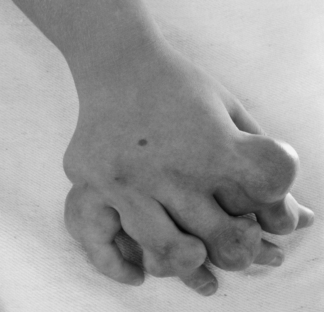
- Sporadic defect of enchondral ossification
- Unilateral involvement --> LLD
- Angular deformity
- DDx: polyostotic fibrous dysplasia, OI
Q2What is Maffucci syndrome and what are its XR features?▸
- Maffucci = Ollier's + hemangioma
- Expanded bone, angular deformity, cutaneous haemangiomata
- XR: Ollier changes + small rounded calcified phleboliths
Q3What are the rates of malignant transformation in enchondromatosis?▸
- Solitary enchondroma <1%
- Ollier's 25-30%
- Maffucci 100%
Q4Which four planes of deformity are considered in treatment?▸
- Coronal: valgus/varus
- Sagittal: flexion/extension
- Transverse: torsional
- Longitudinal: LLD
- Aim: well-aligned limbs in all planes
Q5What are the treatment options for deformity in enchondromatosis?▸
- Hemiepiphysiodesis or guided growth
- Osteotomy for acute or gradual correction
- Combination of techniques
Q6How does enchondroma differ from osteochondroma?▸
- Enchondroma: incomplete enchondral ossification, escape of chondroblast to metaphysis (cartilage in IM canal)
- Enchondroma: Rubin physis hyperplasia
- Osteochondroma: benign chondrogenic lesion from the perichondral ring
- Osteochondroma: metaphysis hyperplasia
Q7What are the XR features of Ollier's disease?▸
- Cartilage-containing lesion + irregular translucent area at metaphysis
- Expanded bone
- Polyostotic involvement
Q8Describe the XR findings of multiple hereditary exostosis at the knee.▸
- Multiple bony protrusions of the metaphyseal region of distal femur/proximal tibia with metaphyseal tubulation
- Pedunculated, well defined, narrow zone of transition, pointing away from the physis
- Trabeculae within lesion continuous with the rest of the bone
- No cortical destruction, no soft tissue calcification
Q9How do you differentiate MHE from Trevor disease?▸
- Clinically: Trevor is younger
- Radiologically: Trevor arise from epiphysis
- Histologically: Trevor cartilage cap has bands of cartilage separating cancellous bone
- Genetics: normal EXT levels in Trevor
Q10What is the risk of malignant transformation in MHE?▸
- 5-10% lifetime risk of malignant transformation
- Versus normal lifetime risk of any malignancy of 1 in 3
- 1% lifetime risk per lesion, chondrosarcoma at around the 5th decade
- Cartilage cap >2cm suspicious (normal 2-3mm)
Q11Which clinical and radiological features suggest malignant transformation?▸
- Increase in size after puberty
- Acute onset of pain in adults
- Cortical destruction, soft tissue mineralization >2cm
- MRI cartilage cap >2cm, sessile, central lesions more malignant
Q12What are the indications for surgery in MHE?▸
- Pain from bursitis
- Deformity (forearm bowing, genu varum and valgum)
- Malignant transformation into chondrosarcoma
- Excise lesion including cartilaginous cap to reduce recurrence; corrective osteotomy +/- lengthening
Q13What is the genetics and pathogenesis of MHE?▸
- AD; EXT1 (Chr 8), EXT2 (Chr 11), EXT3 (Chr 19); EXT1 worst
- 10% spontaneous mutation
- Defect in the groove of Ranvier with aberrant cartilage from the perichondral ring
- Mutations affect prehypertrophic chondrocytes; Rubin: metaphyseal hyperplasia
Q14What investigations are required in MHE?▸
- Further XR of the scapula and pelvis (regions that cannot be palpated clinically)
- Scannogram for lower limb alignment
- Check the forearms clinically for bony exostosis and associated radial head dislocation
Q15What complications must be ruled out in MHE?▸
- Local pressure symptoms (nerve, bursitis, pseudoaneurysm)
- Deformity/growth disturbance
- Malignancy
Q16What is the growth pattern and natural history of MHE lesions?▸
- Sessile or pedunculated growth from the physeal plate away from the joint
- Lesions are benign and stop growing at maturity
Q17Describe the XR findings in this skeletally immature forearm.▸
- Shortening of the ulna with ulnar negative variance and apex radial bowing
- Abnormal bony enlargement of the ulnar head with exostosis
- Dislocated radial head
- Ulnar shift of the carpus
Q18What are the surgical indications from the deformity point of view?▸
- Ulnar shortening >1.5cm
- Radial articular angle >30 degrees
- Carpal translocation >60%
- Painful subluxation of the radial head
- Pronation <60 degrees
Q19What are the surgical indications from the MHE point of view?▸
- Enlarging mass after puberty
- Nerve or skin impingement, pain
- Radiologically: cortical erosion
- Cartilaginous cap >2cm, soft tissue mineralisation >2cm
Q20What are the options for radius reconstruction?▸
- Proximal radius: unstable - one bone forearm; stable - radial head excision
- Distal: osteotomy/ hemiepiphysiodesis
Q21What does the 2018 systematic review conclude about ulnar lengthening?▸
- Restores radiologic anatomy, improves appearance and to a lesser extent objective clinical parameters short to intermediate term
- Poor evidence that gains are maintained long term
- Impact on QOL and function not adequately investigated
- Considerable evidence surgery minimally impacts pre-op function
Q22What counselling points are given to the family?▸
- Risk of malignancy
- Risk of further deformity
- Functional impairment may not improve with surgery - it mainly improves cosmesis
Q23What is the differential diagnosis of the forearm deformity?▸
- Tumour
- Malunion
- Trevor disease (dysplasia epiphysealis hemimelia)
Q24How do you confirm the diagnosis and assess the patient?▸
- Look for other lesions -> ?HME
- Rule out malignancy
- Assess other complications: ROM, nerve compression
- Assess current function
Q25What are the details of surgical treatment?▸
- Excise the exostosis to stop differential growth
- Ulnar lengthening (acute vs gradual depends on length)
- Timing of surgery is controversial
Fact check
Malignant transformation occurs in 100% of Maffucci syndrome patients — Overstated - published rates vary widely; not all patients transform — Reported rates range from about 15-40% up to 52-57%; the JBJS 1987 life-table analysis says degeneration is almost a certainty, not 100% — (medium confidence) — source