Spine Non Trauma
Spinal tumour and infection imaging
Radiographs and MRI of spinal tumours and infection; primary spine tumours
49 questions
4 source pages
3 images
1 fact-check flags
Images appear with the first question taken from each source page — tap a question to open it.
Q1What did the plain X-rays show in this case of spinal TB?📷▸
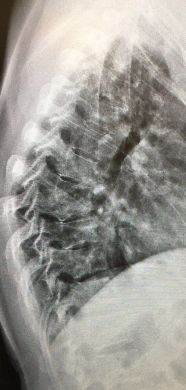
- AP T spine: soft tissue fusiform swelling
- Lateral T spine: collapse of T7 and T8, obliterated disc space, acute kyphosis
- Gibbus = acute structural thoracolumbar kyphosis
- No skipped lesion
Q2What are the typical X-ray and MRI features of spinal tuberculosis?▸
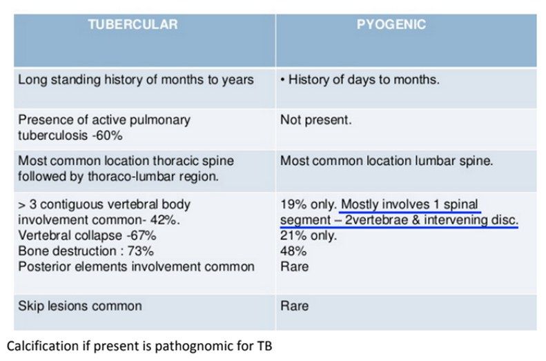
- X-ray: anterior vertebral body collapse, disc sparing, multiple levels +/- skip lesions, kyphosis, +/- calcification
- MRI: skip lesions 15%, paravertebral abscess 50%, disc sparing (multicentric and multilevel), subligamentous spread
- Ddx: infection (TB > pyogenic > fungus) and tumour (lymphoma)
Q3What is the clinical assessment in suspected spinal TB?▸
- Symptoms: deformity, pain, neurology
- Risk factors of infection: immunocompromised, previous TB infection, contact history
- Red flag signs
- Rule out other differentials: pyogenic infection, carcinoma, lymphoma
Q4How is spinal TB diagnosed?▸
- Morning urine and sputum TB PCR
- CXR abnormal in 2/3 of cases
- Biopsy (low sensitivity)
- MRI with gadolinium: low T1, bright T2
Q5What are the aims of management in spinal TB?▸
- Control infection
- Control back pain
- Salvage neurology
- Prevent/correct deformity
Q6How does the spinal level affect the management of TB spine?▸
- C spine: low threshold for surgery
- Lumbar spine: conservative first
- T spine: depends on neurology
Q7How is TB spine managed when there is no neurology and no instability?▸
- Isoniazid + rifampicin, +/- streptomycin
- Expected kyphosis = total vertebral involvement x 30.5 + 5.5 (Rajasekaran)
Q8How is TB spine with neurological involvement or instability managed?▸
- Debridement only: expected kyphosis 10 degrees
- HK operation: anterior radical debridement (extirpation) + fusion with strut graft
- No instrumentation if <3 levels; instrumentation if >=3 levels
- Advantages: direct visualisation of the lesion, fusion under compression anteriorly
Q9What is the epidemiology of TB spine?▸
- Extrapulmonary TB in 3% of cases
- Skeletal TB in 10% of extrapulmonary TB
- Spinal TB in 50% of all skeletal TB
- Site: TLJ > T > LS > C
Q10What is the pathophysiology of spinal TB?▸
- Granulomatous spondylitis with a delayed hypersensitivity reaction
- Starts in the metaphysis of the vertebral body
- Subligamentous spread under the ALL: contiguous multilevel, skip lesions, paraspinal abscess
- Disc is a natural barrier and is usually spared
- Acute kyphosis may progress in children due to growth spurt
Q11What is Pott's paraplegia and what are its early causes?▸
- Neurological involvement found in up to 10% of patients
- Inflammatory tissues/sequestrum
- Epidural abscess
- Prolapsed disc/retropulsed fragment
- Pachymeningitis (poor prognosis)
- Spinal subluxation due to non-union level
Q12What are the Rajasekaran types of paediatric spine deformity?▸
- Retropulsion
- Lateral translation
- Toppling
- Facet separation
- More than 2 = unstable and should be fused
Q13What are the features of healed TB disease?▸
- Direct compression by a transverse bone ridge
- Kyphus: sequestrum >5mm, adjacent segment stenosis
- Cord atrophy (syringomyelia)
- Reactivation
Q14What was the design of the MRC trial of TB spine?▸
- Series of studies since 1965 in multiple centres worldwide; follow-up up to 15 years
- Korea - Masan: inpatient 6 months bed rest vs outpatient; Pusan: POP jacket vs no jacket
- Hong Kong: radical debridement + graft vs debridement; Bulawayo: nonoperative vs operative debridement
- Inclusion: clinical and radiological TB, T1-S1 disease
- Exclusions: significant neurological deficit (unable to walk room length), significant extraspinal disease, >3 levels of destruction, C spine, anti-TB drugs >1 year
- All had anti-TB drugs (PASINAH)
Q15What was a favourable outcome in the MRC trial and what did it conclude?▸
- Favourable outcome: no symptoms, full physical activity, no CNS involvement, no sinus/abscess, radiological healing; assessed at 5, 10, 15 years
- POP jacket not required (Pusan)
- Outpatient antibiotics without bedrest are OK (Masan)
- HK procedure: anterior decompression + radical debridement + uninstrumented autogenous strut graft fusion > debridement alone
- Quicker pain relief, earlier abscess/sinus resolution, higher fusion rate (85% at 5y, 95% at 15y), Kyphosis (3deg@5 maintained @15)
- Nonoperative vs operative: all had favourable outcomes
Q16What did Prof Luk criticise about the MRC trial?▸
- Patients had mild disease
- Only 15-year follow-up
- Surgery gave quicker resolution of sinus and abscess, higher fusion rate, less late deformity (kyphosis ~10 degrees)
Q17What are the absolute indications for surgery in spinal TB?▸
- Failed conservative treatment (3-4 weeks)
- Neurological involvement
- Instability/deformity: kyphosis more than 60 degrees causes significant morbidity, or spinal instability score >2
- Non-compliance or non-diagnostic biopsy
Q18What are the late complications of spinal TB?▸
- Gibbus due to non-union after central necrosis
- Pott's paraplegia: reactivation, adjacent segment disease, cord atrophy
- Kyphus with sequestrum >5mm and adjacent segment stenosis
- Cord atrophy/syringomyelia
Q19What are the surgical approaches and deformity correction options in TB spine?▸
- Approach - anterior: above T4 equal from either side (usually from the side with the large abscess); below T4 from the left; thoracotomy with rib resection 2 levels above
- Decompression: mild - posterior decompression; severe - costotransversectomy + internal kyphus excision
- Stabilisation: anterior fusion with strut graft
- Deformity correction: mild - closing wedge osteotomy; severe - halo pelvic traction
Q20What are the spine-at-risk signs of Rajasekaran?▸
- Spine toppling sign
- Lateral translation
- Posterior facet widening
- Retropulsion
- More than 2 signs = unstable and should be fused
Q21Describe the X-ray findings in spondylodiskitis.📷▸
- L4 lower and L5 upper endplate destruction
- Decreased disc height and decreased lordosis
- Ddx: spondylodiskitis (pyogenic/TB) and malignancy
- Less likely TB because the disc is usually spared
Q22What are the risk factors and assessment findings in spondylodiskitis?▸
- Immunocompromised: IVDA, DM, steroids, malnutrition
- Recent systemic infection: UTI, GI, respiratory; recent spine operation
- Neurology and pain
- Femoral stretch and psoas stretch = psoas abscess
- Constitutional symptoms suggest malignancy
Q23What investigations establish the diagnosis and extent of spondylodiskitis?▸
- CBC, ESR, CRP
- Blood cultures positive in ~33%; if positive, ~85% accurate for isolating the correct organism
- CT-guided biopsy sent for aerobic, anaerobic, fungal and acid-fast culture
- Contrast MRI: loss of T1 body contrast, T2 disc enhancement, contrast-enhancing paravertebral exudate, epidural/psoas abscess
Q24What MRI features favour infection over tumour?▸
- Disc space involvement
- end plate erosion
- Significant inflammation
- Look for epidural abscess, paraspinal collection and psoas abscess
Q25What are the goals of management in spondylodiskitis?▸
- Optimise host
- Establish microbiological diagnosis
- Control of infection
- Monitor and prevent neurological deficit, instability and late deformity
Q26What are the goals and indications for surgery in spondylodiskitis?▸
- Operative goals: eliminate infection (debridement), prevent/improve neurologic deficits (decompression), maintain stability (instrumentation +/- fusion)
- Failure to obtain microbiological diagnosis from closed needle biopsy (e.g. C spine)
- Poor response to conservative treatment, abscess, neurological deficit
- Extensive disease: 2 adjacent VB or single VB with more than 50% collapse, +/- deformity
Q27What is the nonoperative management and expected success rate in spondylodiskitis?▸
- Nonoperative treatment is the mainstay
- IV antibiotics + corset if collapse
- Success rate 60-90%
Q28What is the pathophysiology of spondylodiskitis?▸
- Haematogenous seeding of the endplates (low-flow vascular anastomosis) -> direct extension into the disc -> second endplate
- Arterial: diffusion (children/adolescents via intermetaphyseal artery, adults via metaphyseal artery) and septic emboli
- Venous via the Batson plexus
- Contiguous spread from local infection (retropharyngeal and retroperitoneal abscesses)
- Direct inoculation
Q29What is the anatomical distribution of spondylodiskitis?▸
- Lumbar spine 60% > thoracic 30% > cervical 10%
Q30Which organisms cause spondylodiskitis and how long are antibiotics given?▸
- Staphylococcus aureus and epidermidis; Gram-negative organisms
- E. coli, Pseudomonas in immunocompromised patients
- Salmonella in sickle cell anaemia
- Duration: 6 weeks vs 12 weeks - no difference (Bernard, Lancet 2015)
Q31What are the operative approaches and strut graft options in spondylodiskitis?▸
- Single stage: anterior - debridement + reconstruction with strut graft
- Two stage: delayed posterior instrumentation and fusion after anterior debridement
- Epidural abscess: laminectomy
- Strut graft: autogenous tricortical iliac crest, rib or fibula are safe and effective in acute infection
- Titanium mesh cages filled with autograft improved deformity correction (Carragee, Spine 2008), followed by posterior instrumentation)
Q32What is the role of instrumentation in active spinal infection?▸
- Spinal instrumentation in active infection is controversial
- Studies show stability of the spine is important for suppression and eventual elimination of infection
- Titanium is preferred over stainless steel
Q33What X-ray signs suggest spinal metastasis?📷▸
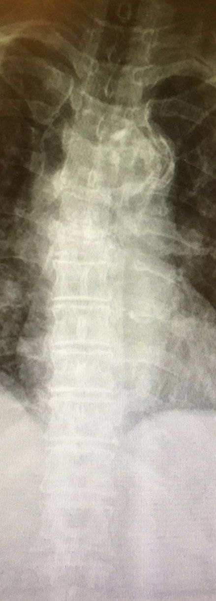
- Collapse and kyphosis
- Winking owl sign (pedicle destruction)
- Fusiform swelling
- Lung mass
Q34What history is important in suspected spinal metastasis?▸
- Premorbid status
- History of malignancy
- History of HIV, IVDU, travel to endemic countries
- Nature of pain: tumour infiltration, cortical expansion, instability/fracture, neurocompression
- Neurology: direct invasion, epidural seedling, fracture, deformity; also expectations
Q35What examination is needed in suspected spinal metastasis?▸
- Full neurological examination
- Systems exam: lungs, abdomen, PR, breast, thyroid
Q36What is the Bilsky ESCC score used for?▸
- Grading epidural spinal cord compression on MRI
- 0: bone only
- Ia/b/c: out of bone, deformation of thecal sac, touching cord
- II: cord compression with CSF still visible; III: cord compression with CSF not visible
- Determines high grade (2,3) vs low grade (1) compression
Q37What imaging and classification systems are used for metastatic spine disease?▸
- Whole spine MRI with the Bilsky ESCC score
- Weinstein, Boriani, Biagini (WBB) classification
Q38What are the treatment goals and principles in metastatic spinal disease?▸
- Treatment is palliative in intent: salvage neurology, stabilise spine, maintain ambulation, pain relief
- Always biopsy if no confirmed bone met
- Look for other spinal/skeletal mets, other primaries and other ddx (infection, TB, benign tumour, MM)
- Manage by the NOMS framework with a multidisciplinary approach
Q39What medical treatment is given for metastatic cord compression?▸
- Dexamethasone for neurocompression (50mg loading then 4mg Q6H, with PPI coverage)
- Treat hypercalcemia
- Bisphosphonate can reduce skeletal events: Zometa 4mg IV every month
Q40What is the evidence for surgery plus radiotherapy in metastatic cord compression?▸
- Patchell, Lancet 2005: better outcomes with surgery + RT vs RT alone
- Adjunct treatment includes preoperative embolisation for vascular tumours
Q41What is the epidemiology of spinal metastasis?▸
- Bone metastasis is common after lung and liver primary
- Spine is the most common site, then proximal femur and humerus (Batson's valveless venous plexus allows seeding like red marrow)
- Primary sites: breast > lung > prostate/renal/thyroid; liver also common
- Survival is variable (6 months to 5 years)
Q42What is the difference between osteolytic and osteoblastic metastases?▸
- Osteolytic: osteolysis caused by tumour-induced activation of osteoclasts (RANK, RANKL and OPG)
- Osteoblastic: tumour secreted endothelin 1 (90% prostate, 60% breast)
Q43How is hypercalcaemia of malignancy managed?▸
- A metastatic emergency
- Hydration
- Loop diuretics
- Bisphosphonate
Q44What are the components and interpretation of the Tokuhashi score?▸
- General condition
- Primary tumour
- Number of vertebral body foci
- Number of extraspinal foci
- Major organ involvement and neurology (palsy)
- Score 0-15; 9-11 predicts >=6 months, >=12 predicts >1 year survival for excisional surgery; PF FONG mnemonic
Q45What are the components of the Tomita score?▸
- Primary tumour
- Mets to vital organs (treatable/untreatable)
- Bone mets
- Score 2-10; >6 = palliative decompression/no surgery
Q46What is the principle of tumour separation surgery?▸
- Circumferential decompression to allow a safety margin for tumoricidal radiation doses without toxicity to the cord
Q47How are benign primary spine tumours classified by Enneking stage?▸
- Enneking 1 (self-limiting) = osteoid osteoma, ABC, haemangioma
- Enneking 2 (active) = osteoblastoma
- Enneking 3 (aggressive) = giant cell tumour
Q48What are the malignant primary spine tumours?▸
- Chordoma
- Osteosarcoma
- Chondrosarcoma
- Ewing's sarcoma (EWS)
Q49Which other classification systems are used for spine tumours?▸
- Tomita
- Weinstein, Boriani, Biagini (WBB)
Fact check
Dexamethasone 50mg loading dose for metastatic spinal cord compression (lecture notes) — non-standard dose, likely error — Guidelines recommend an initial 10mg IV loading dose followed by 4mg (or 6-10mg) every 6 hours; a 50mg bolus is not a recognised regimen — (medium confidence) — source