Spine Non Trauma
Vertebral osteoporotic collapse
Vertebral collapse deformities including osteoporotic and vertebral plana patterns
28 questions
3 source pages
2 images
Images appear with the first question taken from each source page — tap a question to open it.
Q1Describe the X-ray findings of an osteoporotic vertebral collapse and the initial workup.▸
- Anterior wedge collapse of T12 with no retropulsion
- Request AP X-ray to look for widening of the interpedicular distance
- Ask about mechanism of injury, cancer history, features of infection/TB, neurological deficit
- Bloods: bone profile, tumour markers, SEP and inflammatory markers
Q2What are the aims of management in osteoporotic collapse?▸
- Pain relief and early mobilisation
- Prevent deformity and neurological deficit
- Prevent future collapse
Q3How is osteoporotic collapse managed systemically?▸
- Assess osteoporosis severity and calculate future fracture risk
- Quit smoking and alcohol, give calcium and vitamin D, weight-bearing exercise
- Anti-osteoporotic medication
Q4How is osteoporotic collapse managed locally?▸
- Analgesics, calcitonin +/- brace
- If failed conservative management or non-union, consider kyphoplasty
Q5When is vertebroplasty considered for an osteoporotic collapse?▸
- Ongoing pain more than 2 weeks after occurrence of a new fracture despite treatment
- Progressive compression fractures of one or multiple vertebrae with subsequent loss of posture
- Non-union with persisting instability
Q6What are the prerequisites for vertebroplasty?▸
- Intact posterior wall
- Neurologically intact
Q7What are the complications of vertebroplasty?▸
- Anaesthesia-related complications
- Cannula placement: infection, spinal cord injury, fracture of pedicles/transverse process, lung injuries
- Cement injection: cement leakage into the spinal canal causing neurological deficits, pulmonary cement embolism, fat embolism
- Late adjacent segment collapse
Q8Why is kyphoplasty preferred over vertebroplasty?▸
- Better evidence on pain relief and potential kyphosis correction
- Fewer complications than vertebroplasty due to decrease in insertion pressure
- Mechanism: reduces kyphosis, low-pressure injection, increased viscosity of injection
- Greater reduction in kyphotic angle and less extraosseous cement leakage than vertebroplasty
Q9What did the two NEJM 2009 RCTs show about vertebroplasty and how were they criticised?▸
- Buchbinder et al.: pain, QoL and function same as placebo surgery up to 6 months
- Kallmes et al.: pain similar to placebo surgery at 3 months
- A JBJS 2009 publication criticised their methodology, mainly the cement volume
Q10What did the VAPOUR and VERTOS IV trials show about vertebroplasty?▸
- VAPOUR trial (Lancet 2016, Clark): vertebroplasty vs sham - great reduction in pain at 3 and 6 months
- It is the only RCT to support vertebroplasty; patients had more severe pain, fracture age <6 weeks, and more PMMA cement was used
- VERTOS IV (BMJ 2018): no significant pain relief vs sham
- AAOS changed stance in 2014, suggesting limited evidence
Q11What is the AAOS 2010 guideline on treating osteoporotic collapse?▸
- Moderate evidence: calcitonin for pain relief
- Weak evidence: ibandronate and strontium, kyphoplasty
- Inconclusive: bed rest, brace, exercise programme, electrical stimulation
- Not advised: vertebroplasty
- Avoid in fractured posterior wall and neurologically compromised patients
Q12How can adjacent level collapse and non-union be decreased after augmentation?▸
- Treat osteoporosis
- Watch for fracture patterns that increase non-union: split type fracture / large angular motion
- Beware of endplate deficit leading to intradiscal cement leakage
- Avoid aggressive restoration of vertebral height
Q13What other options exist apart from vertebroplasty/kyphoplasty and what are the problems with them?▸
- Problems with more extensive surgery: medically fragile patients, poor bone quality at the bone-implant interface
- Approach: anterior vs A+P vs posterior
- Decompression; reduction?
- Instrumentation: short or long, construct type (screw-rod, plate, sublaminar hooks and wiring), augmentation
- Fusion and rehabilitation; removal of implant: no
Q14How can failure at the bone-implant interface be reduced?▸
- Preop: treat osteoporosis with a bone-forming agent such as teriparatide
- Intraop: screw type (HA coated, PMMA augmented), diameter, length - pullout strength 60% depends on cortical bone of the pedicle, 20% on trabecular bone of the body; advancing to 80% of the vertebral body is adequate
- Self tap screws (stronger pullout force), or undertap (self-tapping screws); diverging screws aimed towards subchondral bone, straightforward technique better than anatomical trajectory
- Expandable pedicle screws; circumferential fusion
- Postop bracing
- Crosslink and increasing the length of fixation only increase construct stiffness, they do not decrease failure at the bone-implant junction
Q15What is the mechanism of calcitonin for pain relief?▸
- Binds receptors in the CNS/PNS
- Releases endorphin and decreases prostaglandins
Q16What is Kummel disease and what are its imaging features?▸
- Post-traumatic osteonecrosis of a vertebral body
- XR: sclerosis of cleft margin and vertebral body, pedicle ossification, stress fracture of spinous process, paravertebral callus
- MRI: double line sign
- Vertebroplasty more likely to fail due to lack of interdigitation of cement
Q17What is the initial X-ray finding in this case?📷▸
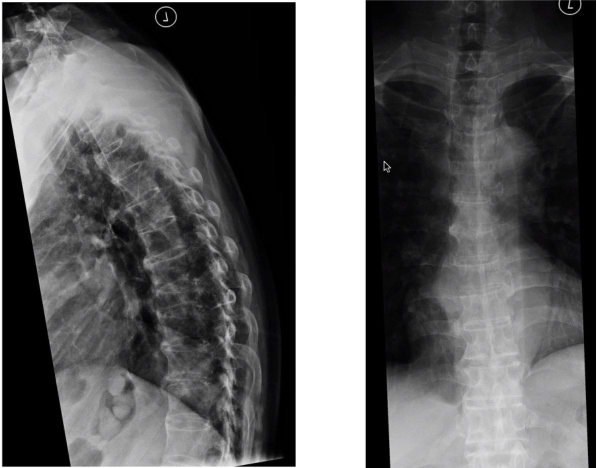
- T9 vertebral collapse
Q18What history and examination points are important in a patient with a collapsed vertebra?▸
- PMHx and premorbid mobility
- Neurology and sphincter function
- Costal margin pain
- PE: neurological examination
Q19What is important about the pain history in a collapsed vertebra?▸
- Nature of pain - rest/nocturnal vs mechanical
- Onset, progression and alleviating factors
Q20What investigations are used for suspected TB spine (T9 collapse)?▸
- Routine lymphocytosis
- Bone profile and tumour markers
- Urinary TB and sputum TB
- CT to see the extent of bony destruction
- Tissue biopsy: caseating granuloma, AFB, giant cells
Q21How is an open biopsy of the spine performed?▸
- Transpedicular approach
- Costotransversectomy
Q22What are the indications for surgery in TB spine and what is the medical regimen?▸
- Indications: debilitating pain, deformity, instability
- Nonoperative: TLSO brace
- DOT: isoniazid, rifampicin, ethambutol, pyrazinamide (intensive phase and continuation phase)
- Follow-up to monitor for complications; MRC trial
Q23What are the X-ray findings in this case?📷▸
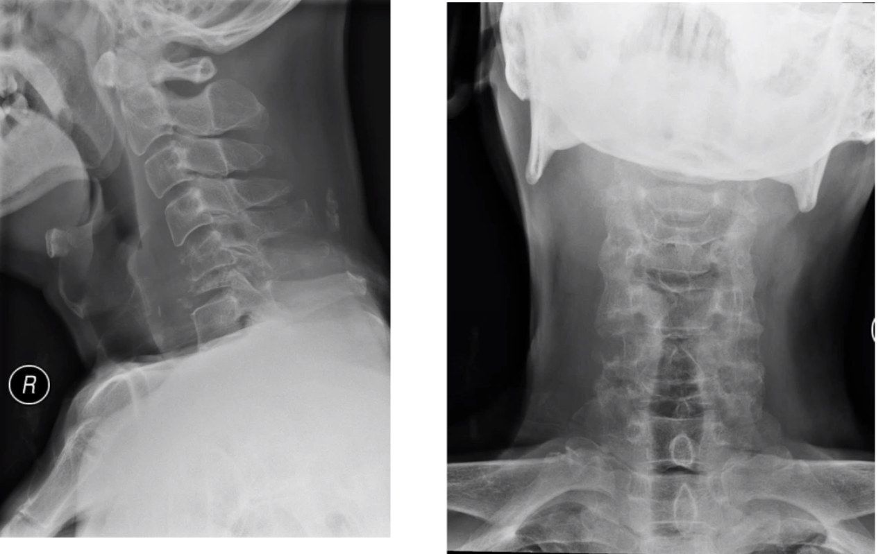
- C6 collapse with vertebral plana deformity
- Acute kyphosis
- Increase in vertebral shadow
Q24How do you differentiate infection from malignancy on X-ray of the spine?▸
- Infection starts in the disc, malignancy in the vertebral body
- More endplate erosion in infection
- Posterior elements less commonly involved in infection
Q25What history, examination and blood tests are needed for a C6 collapse?▸
- Red flags, recent infection (e.g. UTI), risk factors for infection, pain, neurology
- Systems screening: breast, abdomen, thyroid, chest, PR
- Local tenderness and neurological examination
- Bloods: WCC, CRP, CaPO4, ALP, tumour markers
Q26What staging is required once malignancy is confirmed?▸
- Local and systemic staging
- MRI and CT
- Managed in a multidisciplinary team according to the NOMS framework
Q27What are the four components of the NOMS framework as applied to this case?▸
- Neurology: clinical motor deficits; radiological Bilsky stage on MRI - high or low grade cord compression
- Oncologic: radioresistant or radiosensitive tumour
- Mechanical stability: SINS
- Systemic: traditionally Tokuhashi score, now NESM or SORG nomogram
Q28What are the components of the Tokuhashi score?▸
- General condition
- Number of spine metastases
- Number of extraspinal metastases
- Internal organ metastases
- Tissue of origin and neurology
- Score 0-15; >9 predicts surgery