Spine Non Trauma
Spine radiography and sagittal balance
Spine X-rays, lumbar AP views and measurements of spinal sagittal balance
22 questions
4 source pages
1 images
Images appear with the first question taken from each source page — tap a question to open it.
Q1Which conditions are listed under 'Spine - non trauma'?▸
- RA – C1/2 instability, subaxial involvement, cranial settling
- AS – pseudoarthrosis; DISH
- Cervical myelopathy, OPLL, PID, dural tear
- Spinal stenosis, degenerative scoliosis, spondylolysis, sagittal balance
- Vertebroplasty, TB spine, pyogenic spine, spine metastasis, failed back syndrome
Q2What spinal manifestations of RA are listed on this section?▸
- C1/2 instability
- Subaxial involvement
- Cranial settling
Q3Which cervical spine conditions are listed?▸
- Cervical myelopathy
- OPLL
- PID
- Dural tear
Q4Which degenerative/structural topics are listed?▸
- Spinal stenosis
- Degenerative scoliosis
- Spondylolysis
- Sagittal balance
Q5Which infective, neoplastic and miscellaneous topics are listed?▸
- Vertebroplasty
- TB spine
- Pyogenic spine
- Spine metastasis
- Failed back syndrome
Q6Describe the AP X-ray findings in ankylosing spondylitis.▸
- Squaring of vertebrae with marginal syndesmophytes
- Bamboo spine appearance; trolley-track and dagger sign
- Bilateral SIJ ankylosed, hips okay, no discovertebral lesion
- Frontal alignment good, no osteopenia; need lateral XR for facet joint ankylosis
Q7What are the history and examination findings in AS, and how is it diagnosed?▸
- Hx: PMHx, FHx, morning stiffness, SOB, pain, other joint or heel pain
- PE: gait, loss of lumbar lordosis with increased thoracic kyphosis, horizontal gaze, chin-brow vertical angle, wall-to-occiput, Schober's test, chest expansion
- SIJ: Faber and Gaenslen tests; neurological exam of LL; listen to heart and lungs, look at eyes and nails
- Diagnosis: modified New York criteria - at least 1 clinical + 1 radiological criterion
- Clinical: inflammatory back pain >3 months improved by exercise/worse with rest, reduced spine ROM, reduced chest expansion
- Radiological: unilateral grade 3-4 or bilateral grade 2-4 SIJ changes
Q8What are the differentials of multiple syndesmophytes and of SIJ sclerosis?▸
- Multiple syndesmophytes/ossification: DISH - non-marginal syndesmophytes, disc and SIJ spared, more common in older patients (DM)
- SIJ sclerosis: osteitis condensans ilii (young women, iliac side)
- Fibrodysplasia ossificans progressiva
Q9What is ankylosing spondylitis and what is the role of HLA-B27?▸
- A systemic seronegative arthritis mainly affecting the axial skeleton
- More common in young male patients with family history (HLA-B27)
- Presents with back pain not relieved by rest, spine/hip stiffness, stooped posture, fracture
- HLA-B27 is an MHC presenting intracellular protein to T cells, initiating an immune response through TNF
- Patho hypotheses: misfolding -> homodimer triggering a pro-inflammatory response; arthritogenic peptide presented to autoreactive cytotoxic T cells
Q10What are the discovertebral lesions in AS and the causes of pseudoarthrosis?▸
- Romanus lesion (50%): erosion at the anterolateral corner with surrounding sclerosis, posterior column spared
- Andersson lesion (4%): erosion at the mid-endplate (anterior + middle column), posterior column spared
- Pseudoarthrosis: secondary to fracture/Andersson lesion with posterior column involvement - all 3 columns
- Causes (D Fang): acute fracture through fused segment, stress fracture, escape fusion -> need circumferential fusion
Q11How are deformities in ankylosing spondylitis managed?▸
- Aim: posture (horizontal gaze), visceral function, prevent complications (fracture, pseudoarthrosis)
- Medical: physio, NSAIDs, TNF-alpha antagonists (infliximab, adalimumab, etanercept)
- Preop: anaesthetic/airway, biologics, lung function test, echo, steroid coverage, PPI; osteotomy site usually lumbar (wider canal, end of conus, no ribs, large correction power)
- Osteotomies: SPO, Ponte, PSO, VCR; cervical osteotomy for chin-on-chest at C7/T1, aim CBVA within 10 degrees; PSO closing wedge preferred over SPO opening wedge
- Complications: neurologic injury, SMA, aortic rupture, non-union, residual deformity
- Instrumentation: U3L3; C8 root decompression; C7 laminectomy + partial lateral mass resection +/- C6 and T1 partial laminectomy
Q12How does SIJ involvement progress radiologically in ankylosing spondylitis?▸
- X-ray SIJ progression: erosion, sclerosis and ankylosis
- Assess with the Ferguson view (~20 degrees cranial)
- First radiological sign is squaring of the vertebrae
Q13What blood tests and further imaging are requested in suspected AS?▸
- Bloods: CBC, ESR, CRP, HLA-B27
- XR of the thoracic and cervical spine; whole spine sagittal alignment
- Need lateral XR for facet joint ankylosis; watch for vertebral collapse (osteoporosis)
Q14What types of spinal osteotomy are used for AS deformity correction?▸
- SPO: laminectomy + pars
- Ponte: SPO + facets
- PSO: posterior elements + pedicle
- Vertebral column resection (VCR)
- Site usually lumbar: wider canal, end of conus, no ribs, large correction power
Q15What are the key points of cervical spine osteotomy in AS?▸
- Indications: chin-on-chest; residual upper cervical kyphosis causing downward gaze after lumbar osteotomy
- Aim to correct CBVA to within 10 degrees
- Level C7/T1: wide canal, mobile root (C8), vertebral artery not entering lateral mass
- PSO closing wedge better than SPO opening wedge: instability; oesophageal stretching causing dysphagia
- Instrumentation U3L3; C8 root decompression; C7 laminectomy + partial lateral mass resection +/- C6 and T1 partial laminectomy
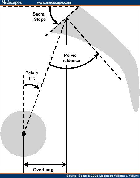
Q16How is pelvic incidence (PI) measured?▸
- Angle between a line from the centre of the sacrum to the femoral head and a line perpendicular to the S1 endplate through its midpoint
- PI = PT + SS
Q17What is the relationship of pelvic incidence to spondylolisthesis?▸
- Direct relationship to the severity of spondylolisthesis
- High PI: more shearing across L5/S1 and less lumbar lordosis
- Body tries to keep sagittal balance by increasing lumbar lordosis
Q18How does the body compensate when lumbar lordosis reduces?▸
- Pelvis rotates posteriorly, reducing the sacral slope
- When the sacrum is vertical, the knee flexes to further compensate
Q19How is sagittal vertical axis (SVA) measured?📷▸
- On standing long film X-ray
- Line from the centre of C7 vertebral body perpendicular to the ground; measure horizontal distance from the posterosuperior corner of S1
- Normal = within 5cm
Q20What is the relationship between sacral slope, pelvic tilt and pelvic incidence?▸
- Sacral slope + pelvic tilt (positional) = pelvic incidence (morphology)
Q21What are the target values for spinopelvic harmony?▸
- Lumbar lordosis = PI +/- 9 degrees
- PT < 25
- SVA < 50mm
- Spinal curvature (spinopelvic harmony / cone of economy)
Q22Why does sagittal imbalance matter?▸
- Imbalance leads to pain and progressive deformity