Spine Non Trauma
Spine imaging modalities
Role of CT, contrast-enhanced MRI and myelography in spinal assessment
19 questions
3 source pages
1 images
Images appear with the first question taken from each source page — tap a question to open it.
Q1Describe the CT/XR findings in cervical myelopathy.📷▸
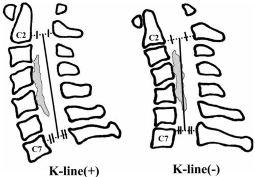
- Degenerative changes with osteophyte formation and mixed-type OPLL; kyphosis (K line/ C2-C7 angle)
- Canal stenosis: absolute <10mm, relative 13mm
- Pavlov ratio <0.8; Penning's jaw diameter <12mm suggests a congenital stenosis component
- Occupancy ratio >0.4 = poor prognosis
- Mizuno XR classification for OPLL: segmental, continuous, mixed, circumscribed/localized (at disc)
Q2Describe the MRI findings in cervical myelopathy.▸
- Multi-level stenosis with compression from both anterior and posterior (disc, bulging, ossified PLL/yellow ligament)
- Kyphosis
- Cord edema
Q3What are the causes of cervical myelopathy and its poor prognostic factors?▸
- Causes: spondylosis (anterior PID, calcified/buckled PLL, Luschka osteophytes; posterior calcified/buckled YL), OPLL, instability (e.g. C1/2), worsened by congenital narrowing (Pavlov <0.8)
- Poor prognosis: age >70, chronic symptoms >6 months, JOA <9, multiple levels, canal <30mm2, cord signal (T1 hypo/T2 hyper), compression ratio <0.4
Q4What is the natural history of cervical myelopathy and OPLL?▸
- Clark: 70% stepwise, 20% gradual, 10% sudden deterioration then plateau
- Matsunaga Spine 2012 OPLL: 20% myelopathy at presentation, 20% develop symptoms, 70% asymptomatic
- Risk of myelopathy: >60% canal stenosis by OPLL and laterally deviated-type OPLL on CT
- Bad CT: C sign = high risk of dural tear
Q5How do you choose the operative approach (PAINS)?▸
- Assess: axial neck pain/ radiculopathy, alignment (rigid kyphosis, K line), instability, number of levels, site of pathology
- 1-2 levels: ASF or ASD if no instability
- >3 levels without rigid kyphosis: posterior (laminectomy + fusion if axial neck pain/instability, else laminoplasty)
- >3 levels with kyphosis: anterior + posterior (anterior to realign/decompress, posterior to decompress/fuse)
- K line: K+ no instability = laminoplasty; K+ instability = laminectomy + fusion; K- = A+P
- Conservative: analgesics for neck pain, physio; advise operation if JOA <=13 as symptoms usually progress
Q6What are the complications of laminoplasty, and how does it compare with laminectomy and fusion?▸
- Complications: fracture hinge, spring back of hinge, C5 palsy, axial neck pain, late kyphosis
- Laminoplasty advantages: motion preserving (30-40% motion loss quoted), less morbidity, no instrumentation, shorter rehab
- Laminectomy + fusion advantages: may relieve neck pain; OPLL may resolve after stabilisation
Q7What history is taken in cervical myelopathy?▸
- Age, demand, history of DM, chief complaint and progress
- Axial neck pain and radicular pain
- Function by JOA: use of chopsticks/spoon, walking on level ground/stairs/walking aids, sphincter condition, numbness of UL, trunk and LL
- Trauma and red flag signs
Q8What does the physical examination include in cervical myelopathy?▸
- Power, sensation, jerks, myelopathy hand signs, long tract signs
- Lhermitte, Spurling, Romberg/tandem walking
- PR examination and jaw jerk
Q9What investigations are performed in cervical myelopathy?▸
- XR oblique views and flexion/extension views
- CT for extent of OPLL and the double layer sign
- MRI for cause, level and severity of stenosis
Q10What is the pathology of OPLL?▸
- Mass of woven and lamellar bone plus fibrocartilage
Q11What is the neurological recovery after surgery for cervical myelopathy (Cheung 2008)?▸
- Cheung WY Int Orthop 2008: neurological recovery in 71% of patients
- Plateau at 6 months
- Best in upper limbs, then lower limbs; worst with sphincter dysfunction
Q12What is the pseudoarthrosis rate after anterior cervical surgery?▸
- Without anterior plate: one level 0-5%, two level 10-20%, three level 30-60%
- With anterior plate: one level 0-5%, two level 0-3%, three level 0-7%
Q13What is the incidence of adjacent segment disease and the evidence for ADR?▸
- Adjacent segment disease: 25% within the first 10 years
- BJJ Findlay 2018: meta-analysis of 14 RCTs, follow-up 2-10 years
- TDR as effective as ACDF and superior for some outcomes; TDR > ACDF at 2 years and also 4-7 years
- Reduces the risk of adjacent segment disease (ACDF ASD 1.6-4.2% per year)
Q14What are the criteria, technique, advantages and disadvantages of ADR?▸
- Strict criteria: no significant neck pain, no facet arthritis, anterior compression only, no instability, good bone stock (no osteoporosis)
- Technique: alignment of COR in coronal and sagittal planes, accurate sizing and endplate preparation
- Advantages: preserve motion, prevent ASD
- Disadvantages: implant failure/migration; easy to overstuff -> posterior neck pain (increased facet loading)
Q15What is the role of gadolinium contrast MRI in spinal infection?▸
- Differentiate infection from tumour
- Differentiate epidural abscess from CSF - both are high signal on T2
- Gadolinium enhance pus in T1, CSF remains low signal
Q16How is an epidural abscess managed?▸
- Laminectomy and drainage because pus is predominantly posterior
- Anterior and posterior decompression if concomitant vertebral osteomyelitis
Q17How is a myelogram performed?▸
- Under fluoroscopy
- Spinal needle at L3/4, withdraw CSF, inject contrast
- Table tilt to allow contrast to fill up and down
- Then XR/CT
Q18How do you interpret an extradural space-occupying lesion on myelogram?▸
- Examples: PID, metastasis, TB abscess
- Paintbrush appearance
Q19How do you interpret an intradural, extramedullary lesion on myelogram?▸
- Examples: neurofibroma, meningioma
- Meniscal sign (can outline the SOL)