Others
Mortality and post-operative outcomes
Timing of deaths after major trauma and the frequency and timing of post-operative complications.
20 questions
3 source pages
2 images
1 fact-check flags
Images appear with the first question taken from each source page — tap a question to open it.
Q1How are trauma deaths distributed over time according to the source?📷▸
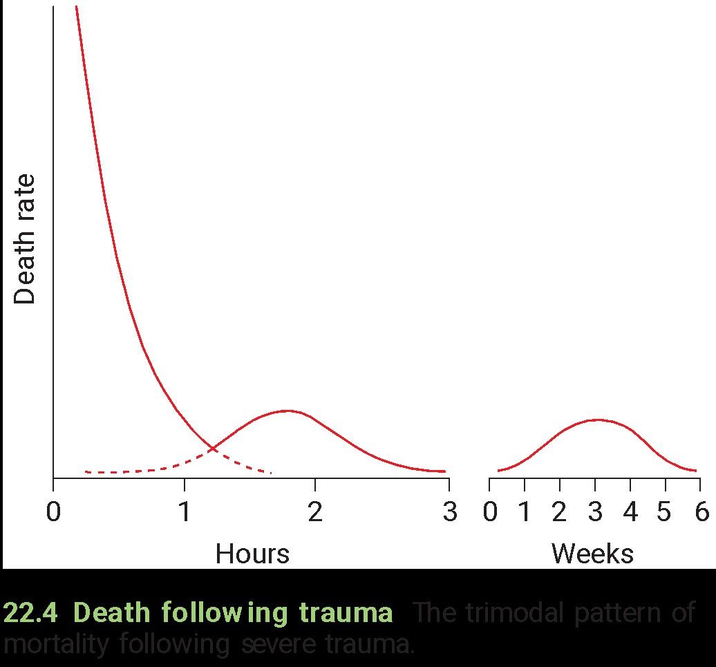
- 50% - die from non-survivable injuries immediately, or within minutes
- 30% - survive the initial trauma, but die within 1-3 hours
- 20% - die from complications at a late stage during the 6 weeks after injury
Q2What characterises the first peak of trauma deaths?▸
- 50% of deaths
- Due to non-survivable injuries, immediately or within minutes
Q3What characterises the second peak of trauma deaths?▸
- 30% of deaths
- Survive the initial trauma but die within 1-3 hours
Q4What characterises the third peak of trauma deaths?▸
- 20% of deaths
- Die from complications at a late stage, during the 6 weeks after injury
Q5Describe the X-ray findings and clinical presentation in this infected non union.▸
- Infected nonunion: previous fracture of the tibia, fibula and syndesmosis, fixed with plates + syndesmosis screw
- Tibia reduction acceptable; syndesmosis widening (reduced tibiofibular overlap, increased clear space)
- Periosteal elevation over the lateral tibia, no lucency around implant, fracture not healed
- Suspected infected implant
- At 1/12 post op: pain, redness
Q6What causes of non union are listed (diamond concept)?▸
- Mechanical instability: inadequate fixation, Bone loss, Poor bone quality
- Inadequate vascularity: severe soft tissue injury with periosteal stripping, malposition or malalignment, bone loss, distraction
- Poor bone contact: soft tissue interposition, malposition or malalignment, bone loss, distraction
Q7What are the risk factors for infected non union?▸
- Patient: old, smoking, DM, PVD, previous injury
- Injury: open fracture, wound comminution
- Surgeon: multiple operations, stripped periosteum, bone necrosis during drilling
Q8What investigations are used in suspected infected non union?▸
- Blood: ESR, CRP, albumin
- CT and USG for collection
- Gallium scan
- Intraoperative deep cultures are most reliable
Q9What are the general principles for managing an infected non union with an implant?▸
- Confirm infection, assess implant stability and bone union
- Healed -> remove implant
- Not healed + unstable -> remove and keep stable
- Not healed + stable -> antibiotics
Q10What is the role of ESR/CRP and imaging in acute infected non union?▸
- ESR, CRP: 100% PPV, 80% NPV
- CT and USG for subperiosteal and intramedullary collection
- Debridement and intraoperative culture, start antibiotics afterwards
Q11How is an infected non union with a stable implant managed?▸
- Retain the implant + debridement
- PMMA antibiotic to fill defects and coat the implant
- Antibiotics x 3 months (BJJ 2021 Shen) + soft tissue coverage
- Second stage after 6-8 weeks: remove cement/implant, revision fixation + autograft
Q12How is an infected non union with an unstable implant managed and how is infection timing classified?▸
- Remove implant + debridement + external fixation
- Gentamicin beads/cement
- Acute <2 weeks, delayed 2-10 weeks, chronic >10 weeks
- Low grade infection can coexist with a healing fracture
Q13How does infected non union differ from prosthetic joint infection in terms of implant retention?▸
- Aim in fracture-related infection: stability until union
- Retention is successful in 50-90% of infected fracture implants
- Joint replacement: only 15-50% successful (aimed at long-term function)
- The concept is different from joint replacement infection
Q14What are the principles of management in infected non union?▸
- Optimise host factors
- Identify the microorganism involved
- Debridement + antibiotics
- Reconstruction: soft tissue coverage + bone defect management (antibiotic beads for small defects, Masquelet for larger)
Q15What is the Masquelet technique?▸
- Antibiotic-impregnated cement beads/spacer deliver local high-dose antibiotics
- Reaming also debrides; circular external fixator allows shortening of any defect
- Soft tissue envelope reconstructed with a vascularised flap
- Second-stage bone grafting at ~8 weeks to the pseudomembranous tube (prevents graft resorption)
Q16What is the likely timing and reported incidence of this periprosthetic fracture?📷▸
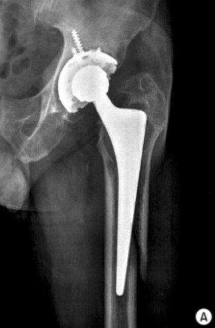
- Day 1 post op; likely an intra-operative fracture
- Incidence 4% cementless, 0.4% cemented, 0.1% post op
- Stem appears stable, bone stock adequate
Q17Describe the Vancouver classification as given for periprosthetic fractures.▸
- Type A diaphyseal
- Type B metaphyseal
- Type C distal to stem (not amendable to longest stem)
- Subtypes: cortical perforation, undisplaced, displaced
Q18How is a periprosthetic femoral fracture managed according to stem stability?▸
- Stable stem -> fix
- Unstable stem -> revise with a long stem
- Lateral plating: at least 3 screws and 3 cables (cables resist bending; screws resist rotation and control length)
Q19What are the risk factors and management of acetabular-side periprosthetic fractures?▸
- Patient: osteoporosis, radiotherapy, dysplasia/fusion, revision surgery
- Surgeon: underream
- Implant: cementless, elliptical cup
- Stable -> protected WB 12 weeks; unstable -> add screw/jumbo cup/fix + protected WB 12 weeks
Q20How can periprosthetic fractures be prevented?▸
- Pre-op planning
- Careful exposure
Fact check
Vancouver classification types are A diaphyseal, B metaphyseal, C distal to stem — non-standard mapping — Standard (postoperative) Vancouver classification: A = trochanteric region (AG/AL), B = around/just below the stem (B1 well-fixed, B2 loose, B3 loose + poor bone), C = well below the stem — (medium confidence) — source