Trauma
Elbow dislocation and chronic instability
Simple elbow dislocation, origins of instability, Morrey staging, posterolateral fracture dislocation.
26 questions
4 source pages
2 images
Images appear with the first question taken from each source page — tap a question to open it.
Q1What is the acute management of a simple elbow dislocation?📷▸
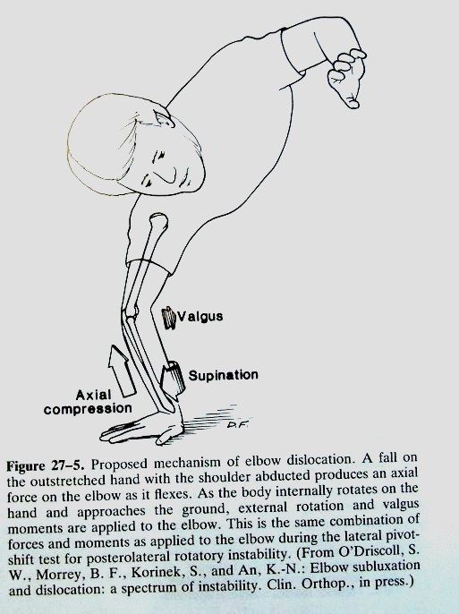
- Stabilise patient and rule out other injuries
- Rule out local complications: open wound, compartment syndrome, NV injury
- Analgesics, immobilise
- Reduction + immobilise +/- operation
Q2Describe the reduction technique for elbow dislocation.▸
- Under conscious sedation: traction, supination, direct manipulation
- Align forearm and humerus; traction with elbow flexed 90 degrees; direct pressure over olecranon; +/- supination
- Parvin's method: prone, traction over wrist for a few minutes, then lift arm
Q3What do you check on post-reduction fluoroscopy?▸
- Joint congruity - drop sign = pseudosubluxation >=4mm ulnohumeral displacement (interposed soft tissue/instability)
- Range of stable reduction
- Collaterals - varus/valgus stress; pMCL only stable in pronation, aMCL+pMCL unstable in pronation; repeat in full extension and 30 degrees flexion
- Rotatory instability - lateral pivot shift, PL drawer, chair sign, table to relocation test
Q4What is the management if the joint is congruent and stable?▸
- Immobilise for 2 weeks + early mobilisation
- Recurrence <2%, loss of terminal extension 5-15 degrees
- Stability is better in flexion and pronation
- Rehab: early ROM within 10 days, avoid immobilisation >3 weeks, aim full extension by 8 weeks
Q5Describe PLRI and PMRI.▸
- Both involve axial load + forearm supination, with valgus stress in PLRI and varus stress in PMRI
- PLRI sequence: LUCL then pMCL then aMCL (Hori circle) +/- bone injury (coronoid tip, radial head)
- PMRI sequence: LUCL, aMCL +/- coronoid base/AM facet; radial head usually intact
- CT/MRI is the gold standard for diagnosis
Q6When do you proceed to open reduction and what are the complications?▸
- Open reduction +/- repair if failed congruent reduction or unstable (dislocates in splint, dislocates with <30 degree flexion + pronation)
- Complications: HO, ulnar nerve irritation, OA, instability
Q7How is the lateral pivot shift test performed?▸
- Forearm supinated, valgus and axial load
- In extension the radial head is dislocated
- Toward 40 degrees flexion the triceps reduces the radial head with a clunk
Q8What physical examination tests suggest PMRI?▸
- Varus/valgus stress with the shoulder in max IR/ER
- Gravity assisted varus stress: shoulder 90 degrees abduction, elbow flexed and extended - pain on flexion due to lack of medial buttress by LUCL and coronoid
- Chair test
- Table top relocation test: radial head subluxes as the forearm presses the table in supination - apprehension relieved by posterior radial head pressure
Q9Where does the MCL originate?▸
- Anteroinferior portion of the medial epicondyle
- MCL = medial collateral ligament
Q10Where does the LCL originate?▸
- Inferior portion of a small tubercle on the lateral epicondyle
- LCL = lateral collateral ligament
Q11What is the Morrey staging of instability used for and how does it progress?📷▸
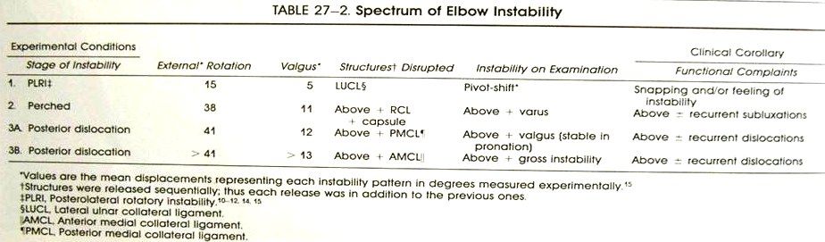
- Used in the chronic condition
- Progresses from lateral to medial
- LCUL --> RCL + capsule --> PMCL --> AMCL
Q12List the structures involved in the Morrey stages of elbow instability, in order.▸
- 1. LCUL
- 2. RCL + capsule
- 3. PMCL
- 4. AMCL
Q13What are the components of a terrible triad elbow injury?▸
- Elbow dislocation
- Radial head fracture
- Coronoid fracture
- XR: fragments over the medial epicondyle and sigmoid notch (likely radial head), no soft tissue gas
Q14What initial assessment and management is described for a fracture dislocation of the elbow?▸
- ATLS protocol, primary and secondary survey, AMPLE history + premorbid status
- Check NV status and compartments; examine one joint above and below (Essex lopresti)
- Analgesia with adequate sedation using midazolam and pethidine
- CR with elbow at 90 degrees flexion + gentle traction and direct olecranon manipulation, then splint
Q15What does Morrey's model describe as the primary static, secondary static and dynamic stabilizers of the elbow?▸
- Primary static = UHJ, LUCL, MCL
- Secondary static = RCJ, CEO, CFO
- Dynamic = anconeus, triceps and biceps
- Indication for surgery = loss of dynamic and static stabilisers
- Aim = concentric stable reduction to allow early mobilisation
Q16What definitive planning is required after initial reduction of a terrible triad?▸
- CT for planning: classify the coronoid fracture (O'Driscoll/Morrey) and radial head fracture (Mason)
- Indicated for surgical repair and ORIF due to loss of dynamic and static stabilisers
- Aim: concentric stable reduction to allow early mobilisation
- Position supine; approach = utility posterior approach
Q17What is the sequence of fixation in operative management of a terrible triad?▸
- Fix the coronoid first through the radial head fracture window - small, use lasso transosseous repair
- Fixation or replacement of the radial head or neck
- Repair the LUCL with suture anchor or fibre tape
- Test stability; repair MCL via a medial approach if still unstable
- If still unstable, prepare radial head replacement or apply a dynamic joint distractor (DJD)
Q18Which approach is chosen in a terrible triad and what are the options?▸
- Posterior utility approach, raising thick flaps on the medial and lateral sides to address the respective pathologies
- Laterally: Kocher interval (ECU-anconeus), Kaplan interval (ECRB-EDC) or Boyd interval (anconeus and FCU)
- Kaplan may preserve the remaining lateral ligamentous complex but has more PIN palsy
- Boyd if there is also an olecranon fracture
- Key point: watch out for the PIN and radial nerve
Q19How can exposure be improved during the posterior utility approach?▸
- Release the ECRB along the supracondylar ridge
- Release some of the supinator distally
Q20What medial approaches are available in a terrible triad and what are their pros and cons?▸
- Hotchkiss (flexor pronator mass, PL-FCU), split FCU, or Taylor Scham (elevate the entire flexor pronator mass from posterior to anterior)
- Watch the medial antebrachial cutaneous nerve on skin incision; deep, watch the ulnar nerve
- Hotchkiss: good access to the anterior elbow capsule, BUT limited access to the medial facet of the coronoid and poor access to its base
- Split FCU probably most versatile in terrible triad (addresses base and anteromedial facet)
- Taylor Sham best for accessing the base
Q21How can PIN injury be avoided during the Kocher approach in terrible triad surgery?▸
- Fully pronate the forearm
- Avoid incising the capsule too anteriorly (PIN lies over it)
- Avoid dissection distal to the annular ligament (PIN lies within supinator)
- Do not place retractors around the radial neck
Q22When is radial head ORIF preferred over replacement, and when is replacement indicated?▸
- ORIF best reserved for non-comminuted fractures with 3 or fewer fragments
- Attempted fixation of more comminuted fractures is prone to fixation failure and nonunion (Ring and Jupiter JBJS 2002)
- Replacement indications: Mason III + >3 articular fragments
- Replacement also for an unstable elbow (Mason IV / Essex-Lopresti injury)
- Prepare radial head replacement if still unstable after MCL + LUCL repair and ORIF
Q23What did the Sun 2016 meta-analysis find comparing arthroplasty with ORIF for radial head fractures?▸
- Metaanalysis by Sun 2016 Eur J ortho trauma: arthroplasty yielded higher satisfaction, better elbow scores, shorter surgical time and lower incidence of recurrent instability
- Replacement has better ROM and is a faster operation
- ORIF has a higher reoperation rate and higher complication rate
- BUT functional score is similar
Q24Describe the safe zones for applying a dynamic joint distractor (DJD).▸
- Distal 1/3 humerus: anterolateral or posterolateral
- Middle 1.3 humerus: anterolateral (middle third of the humerus)
- Proximal forearm: lateral/posterolateral proximal and middle 1/3 ulna, neutral rotation
- Axis guidewire: lateral just distal to lateral epicondyle, medial slightly anterior and distal to medial epicondyle; place hinge over the guidewire
Q25What rehabilitation and prophylaxis follow fixation of a terrible triad?▸
- Long arm slab in pronation for 2 weeks
- Then progressive ROM exercises, starting with active assisted FE and SP
- HO prophylaxis
Q26For a transolecranon fracture dislocation, which radiographic parameters must be restored?▸
- PUDA (proximal ulnar diaphyseal angle): 8-10 degrees
- OW (olecranon width)
- ODA (olecranon diaphyseal angle): 23 degrees between the olecranon tip and the diaphysis