Trauma
Elbow fractures - radial head and olecranon
Radial head fracture and replacement, capitellum and olecranon fracture, Monteggia injury.
42 questions
7 source pages
5 images
1 fact-check flags
Images appear with the first question taken from each source page — tap a question to open it.
Q1Why is metal preferred over silicone for radial head replacement?▸
- Better biomechanics
- Decrease wear debris
- Decrease synovitis
Q2What implant options are available for radial head replacement?▸
- Stems: loose, press fit or cemented
- Head: monopolar, bipolar or anatomic
- Modularity or monoblock
Q3How is the correct length of a radial head prosthesis determined?▸
- Articular surface of the prosthesis at or slightly proximal to the lateral edge of the coronoid articular surface
- Check the DRUJ
Q4How is the correct size checked and what is assessed intra-operatively after radial head replacement?▸
- Size should resemble the radial head fragment
- Check ROM in flexion/extension/supination/pronation
Q5Describe the Mason classification of radial head fractures.📷▸
- Type 1: undisplaced (<2mm)
- Type 2: >2mm displacement
- Type 3: comminution
- Type 4: with dislocation (modified by Hotchkiss)
Q6When is operative treatment indicated for a radial head fracture?▸
- Mechanical block to movement
- >30% articular surface involved
- Unstable elbow
Q7Describe the safe zone for fixation of a radial head fracture.▸
- Keep hardware within a 100 degree arc centred on the dorsal aspect of the neutrally rotated forearm
- Safe zone lies between the radial styloid and Lister tubercle
- Hotchkiss: the posterior limit of the safe zone lies halfway between the reference marks made with the forearm in neutral rotation and full pronation; the anterior limit lies nearly two thirds of the distance between the neutral mark and the mark made in full supination
- Confirm by direct visualisation (thicker cartilage) or direct lateral XR until the radial tuberosity is tangential
Q8What replacement options exist for a radial head fracture?▸
- Metallic vs silicone
- Bipolar vs monopolar
- Modular vs one piece
- Cemented vs non-cemented
Q9What is the outcome of radial head excision?▸
- Good pain relief in Mason II and III (Antuna JBJS 2010)
- In patients <40 years old, >90% have good results
- OA but not functional deficits
- Can cause instability in varus, valgus and longitudinal directions
Q10What are the functions of the radial head?▸
- Blocks proximal migration of the radius
- Stabilizes the elbow
- Reduces valgus stress
- Blocks PLR movement
Q11What are the key anatomical relations of the coronoid process?▸
- Pathognomonic for elbow fracture (no attachment to the tip - shearing)
- AMCL attaches at the sublime tubercle, 18mm distal to the tip
- Anterior capsule attaches 6mm distal to the tip
- Midpoint = line from olecranon tip parallel to the ulnar longitudinal axis
Q12Describe the Regan and Morrey classification of coronoid fractures.▸
- Type 1: tip
- Type 2: <1/2
- Type 3: >1/2
- O'Driscoll: address the anteromedial facet fracture - varus posteromedial rotatory instability
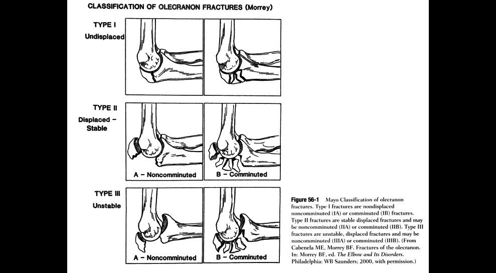
Q13Describe the Mayo classification of olecranon fractures and the indications for surgery.▸
- Type 1 undisplaced, type 2 displaced stable, type 3 unstable
- Subtype A simple, B comminuted
- Operate for a disrupted joint or disrupted extensor mechanism
- Options: fix, or excise and advance triceps
Q14How do capitellum fractures present and why do they displace?▸
- Coronal plane fracture after a fall on outstretched hand
- Little/no soft tissue attachment - often displaced
- Lateral X-ray shows the fragment displaced anteriorly; obtain AP view and CT
Q15Describe the Bryan and Morrey classification of capitellum fractures with eponyms.▸
- Type 1: large osseous fracture (Hahn-Steinthal)
- Type 2: one cartilaginous piece with little osseous component (Kocher-Lorenz)
- Type 3: comminuted (Broberg-Morrey)
- Type 4: extension to the trochlea (McKee)
- Laugier - isolated trochlea fracture
Q16Describe the Dubberley classification of capitellum fractures.▸
- Type 1: capitellum +/- lateral trochlear ridge
- Type 2: capitellum and trochlea as one single piece
- Type 3: capitellum and trochlea as separate pieces
Q17How are capitellum fractures managed?▸
- Undisplaced: splint x 3 weeks
- Displaced: fix - Kaplan approach for type 4, posterior approach for type 1
- May consider excision (type 2) or arthroplasty
Q18What determines the olecranon fracture pattern and what injuries are associated?📷▸
- Pattern: displacement, articular comminution, proximal fragment size
- Associations: UHJ dislocation, radial head dislocation, coronoid fracture
Q19Describe the Mayo classification of olecranon fractures.▸
- Type 1 undisplaced, type 2 displaced and stable, type 3 displaced and unstable
- Subtype A = simple, B = comminuted
Q20What are the aims of olecranon fracture fixation?▸
- Anatomical reduction, rigid fixation
- Restore the width of the trochlear notch
- Restore the proximal ulnar diaphyseal angle and olecranon diaphyseal angle
Q21What is a tension band and what are the prerequisites for its use?▸
- A device that converts a tensile force into a compression force
- Bone must be eccentrically loaded; opposite cortex intact to withstand compression
- Fracture must be transverse and simple; construct applied on the tension side
- Implant must be able to withstand tensile force
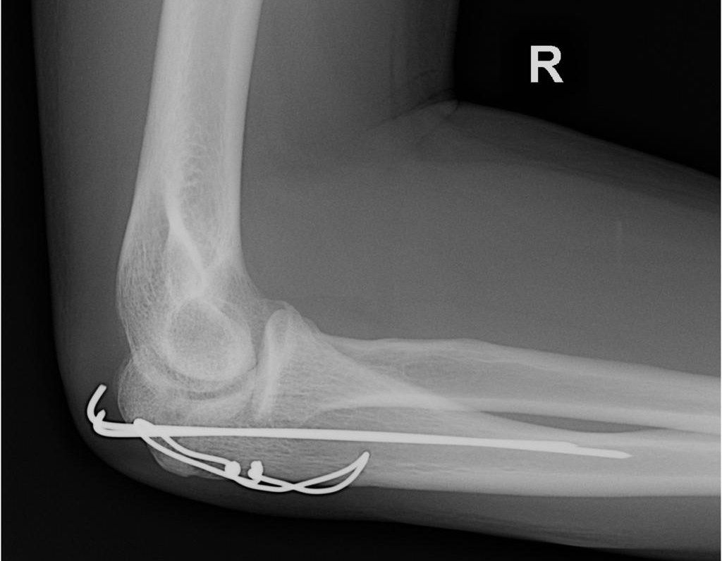
Q22Describe the tension band construct shown on these radiographs.▸
- AP and lateral radiographs of the right elbow
- Olecranon fracture fixed with two parallel pins and transosseous metal wire in a figure-of-eight configuration
- Restoration of the articular surface
Q23Give other fractures where tension band fixation is used.▸
- Patella, calcaneum, medial malleolus
- Greater tuberosity, femur shaft
Q24Why is the lateral side of the femur the tension side for a tension band plate?▸
- There is no lateral bowing of the femur; the tension side is determined by the anatomical axis
- Shifts the neutral axis from mid axis to the plate-bone junction
- Axial loading produces pure tension in the plate and pure compression across the fracture line
- Creates a bending close situation to reduce the working length of the plate
Q25Describe the X-ray findings in this forearm injury.📷▸
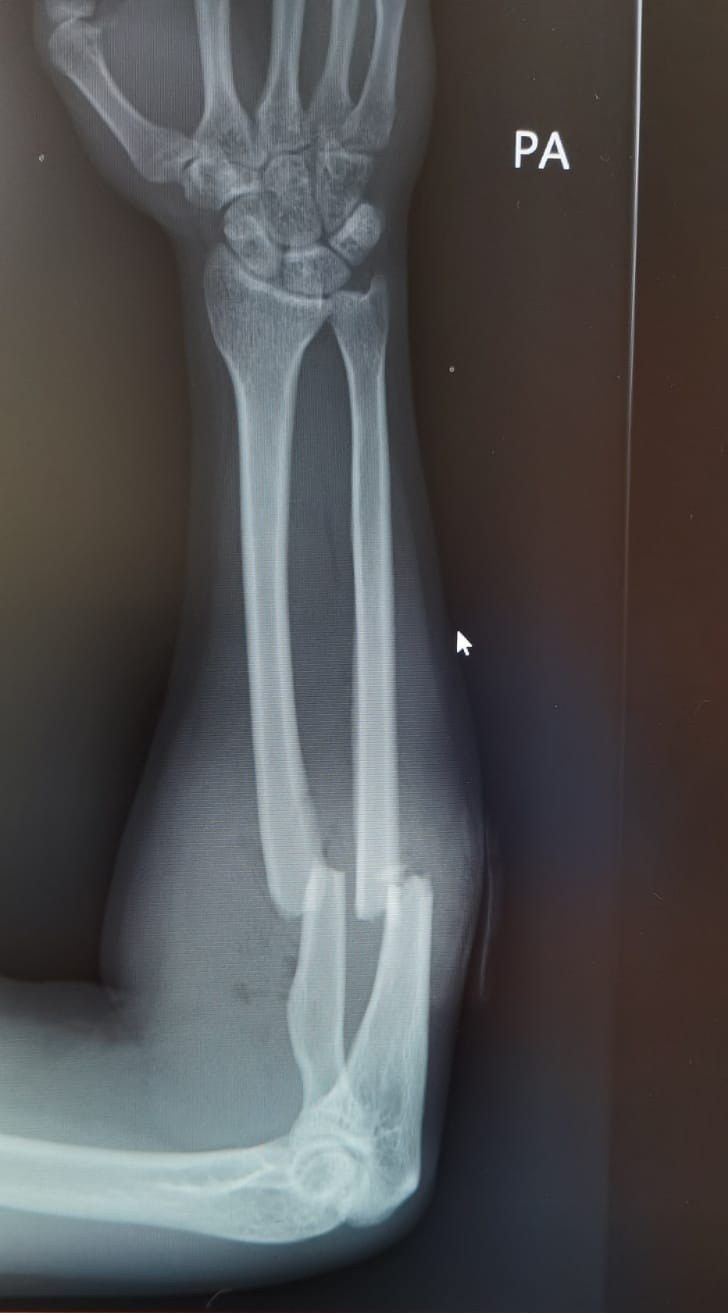
- PA radiograph of the right forearm: fractures of the proximal 1/3 shaft of radius and ulna with translation
- DRUJ and elbow joint congruent
- Gas pocket in the subcutaneous layer - suspicious of open fracture
Q26What history and examination are important in a both-bone forearm fracture?▸
- History: mechanism, associated injury, age, hand dominance, functional demand
- Examination: soft tissue, acute compartment, open wound, neurovascular status
- Examine the joint above and below
- 1mm inside out puncture wound
Q27What is the acute management and aim of treatment for this forearm fracture?▸
- Analgesia, IV antibiotics, temporary stabilization with a long arm slab
- Prepare the patient for ORIF
- Aim: adequate debridement, compression plate fixation, early mobilization
Q28What is the patient setup and skin incision for the approach to the proximal radius?▸
- Supine under GA
- Proximal landmark: biceps tendon
- Distal landmark: radial styloid process
Q29Describe the superficial dissection of the approach to the proximal radius.▸
- Between BR and FCR
- Identify radial artery and SRN beneath BR
- Retract the radial artery medially, ligate recurrent branches to the mobile wad
- Retract the SRN laterally
Q30Describe the deep dissection of the approach to the proximal radius.▸
- Supinate the forearm to displace the PIN away from the field
- Follow the lateral edge of the bicipital tuberosity, incise bursa
- Elevate the medial border of supinator from the radius
- Pronate the forearm (mid part) and partially detach the lateral border of pronator teres
Q31Describe the X-ray findings and diagnosis in this injury.📷▸
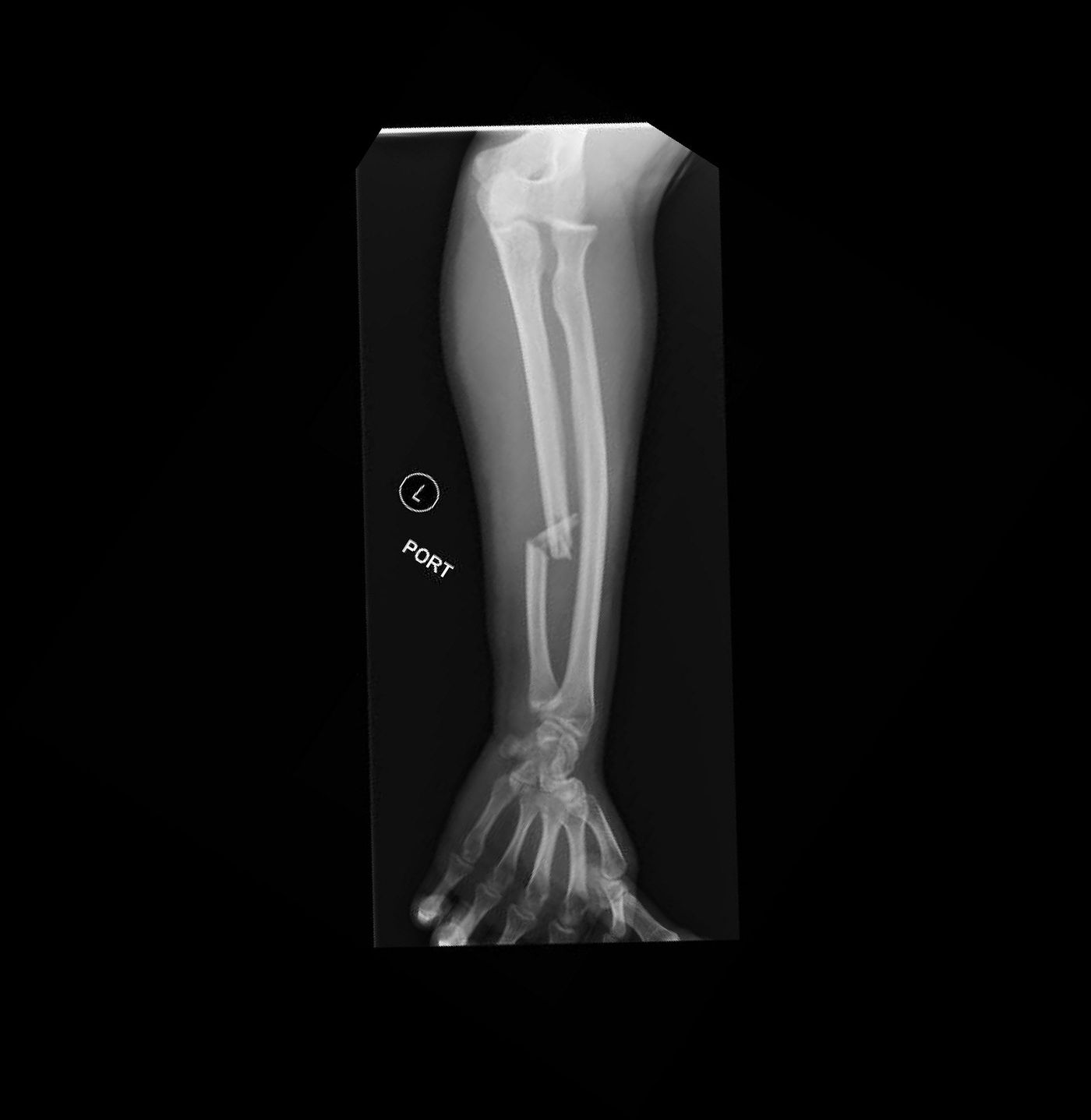
- Fracture of the distal 1/3 ulnar shaft with comminution and displacement
- Anterior dislocation of the radial head
- Diagnosis: Monteggia fracture, Bado 1
Q32What history and examination findings are important in a Monteggia fracture?▸
- History: time and mechanism of injury, associated injury
- P/E: soft tissue condition, open wound, acute compartment
- Palpate along the interosseous membrane and DRUJ for tenderness (Essex-Lopresti injury)
- Neurovascular injury especially PIN; associated injury in the ipsilateral limb
Q33How is a Monteggia fracture temporarily managed before operation?▸
- Fracture dislocation - try CR, but unlikely to maintain reduction with a slab alone
- Temporary stabilisation with a long arm slab
- Prepare for operation under GA
Q34What is the significance of the Bado classification of Monteggia fractures?▸
- Type I anterior: annular ligament
- Type II posterior: LUCL
- Type III lateral: PIN
Q35Describe the Jupiter classification subdivisions of Bado type II fractures.▸
- A: ulnar fracture involves the distal olecranon and coronoid process
- B: fracture at the metadiaphyseal junction distal to the coronoid
- C: diaphyseal ulnar fracture
- D: fracture extends along the proximal third to half of the ulna
Q36What are the aims and technique of ulnar fixation in this Monteggia fracture?▸
- Aim: anatomical reduction of the ulna (length, angulation, rotation), reduction of the RCJ, stable fixation for early mobilization
- Subcutaneous approach to the ulna
- Fix with a 3.5mm LCP in compression mode, aiming for primary bone healing
Q37How is the radiocapitellar joint assessed and managed after ulnar fixation?▸
- True lateral view of the elbow with the forearm in full supination to assess congruency
- If suspicious: recheck the ulnar reduction and prepare open reduction of the RCJ via Kocher
- Look for interposition of soft tissue, e.g. annular ligament
- Persistent radial head subluxation - check for malreduction, capsuloligamentous causes, coronoid or radial head deficiency
- Postop: hinged elbow brace with the forearm in supination
Q38What is the diagnosis and classification of this injury?📷▸
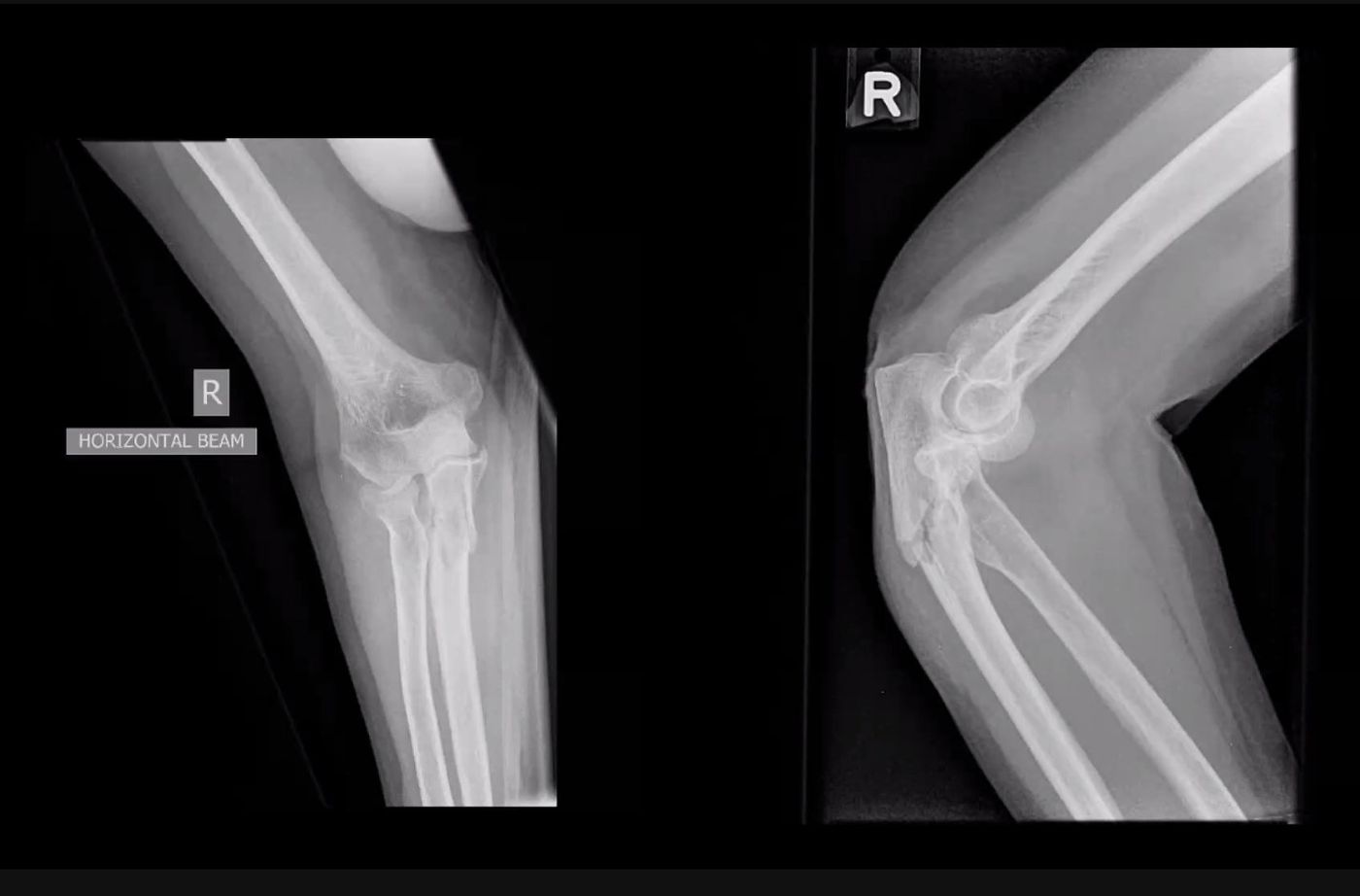
- Fracture of the proximal ulna with posterior radial head dislocation
- Monteggia fracture
- Bado II
- Jupiter IIB
Q39What history and examination are required in this Monteggia Bado II injury?▸
- History: premorbid status, level of activity
- Examination: soft tissue, wound, compartment
- Check PIN neurovascular status
Q40What are the aims of surgery for this Monteggia fracture?▸
- Concentric reduction of the radiocapitellar joint
- Anatomical restoration of ulnar alignment - length, rotation and angulation
- Stable fixation for early mobilisation
Q41Describe the steps of surgical management.▸
- Lateral decubitus position, arm board
- Posterior utilitarian approach, raise thick skin flaps both medially and laterally
- Radial head realigns with the capitellum after anatomical reduction of the ulna
- Temporary K-wire fixation; check PUDA, ODA, OW; then locking plate
Q42How are Monteggia variants with radial head fracture addressed?▸
- Radial head fracture can be addressed through the ulnar fracture or via a Boyd approach
- Address the coronoid fracture to restore the bony buttress of the UHJ
- Then perform ligament repair/reconstruction
Fact check
Mason type 4 radial head fracture (with dislocation) is attributed to modification by Hotchkiss — misattribution — The dislocation type 4 was added by Johnston in 1962 (Mason-Johnston classification); the Hotchkiss modification redefines types 1-3 by operative indication — source