Trauma
Shoulder and acromioclavicular joint injury
Anterior and posterior shoulder dislocation, fracture dislocation, AC joint dislocation.
36 questions
5 source pages
2 images
Images appear with the first question taken from each source page — tap a question to open it.
Q1Which classifications are used for ACJ and clavicle injuries?▸
- ACJ: Rockwood
- Clavicle: Allman & Neer
Q2Which classifications are used for the proximal humerus?▸
- Neer
- Hertel
Q3Which classifications are used for humeral shaft and distal humerus fractures?▸
- Humeral shaft: by morphology
- Distal humerus: AO, Holstein Lewis
Q4Which classifications are used around the elbow?▸
- Elbow dislocation: terrible triad, Cori cycle
- Olecranon: Mayo
- Coronoid process: Regand & Morray
- Radial head & neck: Mason
- Capitellum: Regand & Morray
Q5Describe the radiographic signs of a posterior shoulder dislocation.📷▸
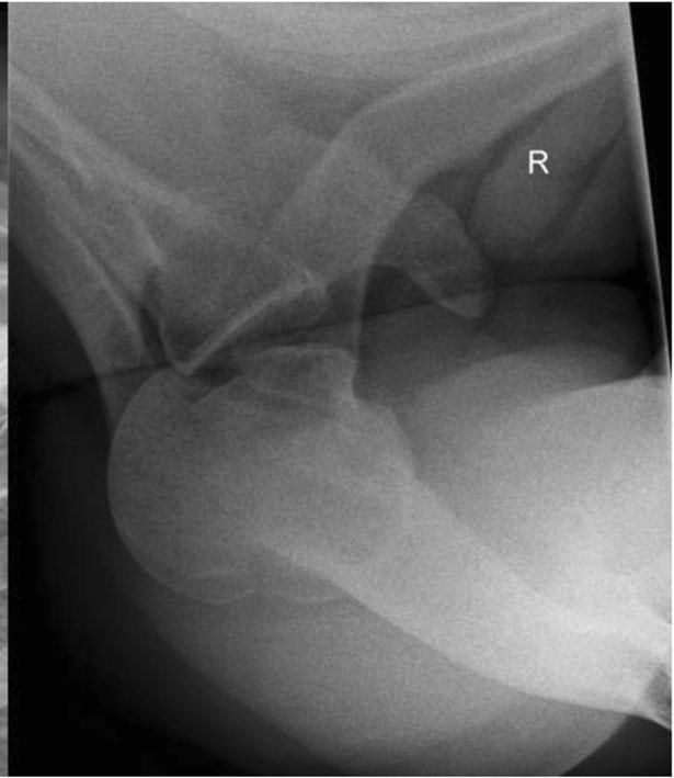
- Light bulb sign
- Trough line
- Positive rim sign (widening of GHJ)
- Absence of half-moon overlap
- Proximal humerus internally rotated and adducted
- No associated fracture in LT or surgical neck
Q6What history and examination findings suggest a posterior shoulder dislocation?▸
- MOI: direct - axial load on arm in flexion, adduction, IR; indirect - electric shock/convulsion
- Loss of shoulder contour, asymmetrical axillary fold, prominent coracoid (not obvious)
- Arm held in IR and adduction; painful reduced ROM particularly ER and FF
- Check NV status esp axillary nerve, brachial plexus, axillary artery; associated trauma (high energy), SSSC
Q7How do you confirm the diagnosis of posterior shoulder dislocation?▸
- Clinical examination
- Axillary view - most useful to assess direction of injury
- Velpeau view - most useful if patient is in pain
- CT - confirm direction of injury and associated fractures
Q8How is an acute traumatic posterior shoulder dislocation reduced?▸
- Closed reduction under GA with an assistant
- Delpalma method: caudal traction in line of the adducted IR deformity (elbow flexed) with lateral lifting
- Recheck NV status post CR; immobilise in ER for 4-6 weeks
- Arrange CT to assess associated occult fracture/bone defect
Q9What are the primary stabilisers against posterior shoulder dislocation?▸
- Posterior band of IGHL - static restraint in IR
- Subscapularis - dynamic restraint in ER
- CH ligament
Q10How is a chronic missed posterior dislocation (>6 weeks) managed?▸
- Reverse HS lesion = loss of normal convexity in the anteromedial part, depression/defect of humeral head
- CR unlikely successful; complete workup with XR/CT to assess degree of reverse HS defect
- Open reduction with subscapularis transfer (McLaughlin) or LT transfer (modified McLaughlin)
- Castagna: modified McLaughlin - significant functional improvement, no intra/postop complications at mean FU 62.8 months
Q11What history points are important in a suspected posterior shoulder dislocation?▸
- Mechanism of injury
- Patient's age and hand dominance
Q12How are the axillary and Velpeau views taken?▸
- Axillary: arm in 90 degrees abduction, plate above the shoulder, beam into the axilla
- Velpeau: patient leans 45deg backward, plate on the floor, beam directed caudally
Q13What is the difference between the classic and modified McLaughlin procedure?▸
- Classic: subscapularis transfer
- Modified: adds transfer of the lesser tuberosity for better bone filling of the defect and more secure subscapularis insertion
Q14What is the terrible triad of shoulder dislocation?▸
- Dislocation
- Cuff injury
- Brachial plexus injury
Q15When should a greater tuberosity fracture be fixed after reduction?▸
- If GT displaced >5mm post reduction
- May be fixed open or arthroscopically
Q16What is the evidence for arthroscopic fixation of the greater tuberosity?▸
- Fixation with a double row of anchors shown to have superior postoperative ROM
- CORR 2016
Q17How does the Rockwood classification guide management of ACJ dislocation?📷▸
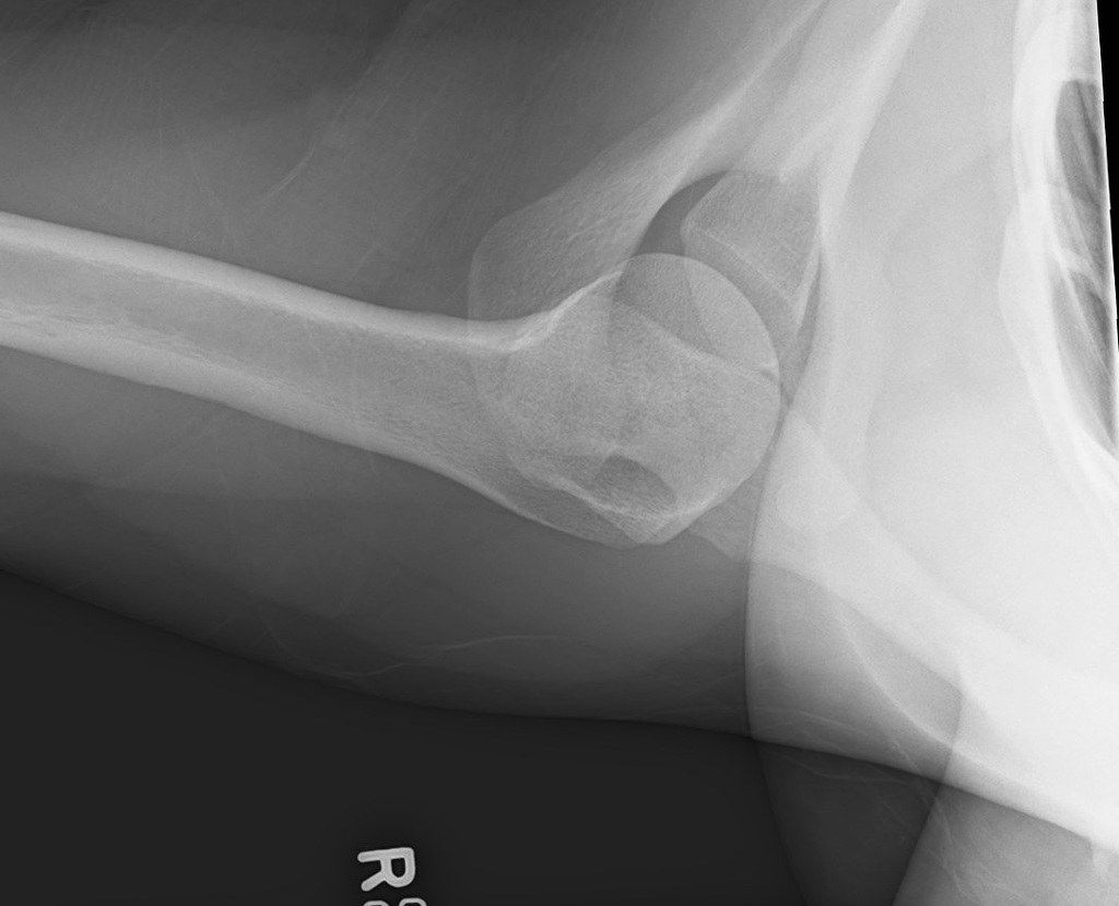
- 1-3: static stabilisers disrupted (AC sprain, AC torn, CC torn) - conservative
- 4-6: dynamic (posterior through trapezius, D+T torn, inferior below coracoid) - surgical
- Type 3: +/- operation
- If untreated the shoulder becomes protracted
Q18What imaging views are used for ACJ dislocation?▸
- Contralateral side for comparison
- Axillary view - AP translation
- Zanca view - 10 degree cephalic tilt, vertical translation
- +/- stress view
Q19What is the Cochrane 2019 evidence for ACJ dislocation?▸
- Low evidence for surgery
- At 10 yr FU, minimal degenerative changes
- Conservative actually has better functional outcomes at 6 weeks
Q20When is distal clavicle resection indicated in ACJ disease and how much bone is removed?▸
- Indicated for ACJ arthritis without instability
- Resect 7-8mm, <1.5cm of distal clavicle - too much jeopardises the ACJ capsule
- Can be performed arthroscopically
Q21What is the role of tendon reconstruction in chronic ACJ dislocation?▸
- Tauber 2009 AJSM: semitendinosus tendon graft for coracoclavicular ligament reconstruction
- Gave superior clinical and radiologic outcomes compared with the modified Weaver-Dunn procedure
Q22Describe the X-ray findings in this ACJ dislocation.▸
- AP: ACJ dislocation with marked superior displacement of the distal end of the clavicle
- No associated fractures; will need further Xray to classify
- Axillary view: posterior dislocation of the distal clavicle
- Summary: Rockwood 3 posterior dislocation of the ACJ
Q23What history and examination points are important in ACJ dislocation?▸
- Mechanism of injury; acute vs chronic
- Associated injury around the shoulder girdle - SSSC, floating shoulder
- Neurovascular status
- Compare with the contralateral side
Q24What are the options for CC fixation in acute ACJ dislocation?▸
- Bosworth screw, CC sling, fibre tape
- Tightrope loop suspensory fixation (A-assisted)
Q25What is the direct ACJ fixation option and its drawback?▸
- Hook plate
- Provides anatomical reduction
- Needs a second operation for implant removal
Q26How is chronic ACJ dislocation managed?▸
- Modified Weaver-Dunn procedure (classic without additional fixation)
- If just ACJ arthritis with no instability: resection of the distal clavicle
- Tendon reconstruction with semitendinosus graft (Tauber 2009 AJSM)
Q27What are the ACJ stabilisers?▸
- Static: AC lig, CC lig, ACJ capsule
- Dynamic: Trapezius, deltoid muscle
Q28How do you assess a patient with a suspected ACJ dislocation?▸
- Hx: mechanism of injury, acute vs chronic
- P/E: associated injury around the shoulder girdle - floating shoulder / SSSC
- Neurovascular status and contralateral side for comparison
Q29What is the evidence from the Cochrane 2019 review?▸
- Low evidence for surgery; most studies used hook plate
- At 10 yr FU, minimal degenerative change; conservative better functional outcomes at 6 weeks
- Late reconstruction results comparable to early surgery; main problem is deformity
- One report quotes 15% reduced bench press strength
Q30What are the options for CC fixation in acute ACJ dislocation?▸
- Loop suspensory fixation (tightrope, A-assisted)
- LARS ligament, Bosworth screw, CC sling, fibre tape
Q31What does the Arirachakaran 2016 meta-analysis show?▸
- Suspensory device fixation gives higher shoulder function scores than metallic fixation (hook plate)
- Suspensory fixation also had lower postop pain
Q32Describe the modified Weaver-Dunn procedure.▸
- Excision of distal clavicle
- Transfer acromial end of CA ligament to distal clavicle
- CC fixation
- Indicated for chronic ACJ dislocation with +ve arthritis
Q33What are the static and dynamic stabilisers of the ACJ?▸
- Static: AC lig, CC lig, ACJ capsule
- Dynamic: trapezius, deltoid muscle
Q34Describe the X-ray findings in this ACJ dislocation.▸
- ACJ dislocation with marked superior displacement of the distal end of the clavicle
- Look for associated fracture, especially floating shoulder or SSSC injury
- Further X-ray needed to classify
Q35What is the direct fixation option for ACJ dislocation?▸
- Hook plate
- Provides anatomical reduction but needs a second operation for removal of the implant
Q36When is resection of the distal clavicle performed and how much bone is removed?▸
- For chronic ACJ arthrosis without instability
- Remove 7-8mm, less than 1.5cm of distal clavicle
- Too much resection jeopardises the capsule
- Can be performed arthroscopically