Trauma
Hindfoot and midfoot fractures
Talus and calcaneal fractures, Broden view, cuboid and navicular fractures, subtalar dislocation.
80 questions
11 source pages
5 images
Images appear with the first question taken from each source page — tap a question to open it.
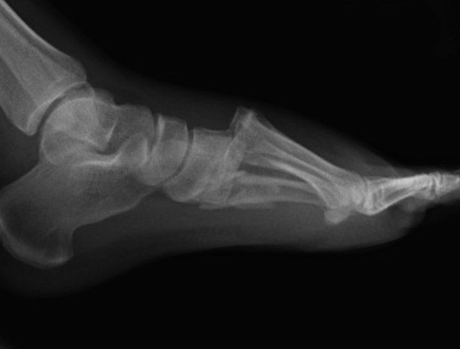
Q1Describe the X-ray findings in this talar neck fracture.📷▸
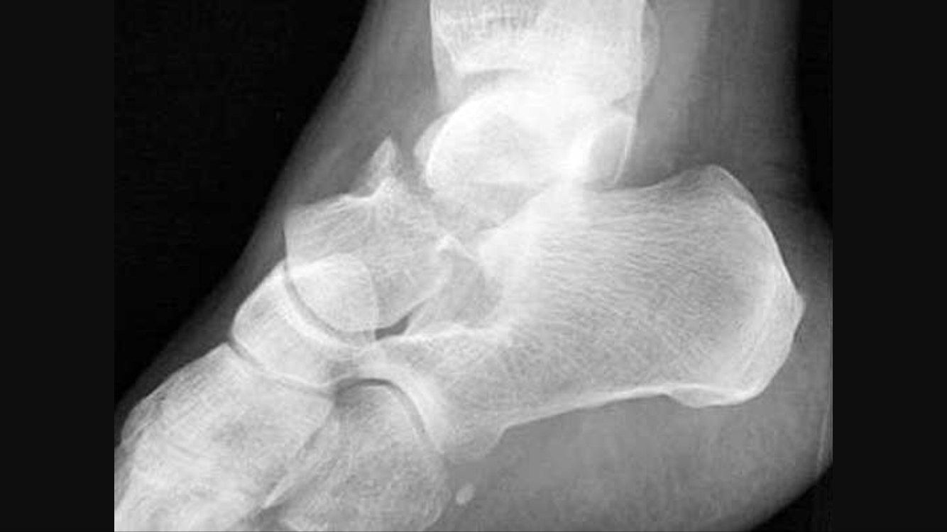
- Hawkin 2 fracture of talar neck with subtalar joint dislocation; tibiotalar and TNJ congruent
- No soft tissue gas (open fracture in 50% of type 3 and 4)
- Need other X-rays - ipsilateral foot fracture in 90%
Q2What is the mechanism of a talar neck fracture?▸
- Dorsiflexion with axial load
- Impingement of the talar neck by the anterior lip of the tibia
Q3Describe the Hawkins classification and AVN risk.▸
- 1: no dislocation, minimally displaced (0-13%)
- 2: subtalar joint dislocation (50%)
- 3: subtalar joint + ankle dislocation (90%)
- 4: subtalar + TN + ankle joint dislocation (100%)
Q4How is a talar neck fracture reduced acutely?▸
- ATLS; assess skin condition, compartment syndrome and NV injury
- Sedation; flexed knee relaxes gastroc, ankle PF relaxes TA; stabilise malleoli, apply longitudinal traction +/- varus/valgus correction
- No multiple attempts; failed CR in 25% -> OR require MM osteotomy to preserve deltoid artery
- Analgesia and backslab after CR; Canale view and CT for fracture personality/medial comminution
Q5How is the Canale view performed?▸
- Ankle in maximum plantarflexion
- Foot pronated 15 degrees
- XR beam 75deg from horizontal
Q6What is the definitive management of a talar neck fracture?▸
- Aim: anatomical reduction, rigid fixation for primary bone healing
- Non-op if CT-confirmed Hawkin I with no articular step off: short leg cast 8-12 weeks
- ORIF: usually AL approach first (beware superficial peroneal nerve, full-thickness flaps, EDB retracted)
- Fix lateral first as foundation or cortical key for reduction, then medial; medial buttress plate to prevent varus
- Posterior: percutaneous headless cannulated screw posterior to anterior
Q7What are the reduction and fixation details of ORIF for talar neck fracture?▸
- Reduction with forceps, direct manipulation, K wire as joystick +/- joint distractor; prelim K wire fixation assessed on Canale view
- Fix with lag screw/positional screw
- Medial approach: medial malleolus to base of 1st MT, plane between TA and TP, protect saphenous nerve and vein; +/- medial malleolar osteotomy + buttress plate/positional screw
- Posterior: percutaneous cannulated headless titanium screw (countersink), posterior to anterior, perpendicular to fracture line, maximal bone purchase (P to A mechanically more stable, does not disrupt TNJ)
Q8What is the post-operative rehabilitation and what are the risk factors for AVN?▸
- NWB walking for 10-12 weeks
- AVN risk factors: Hawkins classification and timing of reduction
Q9What is Hawkins sign and its significance?▸
- Subchondral radiolucency at the talar dome at 6-8 weeks on serial AP XR
- Reactive hyperaemia causing bone resorption - likely no AVN (but not r/o)
- Sclerosis indicates loss of blood supply
- Sensitivity 100%, specificity 58%; MRI if in doubt
Q10What is the blood supply of the talus and the complications of talar neck fractures?▸
- Posterior tibial artery via artery of tarsal canal (inferior body) and deltoid branch (superior body)
- Perforating peroneal artery via artery of sinus tarsi (lateral); anterior tibial artery via dorsalis pedis (head/neck)
- Talus prone to AVN: 80% covered by articular surface, no muscular attachment, tenuous retrograde supply
- AVN overall 30%, subtalar arthritis 50%, ankle OA 33%
Q11What causes varus malunion after talar neck fracture?▸
- Medial comminution -> varus malunion
- Decreased subtalar eversion and weight bearing on the lateral foot
Q12How is AVN of the talus managed?▸
- Differentiate the cause of pain (talus vs ankle joint)
- NWB until creeping substitution
- Usually symptoms are not bad
Q13Describe the anatomy of the talus.▸
- Head, neck and body
- Superior = trochlear surface (articulates tibia and fibula); inferior lateral process articulates with the posterior facet of the calcaneum
- Posterior process: medial and lateral tubercles separated by a groove for FHL; PTFL attaches to the lateral tubercle (Shepherd fracture); medial tubercle fracture = Cedell fracture
- Anterior surface articulates with navicular and sustentaculum tali
Q14How is a medial malleolar osteotomy performed?▸
- AM approach; open capsule to visualise the apex between the medial malleolus and tibial plafond
- Predrill screw tract and pretap
- Osteotomy at 90 degrees to the screw tract (45 degrees from tibial plafond); finish cut with osteotome
Q15Describe the X-ray findings and fracture type.📷▸
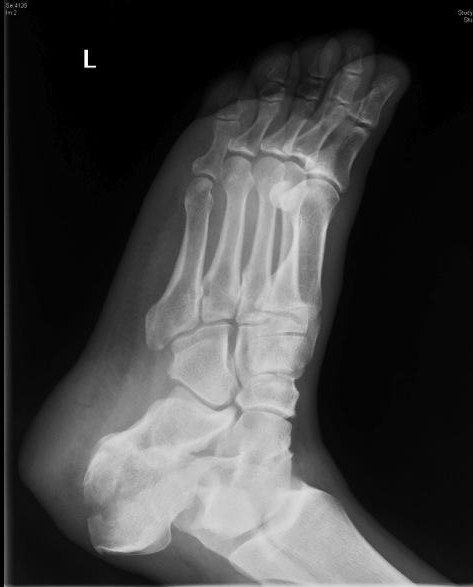
- Intraarticular calcaneal fracture extending into the posterior facet
- Reduced Bohler and increased Gissane angle; no obvious fracture to the CC joint
- Essex-Lopresti tongue type (extends to posterior tuberosity); otherwise joint depression type
Q16What imaging views and classification are used?▸
- Lateral: Bohler, Gissane, calcaneal shortening
- Harris (axial): widening, varus
- Broden: posterior facet
- CT to plan; Sanders classification on Widest 30 deg semicoronal cut (1-4)
Q17What is the initial assessment and management of a calcaneal fracture?▸
- High energy trauma: ATLS, AMPLE, primary survey
- Slab for rest + elevate and ice
- Frequent monitoring for compartment syndrome; unconscious patient needs continuous slit catheter monitoring
- CT to plan management
Q18What associated injuries must be ruled out?▸
- Local: wound, skin impingement, compartment syndrome, NV status
- Systemic signpost injuries: TL/L spine compression fracture 10%, contralateral os calcis fracture 10%
Q19Describe the Sanders classification.▸
- Based on the widest 30-degree semicoronal CT cut
- Grades 1-4 (1: no stepping; 2-4: 2-4 parts)
Q20Which patient and fracture factors affect definitive management?▸
- Patient: DM, PVD, smoking - >90% wound complication rate and poor outcome
- Fracture: articular step >2mm, calcaneal height, width, valgus/varus alignment
- Soft tissue factors such as skin impingement
Q21What is the timing of calcaneal surgery?▸
- Wait until the wrinkle sign is positive and blisters are epithelialized
Q22What are the aims of operative treatment?▸
- OT aim: restore articular surface of posterior facet
- Restore calcaneal height and length (Bohler and Gissane angles) to restore triceps surae lever arm
- Correct varus to prevent late instability (hindfoot in varus locks up CC and TN joints, more lateral foot weight bear, more metatarsalgia)
- Restore width to prevent peroneal tendon impingement
Q23Describe the sinus tarsi approach and reduction sequence.▸
- Incision 2cm distal to fibular tip to base of 4th MT; beware sural nerve, IDCN and peroneal tendons
- Use sustenaculum tali as the keystone for reduction (constant fragment held by surrounding TC ligament); reduce anterior process first, then jig up posterior facet with osteotome or lamina spreader, K wires to sustentaculum tali
- Steinman pin restores length and varus/valgus; Xray to assess reduction for posterior facet by broden's view, calcaneal alignment by Harris view
- Bone graft, subchondral raft screws with 3.5mm cortical screw, lateral wall, low-profile locking plate, Allgower technique closure
Q24How are the Broden and Harris views taken?▸
- Broden: ankle neutral dorsiflexion, 45 IR (45 degrees internal rotation), XR with 40, 30, 20, 10 degrees cephalic tilt
- Harris: ankle maximum dorsiflexion, beam 45 degrees from horizontal caudal tilt
Q25What is the rehabilitation after calcaneal fracture?▸
- NWB for 6-8 weeks
Q26What causes long-term chronic pain after calcaneal fracture?▸
- 40% will have long term chronic pain
- Causes: subtalar incongruity, penetration of screws, lateral impingement, peroneal tendinitis, talar neck impingement
Q27What are the epidemiological associations of calcaneal fractures?▸
- 17% of fractures are open
- 10% bilateral, 10% involve spine fracture
- 63% involve the CCJ
Q28Describe the relevant calcaneal anatomy.▸
- Posterior, middle and anterior facets for talus; anterior facet for cuboid
- Sinus tarsi: groove dividing anterior and posterior subtalar joint
- Sustenaculum tali: medial projection supporting the talar neck, FHL runs below; deltoid and talocalcaneal ligament attachments -> constant AM fragment
- Bifurcate ligament: anterior process to CC and CN, Y shaped
Q29How are Bohler and Gissane angles measured and what are the normal values?▸
- Bohler: angle from highest point of anterior process to highest point of posterior facet and highest point of superior tuberosity; normal 20-40 degrees; reduced = collapse of posterior facet
- Gissane: angle between posterior facet and line joining lowest point of posterior facet to highest point of anterior process; normal 130-145 degrees; increased = collapse
Q30What are the emergency presentations of a calcaneal fracture?▸
- Skin impingement in tongue type/avulsion fracture
- Compartment syndrome (10%) and NV injury
- Open fracture (10%)
Q31Describe the Essex-Lopresti classification.▸
- Tongue type: secondary fracture line exits at the posterior calcaneal surface
- Joint depression type: secondary fracture line exits at the superior calcaneal surface
Q32Describe the primary fracture line and the constant fragment.▸
- Oblique fracture line from anterolateral to posteromedial
- Two fragments: AM & PL
- AM contains the sustentaculum tali, attached to the proximal fragment by strong ligaments (deltoid, medial talocalcaneal, spring) = constant fragment
Q33Describe the extensile L-shaped approach.▸
- Vertical limb between lateral malleolus and Achilles; >100 degree turn; horizontal limb between glabrous and non-glabrous skin
- Protect sural nerve and lateral calcaneal branch of the peroneal artery
- Subperiosteal dissection, elevate full thickness periosteocutaneous flap; extend proximally to body of talus; excise CF ligament; bent K wires as retractors
Q34What are the complications and their management?▸
- Wound complications 25%; FHL irritation from a long screw at the sustentaculum tali; compartment syndrome
- Malunion (Stephen and Sanders classification: talar declination angle <20, talocalcaneal angle)
- Lateral exostosis - resection; subtalar OA - fusion (distraction bone block); varus - valgus osteotomy
Q35What is the evidence and prognosis?▸
- UK heel trial BMJ 2014 and extended Heft trial 2021 BJJ do not support surgery (methodological flaws)
- Buckley JBJS 2002: outcomes similar; surgery may be better when IOD group is excluded
- Overall poor prognosis: >40% long-term chronic pain
- Complications: wound complications 25%, FHL irritation, compartment syndrome, malunion
Q36What are the details and criticisms of the key trials, and what predicts a favourable outcome?▸
- UK heel trial BMJ 2014: wound complication 19%, many excluded as 'obviously needed surgery', postop CT only looked at subtalar stepoff
- Buckley JBJS Am 2002: outcomes similar; when IOD excluded, surgery better
- Favourable prognosis: female, non-IOD, sedentary work, anatomical reduction, less initial disruption of Gissane/Bohler
- Later study: overall cost less with surgical management due to fusion surgery and disability costs in non-op patients
Q37Describe the fracture seen on this lateral X-ray.📷▸
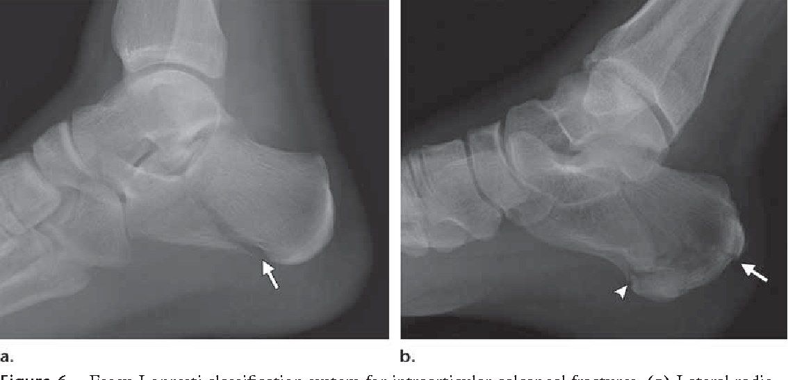
- Intraarticular calcaneal fracture - joint depression / tongue / both JD&T type
- Fracture extends from the posterior facet and exits the inferior/posterior calcaneal surface
- Posterior facet collapsed: decreased Bohler angle, increased angle of Gissane
Q38What is the significance of the changed Bohler and Gissane angles?▸
- Indicates collapse of the posterior facet
- Decreased Bohler angle
- Increased angle of Gissane
Q39What is the Broden view and when is it used?▸
- Used intraoperatively for facet joint reduction
- Ankle in neutral dorsiflexion
- X-ray taken at 45 degrees internal rotation
Q40What is the Harris view and what does it show?▸
- Shows widening of the calcaneum and angulation of the tuberosity fragment
- Ankle in dorsiflexion
- X-ray beam at 45 degrees caudal tilt
Q41What is the mechanism and clinical concern in this fracture?▸
- Avulsion fracture from eccentric contraction of gastrocnemius
- Displaced fracture -> skin tenting, skin necrosis
Q42What is examined in a calcaneal tuberosity fracture?▸
- Soft tissue: skin impingement
- Distal NV status
Q43What is the Beavis classification of calcaneal tuberosity fractures?▸
- Type 1: avulsion (usually osteoporotic insufficiency fracture)
- Type 2: beak (direct blow, EOT)
- Type 3: infrabursal avulsion (superficial fibres involved; suture anchor)
- Type 4 (modified by Lee): only deep fibres involved (conservative treatment)
Q44What is the management and what are the complications?▸
- CR + percutaneous screw fixation with washers
- OR + tension band wire fixation
- Undisplaced: conservative - below knee cast with ankle in plantarflexion
- Complications: skin necrosis, fracture displacement, weak push-off strength
Q45What injury is shown and which radiographic signs confirm it?📷▸
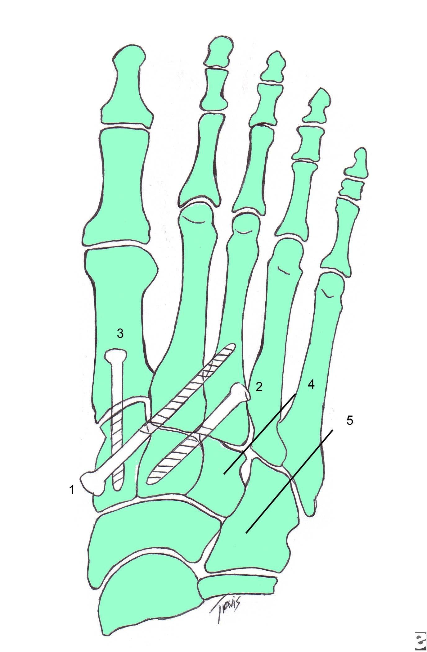
- This is a bony Lisfranc injury
- DP view: widening of the space between the 1st and 2nd MT with a fleck sign
- The line along the medial border of the middle cuneiform and 2nd MT is disrupted
- No other cuboid or navicular fractures seen
Q46What additional views are needed and what do they show?▸
- Lateral: dorsal subluxation of the 1st or 2nd MT base
- Oblique: 4th MT medial line with medial cuboid; 3rd MT lateral line with lateral border of lateral cuneiform
Q47What X-ray views are required in suspected Lisfranc injury?📷▸
- DP: line along medial 2nd MT and middle cuneiform; 1st-2nd MT widening >3mm; fleck sign
- Oblique: TMTJ overlap; medial 4th MT and cuboid
- Lateral: dorsal subluxation of 1st/2nd MT base
- Relevant negatives: cuboid and dorsal navicular avulsion fracture; abduction stress views if delayed presentation
Q48What is the spectrum of Lisfranc injury and the Nunley classification?▸
- Spectrum from sprain to dislocation of the TMT joint complex
- High energy/athletic: axial load through hyperplantarflexed forefoot, associated MT/T fractures
- Nunley (low energy): Stage 1 = undisplaced; Stage 2 = widening >2mm on AP with no dorsal subluxation; Stage 3 = dorsal subluxation on lateral
Q49What structures provide Lisfranc stability?▸
- Lisfranc joint complex = MT, intermetatarsal and intertarsal articulations
- Static bone: roman arch, keystone (mortise created by medial cuneiform and recessed middle cuneiform)
- Static ligaments: Lisfranc ligament, interMT, plantar (C1M2M3) and dorsal TMT ligaments
- Dynamic: plantar aponeurosis, intrinsics, PTT and ATT, peroneus longus
Q50What is the Lisfranc ligament?▸
- Interosseous ligament from the medial cuneiform to the base of the 2nd MT on the plantar surface
- Tightens with pronation and abduction of the forefoot
Q51What is the Myerson classification and what initial management is required?▸
- Myerson: total, partial incongruent, divergent
- Rule out compartment syndrome; if grossly displaced try CR then temporary immobilization + elevate and ice
- Assess soft tissue: plantar ecchymosis is the hallmark feature
- +/- CT (mainly preop planning) / MRI (pure ligamentous injury)
Q52What are the aims and timing of definitive management?▸
- Anatomical reduction of the Lisfranc joint, prevent instability and early OA
- If missed -> rocker bottom deformity
- Emergent if open/compartment/irreducible; else wait 2-3/52
- Options: ORIF for fracture dislocation (medial screw, lateral K wire); primary medial column arthrodesis for purely ligamentous arch injuries
Q53What are the indications for primary fusion?▸
- Ligamentous injury + multiplanar instability
- Delayed presentation
- Mid to advanced age
- Intraarticular comminution
Q54How would you perform an EUA?▸
- Stabilise the midfoot then apply varus/valgus stress, supination/pronation, dorsoplantar force to the forefoot
- Widening with pronation and abduction of the forefoot
- Assess TMTJ dorsal subluxation
Q55Describe the operative details of ORIF.▸
- GA, tourniquet; mark DP before tourniquet
- Dorsal incisions between 1st-2nd MT and over 4th MT (at risk: dorsalis pedis, superficial and deep peroneal nerves)
- Dorsomedial approach: plane between TA and EHL
- Reduce 2nd MT into the keystone, then reduce and fix 1st TMTJ; assess reduction on XR
- Medial column rigid fixation: retrograde 3.5mm cortical positional screw (pocket hole) or 2.4/2.7 VALCP in bridging mode; 2.0 K wire lateral column (4th MT to cuboid)
- Assess the foot for intercunieform instability and fix if needed; short leg cast/splint
- NWB 6 weeks; K wires out at 6 weeks then gradual weight bearing; screws out at 6 months; arch support 6 months
Q56What is the evidence for ORIF versus primary arthrodesis?▸
- JBJS 2006 Ly: primary arthrodesis of 2-3 medial rays - better pain control and function, less hardware removal in pure ligamentous injury
- Henning 2009 / CORR 2016 Smith: ORIF more hardware removal, functional outcome no difference
- ORIF shifting towards bridging plates rather than screw fixation
Q57What is the prognosis of a Lisfranc injury?▸
- 50% posttraumatic OA
- Poor prognosis: IOD, female and young, smoker
Q58What force causes a cuboid fracture and what are the surgical indications?▸
- Abduction force
- Surgery if articular step >2mm or lateral column shortening
Q59How is a cuboid fracture treated operatively?▸
- Simple - plating/screw
- Comminuted/shortening/severe/bone loss - bridge plate or ex fix
Q60What are the poor prognostic factors for a cuboid fracture?▸
- Patient: young age, IOD, high energy/direct trauma
- Fracture: non-anatomical reduction
Q61What is the management of a failed ORIF of a cuboid fracture?▸
- Salvage fusion
Q62Which subtalar dislocation is more common and how does it lock?▸
- Medial dislocation is more common - 70%
- Medial locks in supination; lateral locks in pronation
- Lateral - more high energy, often open
Q63What structures can block reduction in lateral versus medial subtalar dislocation?▸
- Lateral dislocation - medial structures: PT, FDL, FHL
- Medial dislocation - lateral structures: EDB, peroneal tendon, TN dorsal capsule
Q64How is closed reduction performed for a subtalar dislocation?▸
- Knee flexed
- Inversion for lateral dislocation
- Eversion for medial dislocation
Q65What imaging is required in a subtalar dislocation?▸
- CT to rule out fracture
- 40% have an associated fracture
Q66What is the definitive management of a subtalar dislocation?▸
- Most are stable after reduction: NWB with below knee cast
- Unstable with no fracture: ensure no soft tissue interposition, then transfixing K wire
- Failed CR - OR (medial dislocation via AL/sinus tarsi approach and vice versa)
Q67Which side is associated with a fracture in a subtalar dislocation?▸
- Fracture is associated on the displaced side
Q68Which classification is used for navicular fractures?▸
- Sangeorzan classification
- Based on fracture plane, comminution and direction of forefoot displacement
Q69Describe a Sangeorzan type I navicular fracture.▸
- Fracture in the coronal plane
- No angulation of the forefoot
Q70Describe a Sangeorzan type II navicular fracture.▸
- Fracture line from dorsal-lateral to plantar-medial
- Forefoot displaced medially
Q71Describe a Sangeorzan type III navicular fracture.▸
- Comminuted fracture in the sagittal plane
- Forefoot displaced laterally
Q72What are the indications for surgery in a navicular fracture?▸
- >2 mm displacement
Q73What are the complications of a navicular fracture?▸
- Nonunion
- Loss of medial longitudinal arch support
- Painful TNJ (talonavicular joint)
- Post-traumatic OA
- AVN and collapse
Q74Why do you release the foot compartments?▸
- Ischaemia of the intrinsic muscles leads to clawing
- Compartment release is performed to prevent this
Q75How many compartments does the foot have and what are they?▸
- 9 compartments
- Medial and lateral compartments
- 3 central compartments - superficial, mid and deep
- 4 interosseous compartments
Q76What does the medial release of the foot decompress?▸
- Medial foot border
- Central superficial and middle compartments
- Performed through a medial wound
Q77What does the DL release decompress?▸
- Lateral 2 interossei
- Lateral compartment
- Performed through a dorsal wound over the 2nd and 4th MT
Q78What does the DM release decompress?▸
- Medial 2 interossei
- Central deep compartment
Q79What is the evidence for management of foot compartment syndrome?▸
- AAOS 2013 review article: no strong evidence exists to guide management
- Emergent fasciotomy is commonly recommended to prevent pain and deformity
Q80What surgical wounds are used to release the foot compartments?▸
- Dorsal wound over the 2nd and 4th MT
- Medial wound