Trauma
Ankle fracture - patterns and syndesmosis
Intra-articular tibial fracture, syndesmosis complex, Lauge-Hansen and Maisoneuve injury.
32 questions
5 source pages
2 images
1 fact-check flags
Images appear with the first question taken from each source page — tap a question to open it.
Q1Describe the X-ray findings in this ankle.▸
- Comminuted tibial metaphyseal fracture with intraarticular extension, fracture distal fibula
- Articular impaction and comminution
- Any association joint subluxation or dislocation. The talar dome appears intact
Q2What classifications are used for tibial plafond (pilon) fractures?▸
- Ruedi-Allgower: 1 undisplaced, 2 displaced with minimal comminution, 3 comminuted
- AO: A extraarticular, B partial articular, C complete articular
- CT fragment description: Topliss classification
Q3What is the initial assessment for a high-energy pilon fracture?▸
- High-energy trauma - ATLS protocol
- AMPLE history
- Primary and secondary survey, looking for other axial loading type injuries
Q4What local and systemic assessment is needed in a pilon fracture?▸
- Local: circumferential assessment of the limb - soft tissue, compartment syndrome, NV status
- Systemic: secondary survey for axial load injuries - calcaneus, knee, spine
Q5What is the initial Span Scan Plan management?▸
- Analgesia, elevate, ice therapy
- Span: ankle spanning external fixator - delta frame construct; tibial pin >=3cm from fracture site to prevent fracture propagation; +/- plate fibula
- Scan: CT for planning - typically 3 fragments: medial, Chaput (AL), Volkman (PL), central, die punch
Q6Why is the fibula plated alongside the spanning fixator?▸
- According to Robertson's paper
- Maintain length and better soft tissue resting
Q7What are the typical CT fragments and their ligament attachments?▸
- Typically 3 fragments; ligaments usually intact
- Medial, Chaput (AL) - AITFL, Volkman (PL) - PITFL, central, die punch
- Descriptive Topliss classification
Q8What is the Topliss classification?▸
- Coronal: CS, PS, AS, V, Y
- Sagittal: SS, IV, T
Q9What are the definitive management options and timing?▸
- ORIF with plating; definitive external fixator (hybrid/ring); combined ex fix + ORIF
- Consider patient factors: premorbid state, DM, PVD, smoking
- Timing: when wrinkle sign positive and blisters epithelialized (13-28 days)
Q10What are the Ruedi-Allgower principles of fixation?▸
- 1. Restore fibular length and rotation
- 2. Reconstruct the articular surface posterior to anterior (PL -> C -> AL -> M); lag screw A to P
- 3. Restoration of bone defect (bone graft)
- 4. Tibial buttressing (articular block to shaft); plate medial if varus, lateral if valgus
Q11What are the surgical approach and fixation details?▸
- Classic PL + AM approach; beware 7cm skin bridge; fragment-specific (AM for plate, AL for Chaput) if <7cm
- Fix fibula: simple fracture - fix before tibia; complex - fix after
- Reduce articular block first, then connect to metaphysis + compression if possible
- Lag screw anterior to posterior; angular stable plate 2.7/3.5 in buttressing mode (medial), 3 screws above and below; +/- concept of bicolumn plating
- Rehab: NWB walking, resting AFO to prevent equinus
Q12Which approaches are used and what is the evidence for fragment-specific fixation?▸
- Classic PL + AM (AL if small AL fragment, AM if large AL fragment); or anterior approach (EHL/EDC)
- Fragment specific (AM for plate, AL for Chaput): <7cm skin bridge acceptable with good timing and careful soft tissue dissection (Howard JOT 2008)
Q13What adjuncts and checks are used during reduction?▸
- K wire joystick and reduction clamps; distractor as adjunct
- Reduce articular block first, then connect to metaphysis + compression if possible
- + Bone graft for bone defect
- Assess reduction of fibular length by dime sign and talocrural angle (83deg); mortise by Shenton line, overlap, clear space
Q14What is the evidence for ORIF versus external fixation in pilon fractures?▸
- Wang et al J Foot Ankle Surg 2015 meta-analysis: equivalent results
- Equal incidence of complications including arthritis
- ORIF has more severe complications
Q15Describe the anterolateral (AL) approach.▸
- Skin incision centred at the ankle joint proximally, parallel to the 4th MT distally
- Protect branches of the superficial peroneal nerve
- Raise full thickness flaps; incise extensor retinaculum and retract anterior compartment tendons medially
Q16What is the syndesmosis ligament complex?📷▸
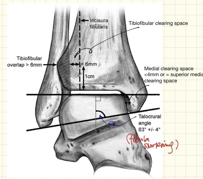
- Consists of AITFL, PITFL, ITL and interosseous ligament
- PITFL is the strongest
Q17How is syndesmosis injury assessed clinically and on X-ray?▸
- Clinical: local tenderness at AITFL, squeeze test, ER stress test, cotton test
- AP: medial clear space <4mm, TF clear space <6mm, TF overlap >6mm
- Mortise: Shenton line, symmetrical joint space (<5 degrees talar tilt), talocrural angle 83 degrees, dime sign
- Lateral: ATFI/TW = 39 +/- 9%; stress views (abd + ER on DF), hook test, CT, arthroscopy
- Ramsey: 1mm lateral talar shift = 42% reduction in tibiotalar contact area
Q18Where is the syndesmosis assessed and how is the TF clear space defined?▸
- Syndesmosis is assessed 1cm above the joint line
- TF clear space: from the medial border of the fibula to the incisural surface of the tibia
- Mortise view: TF clear space <6mm, TF overlap >1mm
Q19What does arthroscopy add in syndesmosis injury?▸
- Detects multi-directional instability, especially sagittal and rotational
- Lui arthroscopy 2005
Q20How do you insert a syndesmosis screw?▸
- X-ray control +/- CT; reduce with IR and anterior drawer, hold with periarticular clamp or K wires
- Keep ankle in neutral position (traditional teaching used DF; Gonzalez Injury 2017 - an anatomically reduced syndesmosis will not be over-tightened)
- 2cm above ankle joint, posterolateral to anteromedial (20 deg from horizontal), parallel to joint line
- One 3.5mm cortical positional screw through 3 cortices (2 for Maisonneuve)
- NWB for 8/52, then remove screw and gradually increase weight bearing
Q21What is the evidence for Tightrope versus syndesmosis screw fixation?▸
- Injury 2015 RCT Kortekangas: similar postop malreduction rates
- Similar functional outcomes
- Similar OA incidence
Q22When would you fix the posterior malleolus?▸
- Traditionally if >25% articular surface, >2mm step off, or unstable syndesmosis
- Recent studies suggest a lower threshold - it is the attachment of the PITFL (contributes 42% of syndesmosis stability)
- Isolated PM fixation restores 70% of syndesmosis stiffness vs 40% with isolated syndesmosis fixation
- 2017 JBJS Solan suggests much lower threshold; POSTFIX trial to be released in 2023
Q23Describe the Lauge-Hansen classification.▸
- Based on foot position and force applied relative to tibia
- SAD: transverse Weber A + vertical medial malleolus +/- impaction
- SER: Weber B + transverse medial malleolus + PM# (AITFL -> fibula -> PITFL/PM -> deltoid)
- PAB: comminuted impacted Weber C + transverse medial malleolus (Transverse MM --> PITFL/ PM --> Fibula)
- PER: spiral Weber C + transverse medial malleolus + PM# (Maisonneuve) (Deltoid/ avulsion from MM --> AITFL --> high fibula --> PITFL)
Q24What is the general fixation for each malleolus?▸
- Lateral malleolus: posterolateral approach - lag screw + neutralization plate, locking plate, or antiglide plate/buttress
- Medial malleolus: anteromedial approach with screw/plate (plate if vertical)
- Posterior malleolus: percutaneous screw
Q25What is the classic SAD pattern and its fixation?▸
- Marginal impaction - reduce and bone graft
- Fix with buttress screw/antiglide plate
Q26How are PAB and PER patterns managed?▸
- PAB: comminuted impaction of Weber B distal fibula - plate; transverse medial malleolus - tension band fixation
- PER: lateral fibula plating to restore length; restore ankle mortise and syndesmosis stability
- PM fixation if >25% articular surface, 2mm step off
Q27What are the poor prognostic factors for ankle fractures?▸
- Smoking/alcohol
- DM
- Osteoporosis
- IOD
Q28Describe the X-ray findings and injury pattern.▸
- AP view of ankle with fractured tibia and fibula
- Lauge-Hansen supination-adduction type injury
- Vertical fracture of medial malleolus + transverse infrasyndesmosis fibular fracture
Q29What other views and relevant negatives do you need?▸
- Ask for lateral X-ray and X-ray one joint above
- Relevant negatives: no talus fracture
- No os calcis fracture
- No MT fracture
Q30What are the definitive management aims?▸
- Anatomical reduction and stable fixation for primary healing
- Restore joint congruency of the tibial plafond
- Bone graft for defects
Q31What is a Maisonneuve fracture and what must you check?📷▸
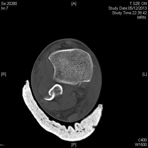
- Proximal fibula fracture
- Check the proximal fibula and compartments
Q32What is a Bosworth fracture and how is it managed?▸
- Fibula trapped behind the incisura fibularis (SER pattern)
- Requires urgent ORIF
Fact check
Posterior malleolus fixation threshold is to be confirmed by the POSTFIX trial (to be released in 2023) — outdated — POSTFIX one-year results (Bone Joint J 2025) found ORIF of medium-sized (5-25%) posterior fragments in AO type B fractures did not improve outcome, so size threshold alone is not validated — source