Trauma
Open and mangled extremity injuries
Gustilo open fracture, mangled limb, soft tissue coverage, flaps, gunshot wound, VAC closure.
47 questions
8 source pages
3 images
2 fact-check flags
Images appear with the first question taken from each source page — tap a question to open it.
Q1Describe the injury and X-ray findings in this mangled leg.📷▸
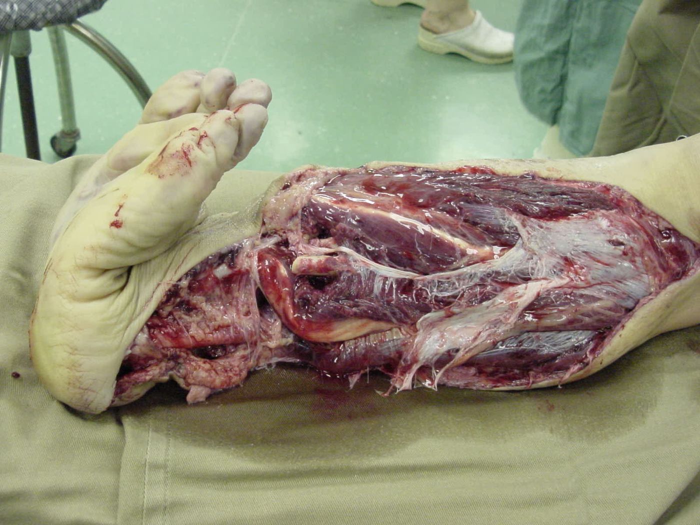
- Open fracture, mangled left leg with vascular compromise (pale looking sole)
- No gross contamination with soil/feces
- X-ray: comminuted, segmental tibial shaft fracture, extraarticular with gas shadow
- Gustilo determined intraop - at least IIIB, probably IIIC
Q2What is the initial ATLS/AED management of this open fracture?▸
- ATLS ABCDs, tranexamic acid, massive transfusion protocol; primary and secondary survey
- Antibiotics within 1 hr (1st gen cephalosporin) + tetanus; BOAST 4: <3hr infection 4.7%, >3hr 7.4%
- Direct pressure to control bleeding; remove gross contamination; sterile saline-soaked gauze dressing
- Temporary stabilization with splintage; AMPLE history incl ischemic time
Q3What is a mangled extremity and how is the amputation decision made?▸
- Consider 4 factors: soft tissue, bone, nerve, vessel; if 3/4 are disrupted it is a mangled extremity
- Consider patient/injury factors and scoring systems e.g. MESS
- Per LEAP, the single most important predictive factor is the patient's self efficacy
- If possible, will try to salvage the limb in acute setting if we are able to revascularize the limb; then buy time to have thorough discussion with patient and relative for delay primary amputation
- Patient need to understand limb salvage operation will likely involve multiple surgery and long rehab
Q4What is the MESS score and what are its components?▸
- MESS 1990 original paper: score >7 had 100% predictive value of amputation in that series
- Patient factors: age, shock
- Disease/injury factors: energy, extremity perfusion
Q5Which factors determine the choice between amputation and reconstruction?▸
- If possible keep the limb first; can do secondary amputation later
- Factors (referenced from MESS): shock, energy of trauma, distal pulse, age, time of injury
- Whether the ipsilateral foot is reconstructable
Q6What are the general indications for amputation?▸
- Dead (non viable)
- Dangerous (life threatening)
- Damn nuisance (non functional)
Q7What did the LEAP study show?▸
- Amputation vs reconstruction: no difference in outcome
- Self efficacy is the single determining factor for long-term outcome
- Satisfaction driven by function and pain; psychosocial factors more important
- Lifetime cost of salvage < amputation; no single score can be used for decision making
- Tibial nerve integrity not reliable/predictive - at 2 years almost 50% regained some plantar sensation
Q8What are the common problems after limb salvage?▸
- Recurrent infection, nonunion, delayed union
- LLD, OA, pain and stiffness
- Second amputation
Q9What is the initial operative plan and early care?▸
- DCO - damage control orthopaedics (staged management)
- Consent: external fixator + debridement + temporary wound coverage +/- vascular repair/shunting +/- amputation
- Early 2nd look in 48 hr; gentamicin beads (high local concentration, high MIC, fill dead space)
- If salvage succeeds: early fix and flap as early as D3, within 1 week, before infection sets in
Q10Which flaps are used for soft tissue coverage in limb salvage?▸
- Medial gastrocnemius flap
- Medial soleus flap
- Reverse sural flap (fasciocutaneous)
Q11How is conversion from external to internal fixation timed?▸
- <2/52: can be one stage
- >2/52: pin tract resting x 1 week (daily pin tract resting + antibiotics)
Q12What injuries does this multiply injured patient have and what are the initial priorities?▸
- Unstable pelvic fracture, open leg fracture and tibial plateau fracture
- ATLS protocol and pelvic binder
- Rule out life-threatening injury
- Manage according to damage control orthopaedics
Q13What is the 'second hit' phenomenon in damage control orthopaedics?▸
- The initial injury is the first hit; surgery is a potential second hit
- Triggers inflammatory, immunological and coagulatory cascades
- May push the patient into an irreversible physiological response (lethal triad) with increased mortality
- Limiting the second hit is the rationale for a staged approach
Q14What are the 3 stages of DCO?▸
- 1. Initial stabilization: life-saving surgery for skeletal and hemorrhagic stabilization, to facilitate nursing care (hemostasis, skeletal stabilization, decompression, decontaminate, revascularize)
- 2. ICU resuscitation and optimization
- 3. Definitive surgery
Q15What is the DCO timeline?▸
- D1 DCO
- D2 second look
- D5-10 window of opportunity
- 3+ can go to OT
Q16When is the patient stable for definitive surgery?▸
- When acidosis, hypothermia and coagulopathy are corrected
- Clinical markers: BP, urine output, inotrope use, temperature
- Biochemical markers: pH (lactate, base excess), coagulation
Q17What is early total care?▸
- Immediate fixation of all fractures
- Aim: early OT, early mobilization, less recumbency, early discharge
- Increased ARDS and multi-organ failure in borderline patients
Q18Which patients are classified as borderline for DCO?▸
- Patients classified: stable, borderline, unstable, extremities
- Borderline: ISS >40 or ISS >20 + chest injury
- Bilateral femoral fracture, chest contusion, pelvic/abdominal injury
Q19Does damage control orthopaedics improve mortality?▸
- DCO is shown to improve mortality compared with immediate definitive care in the unstable patient
Q20What three topics are discussed in this case?▸
- Open fracture management
- Definitive management for bone (bone defect) and soft tissue (flap coverage)
- Mangled extremity management
Q21What are the priorities in managing an open fracture?▸
- 1. NV status
- 2. Prevent infection
- 3. Soft tissue
- 4. Bone
Q22What is the acute AED management of an open fracture?▸
- ATLS, BOA/BOAST guidelines, AMPLE history
- Antibiotic within 1 hr (1st gen cephalosporin; BOAST suggests 1.5g IV cefuroxime + aminoglycoside) + anti-tetanus
- Remove gross contamination, take a photo, dressing with NS gauze + impermeable film
- Direct pressure for bleeding control; temporary reduction + slab
Q23What examination and imaging must be documented in open fracture assessment?▸
- Local: compartment syndrome and NV status
- Systemic: secondary survey to rule out other injuries
- XR of the injured part
Q24What are the operative debridement and irrigation principles?▸
- Early OT within 24 hrs: ex fix + debridement (keep skin/bone, remove all doubtful muscle)
- Irrigation: 3, 6, 9 L at low pressure 8 psi; no evidence for which fluid type
- NPWT if primary closure not possible (WOLLF trial JAMA 2019 Costa: no improvement in self-rated disability at 12 months)
Q25What is the timing of second look, flap cover and antibiotics?▸
- Early 2nd look at 48 hr with gentamicin beads
- Early fix and flap within 72hrs (should not exceed 7 days) per BOAST, before infection sets in
- Antibiotics continued for 72 hrs or until wound closure
Q26What is the evidence for timing of surgery in open fractures?▸
- Within 24 hrs; older studies recommended 6 hrs but based on animal studies
- LEAP: no difference in infection rates when managed within 6 hr or 24 hrs
- Latest BOAST: urgent surgery if vascular injury or heavily contaminated with sewage, marine or agricultural matter
Q27How is a bone defect managed?▸
- Eradicate infection first; depends on size of defect, soft tissue coverage and vascularity of the tissue bed
- Small <6cm: acute shortening, non-vascularized bone graft (Hammon PL bone grafting or Papineau)
- Large >6cm: Masquelet technique (cement spacer 4-5 weeks + staged bone graft), vascularized bone graft (fibula), bone transport
- Large defect + soft tissue/NV defect or unfit patient: consider amputation
Q28What flap options cover proximal, middle and distal third tibial defects?📷▸
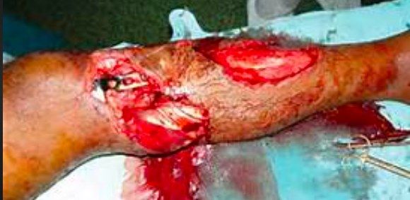
- Proximal 1/3: medial gastroc flap (medial sural artery) + PTSG
- Middle 1/3: soleal flap (posterior tibial artery)
- Distal 1/3: classic free flap; recent reverse hemisoleal (tibial perforators) or reverse sural (superficial sural artery)
Q29What is the BOA initial treatment guideline for adult open fractures?▸
- IV antibiotics within 1 hr + tetanus
- Document distal NV status and compartments
- Re-align the fracture and apply a splint
- Remove gross contamination, dress with saline-soaked gauze + occlusive film, take a clinical photo
Q30How are flaps classified?📷▸
- Location: local, regional, distant (pedicle/free)
- Pedicles: random, axial
- Composite/tissue: cutaneous, fasciocutaneous, musculocutaneous, osseocutaneous
Q31What are the Mathes types of muscle flap pedicles?▸
- I: one dominant pedicle (e.g. gastrocnemius, TFL)
- II: one dominant + one minor pedicle (e.g. gracilis)
- III: two dominant pedicles (e.g. rectus abdominis)
- IV: segmental pedicles (e.g. sartorius)
- V: one dominant + secondary segmental pedicles (e.g. pectoralis major)
Q32Which Mathes pedicle types are most reliable in muscle flaps?▸
- Types I, III and V are more reliable
- Preferred because they have a broad base
Q33What systemic factors are assessed pre-operatively before a flap?▸
- Smoking, obesity, hypertension, PVD, immunosuppression
Q34What must be planned in flap design?▸
- Type of flap, size
- Pivot point, axis, arc of rotation
- +/- Doppler to identify the pedicle
Q35How is a flap elevated and inset intra-operatively?▸
- Elevation: preserve minor pedicles; complete pedicle mobilization not required unless free flap; avoid tension during rotation and inset
- Inset: tunnel size = flap base x 2, avoid tension, ensure haemostasis
Q36What are the causes and management of impaired flap circulation post-operatively?▸
- Consider a systemic haemodynamic problem or local arterial/venous obstruction
- Try releasing stitches and repositioning the flap
- Avoid pressure, constrictive bandage and motion (splint)
Q37What is the mechanism of VAC?▸
- Induces granulation tissue and epithelisation
- Decreases dead space and edema
- Down grade soft tissue reconstruction ladder
Q38What are the common pedicle flaps for the leg?▸
- Proximal: medial gastrocnemius flap (muscle flap; medial sural artery from popliteal artery)
- Middle: soleus flap (muscle flap; branches from posterior tibial and peroneal arteries)
- Distal: distant flap/reverse sural (fasciocutaneous; superficial sural artery depending on perforating branch of peroneal artery)
Q39List common free flap choices and their pedicles.▸
- LD flap: myocutaneous, thoracodorsal artery
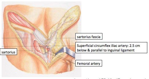
- Groin flap: fasciocutaneous, superficial circumflex iliac artery 2.5cm inferior to inguinal ligament, very consistent
- ALT flap: fasciocutaneous, descending branch of lateral femoral circumflex artery
- Radial forearm flap: radial artery; skin +/- muscle +/- bone; can be retrograde/antegrade
Q40What are the key points in the initial assessment of a gunshot wound?▸
- Fully expose the patient and look for entry and exit wounds
- Look for signs of major bleeding
- Any field tourniquet should be exchanged for a pneumatic tourniquet
- Otherwise manage as per open fracture
Q41What are the key points of OT debridement in a gunshot wound?▸
- Remove shrapnel
- Extend the wound longitudinally and excise necrotic tissue
- Take multiple samples for c/st
- Irrigation with 9L saline
- Explore the entire extent of the injury tract
- Temporary bone stabilization with exfix, slab
Q42What is the staged treatment plan after debridement of a gunshot wound?▸
- Delay staged treatment: second look debridement +/- closure at 48 hrs
Q43How does VAC affect wound size and the reconstruction ladder?▸
- Reduces wound size
- Downgrades the soft tissue reconstruction ladder
Q44How does VAC affect edema and dead space?▸
- Reduces edema
- Reduces dead space
Q45What tissue processes does VAC promote?▸
- Promotes granulation tissue
- Promotes epithelialization
Q46Describe the Tscherne classification of soft tissue injury in closed fractures.▸
- Grade 0: minimal, simple fracture, indirect force
- Grade 1: superficial abrasion, mild fracture pattern
- Grade 2: deep abrasion, skin or muscle contusion, comminuted fracture
- Grade 3: crush/degloving injury
Q47Describe the Tscherne classification of open fractures.▸
- Grades 1-4
- Based on: wound size, degree of contamination, fracture pattern
- Grade 4 = incomplete/complete amputation
Fact check
MESS score >7 has a 100% predictive value of amputation — misleading/outdated — True only in Johansen's original 1990 series; LEAP and later studies could not validate any trauma score, with MESS >7 sensitivity around 44% in recent cohorts — source
BOAST guidelines suggest 1.5g IV cefuroxime + aminoglycoside for open fractures — imprecise attribution — 2017 BOAST-4 recommends IV antibiotics ideally within 1 hour of injury but defers the specific antibiotic choice to local network guidelines; it does not mandate this regimen — (medium confidence) — source