Trauma
Knee and tibial shaft fractures
Floating knee and Fraser classification, Hoffa fragment, tibial plateau and tibial shaft fractures.
43 questions
7 source pages
3 images
1 fact-check flags
Images appear with the first question taken from each source page — tap a question to open it.
Q1What is the Fraser classification for floating knee and what are the surgical considerations?▸
- Type 1 extraarticular
- Femur first; if it becomes unstable can slab the tibia (better than traction on the femur)
- Can immobilise tibia during femoral fixation but not vice versa (need knee flexion for tibial fixation)
- Type 1 can consider retrograde nail, same wound for both fractures
Q2What is the management if there is a vascular injury?▸
- Reduce the fracture, reassess
- CTA +/- exploration
Q3What is a Hoffa fragment and how is it detected?▸
- Coronal fracture of the femoral condyle
- Easily missed - request lateral X-ray/CT in any distal femur fracture
Q4What are the considerations in managing a Hoffa fragment?▸
- Fragment may not be well fixed with a lateral locking plate; needs anatomical reduction and lag screw in AP manner before plating
- Use countersink or headless screw
- Isolated fragment may need posteromedial/posterolateral incision
Q5Describe the tibial plateau fracture pattern and metaphyseal extension on X-ray.▸
- Tibial plateau fracture involving both medial and lateral condyles, articular stepping, metaphyseal involvement, metaphyseal-diaphyseal dissociation
- +/- fibular fracture, joint subluxation
- Schatzker type VI
Q6What is the acute assessment and soft tissue management?▸
- ATLS, primary survey, AMPLE history; full circumferential soft tissue examination, compartments
- NV status: popliteal artery tethered at adductor hiatus and soleus arch; common peroneal nerve
- Soft tissue compromise: Span Scan Plan
- CT to assess the three columns and plan approach
Q7What are the aims of definitive fixation?▸
- Anatomical reduction of the articular surface; primary bone healing with absolute rigidity
- Correct coronal and sagittal alignment
- Buttress plating for the metaphysis
Q8Describe the approaches.▸
- Anterolateral (TA/bone) and posteromedial (medial hamstring/medial gastrocnemius; risk saphaneus nerve (saphenous), MCL)
- Assess articular reduction through submeniscal arthrotomy/scope for meniscus and cruciate status
Q9How is reduction and fixation performed?▸
- Split: periarticular clamp/femoral distractor; depression: bone punch from the split
- Fill void with bone graft (autograft, allograft, synthetic bone graft, injectable bone cement)
- Support articular fragment with 3.5mm raft screw, followed by an angular stable device such as a 4.5mm periarticular locking plate in buttressing mode
- Assess overall alignment with the cable method; too comminuted then ring/hybrid external fixator
Q10What is the prognosis?▸
- Key to good outcome: restoration of joint line and mechanical axis (mechanical axis > articular surface)
- ~7% had TKR 10 years after index operation (Wasserstein JBJS 2014)
- OA incidence up to 30% if axis deviation more than 5 degrees, increasing TKA likelihood 5 times
- Complications ~10%; 1-year mortality 14%
Q11What is the incidence of compartment syndrome in tibial plateau fractures?▸
- 0.7-12%
Q12What is the evidence comparing ORIF with circular external fixation?▸
- Canadian OTS multicentre RCT 2006: functional results similar
- ORIF group had more deep infections and unplanned returns to theatre
Q13Describe the anterolateral approach to the tibial plateau.▸
- Longitudinal incision lateral to patella starting 2cm below Gerdy tubercle
- Incise deep fascia anterior to ITB
- Subperiosteal dissection of the proximal attachment of tibialis anterior
- Submeniscal arthrotomy; +/- lateral epicondyle osteotomy (extended AL approach)
Q14Describe the lateral approach to the tibial plateau.▸
- Skin incision from lateral epicondyle to posterior fibular head
- Protect the CPN
- Interval: ITB and biceps femoris; enter the joint posteriorly between LCL and gastroc LH (gastrocnemius lateral head), or anterior to the LCL
- +/- fibular osteotomy (Lobenhoffer) for better exposure
Q15Describe the posteromedial approach to the tibial plateau.▸
- Skin incision from medial epicondyle to posterior edge of tibia
- Release pes anserinus, retract it anteriorly; medial head of gastrocnemius posteriorly
- May release part of the popliteus for exposure; submeniscal arthrotomy
Q16What are the criteria for non-operative management?▸
- <2mm articular step
- Stable in knee extension
- No varus/valgus malalignment
Q17What is the rehabilitation after tibial plateau fixation?▸
- Protected weight bearing
- Controlled ROM exercises
- Regime tailor made to fixation type, comminution at surgery and associated injuries
Q18What is the evidence for primary arthroplasty in elderly patients?▸
- For elderly patients with tibial plateau fracture and pre-existing knee OA
- EFFORT group 2016; also Injury 2021 Sabaitini
Q19What is the initial assessment and acute management of a tibial plateau fracture?▸
- High energy mechanism: follow ATLS and rule out hip and ankle pathology
- R/O open fracture, compartment Sx and NV injury
- Think possible knee dislocation
- Ext fix +/- compartmental release
- CT to delineate fracture pattern and comminution
- Expect 1-2/52 for soft tissue to settle
Q20What is the aim of operation for a tibial plateau fracture?▸
- Restore articular surface
- Stable fixation
Q21What approaches and fixation are used for a tibial plateau fracture?▸
- AL (anterolateral) and medial approach
- Buttress plate + raft screw +/- bone graft
- +/- submeniscal dissection x articular reduction
- EUA x ligamentous injury
Q22What is the post-operative regimen after tibial plateau fracture fixation?▸
- Post-op ROM 0-90
- NWB
Q23Describe the tibial shaft fracture and associated fibular injury on X-ray.📷▸
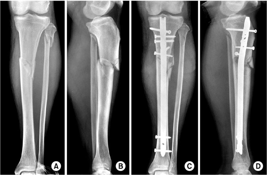
- Fracture of the proximal 1/3 of the tibial shaft with valgus and apex anterior displacement
- Fibula neck fracture
- AO B2
Q24What is the initial assessment and management?▸
- ATLS, primary survey, AMPLE history
- Local: soft tissue, skin impingement, NV status (CPN), compartment syndrome; systemic secondary survey
- XR one joint above and below
- Long leg slab + analgesics; close monitoring for compartment syndrome
Q25What are the principles of definitive management and why use an antegrade IM nail?▸
- Restore tibial length, correct angulation and rotation; achieve secondary bone healing; prevent complications
- Load sharing device that is biomechanically more rigid than a plate
- Preserves biology (not disrupting fracture haematoma); minimally invasive; Have the option of secondary dynamization if delay union
Q26What is the classic deformity and how is it corrected?▸
- Proximal: procurvatum (patellar tendon) + valgus (by ATT - anterior tibial tendon)
- Distal: varus + IR (PTT, FHL, FDL)
- Reduction: traction, manipulation, reduction forceps, unicortical Shanz pins
- Poller screw on concave side in proximal fragment (lateral, posterior), convex side distally; +/- anterior unicortical plate
Q27Describe the entry point and nailing steps.▸
- Lateral to midline, medial to lateral tibial spine (more lateral prevents valgus); just anterior to articular surface, as proximal/posterior as possible without breaking it
- Knee in slightly extended position; awl, guidewire to centre of tibial plafond at physeal scar; serial reaming; measure length; static locked nail
- Choose a reamed nail: bone graft, bigger nail more rigid, better endosteal fit, reduced working length
- SPRINT trial: reaming decreased future bone grafting and nail exchange
Q28What are the non-operative regime and alignment targets?▸
- Long leg cast 4 wk then patellar bearing brace/ Sarmiento brace for 12 week; union rate 90%
- Varus/valgus <5 degrees, AP angulation <10 degrees, rotation 10 degrees, shortening <1cm, joint line parallel
Q29What is the post-operative rehabilitation?▸
- FWB (full weight bearing)
Q30Describe the injury.📷▸
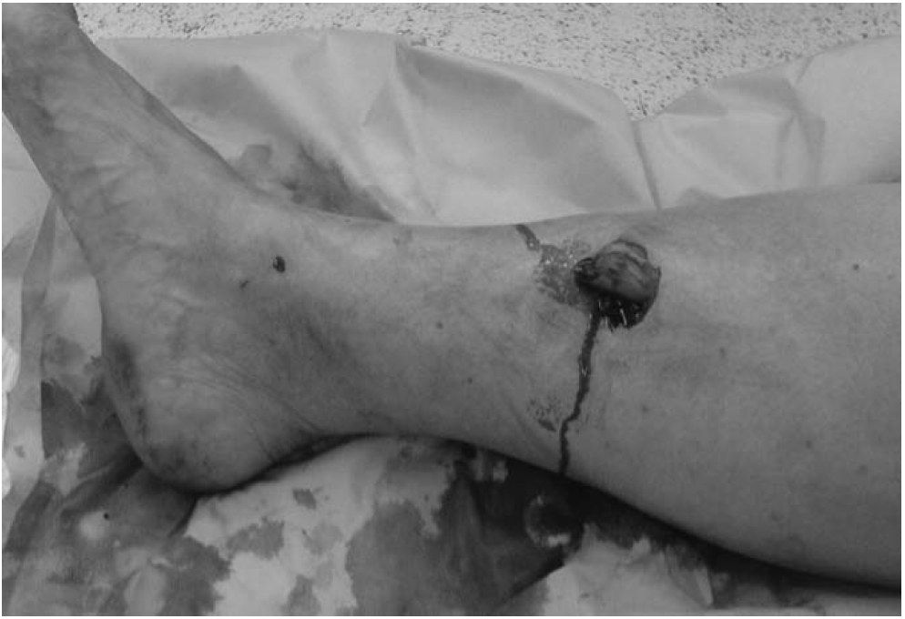
- Gustilo I fracture of the tibial shaft with the proximal fragment protruding out of skin in an inside-out manner
- Leg pink
Q31What is the acute management of an open fracture per BOA guidelines?▸
- Antibiotic within 1 hour (1st gen cephalosporin + aminoglycoside) + anti-tetanus
- Remove gross contamination, take a picture, dressing with NS gauze + impermeable film
- Direct pressure for bleeding control; temporary reduction + slab
- Next step is XR
Q32What is the timing of early total care?▸
- Within 12 hours for solitary high energy open fractures
- Within 24 hours for all other low energy open fractures
- If there is no vascular compromise
Q33Describe the procedure.▸
- GA, supine with triangular support; irrigation with 3L NS under low pressure; remove non-viable bone/soft tissue; assess wound coverage after debridement
- If soft tissue acceptable, go for definitive nailing via a split patellar approach
- Entry site: coronal plane parallel to the canal; sagittal plane just distal to the angle between tibial plateau and anterior tibial metaphysis
- 2 static locking screws proximal and distal; continue antibiotics until wound ok; Closely monitor for compartment syndrome; WBAT
Q34What is the evidence for nailing in open fractures?▸
- Unreamed IMN interlocking showed superior results for Gustilo II/IIIA/B: 1less malunion, 2lower infection rate, 3↓no. of OT needed (Bhandari 2001 JBJS Br)
- Foote from canada published in CORR 2015 network meta-analysis: definitive IM nail once soft tissue settled may be superior independent of Gustilo grade; Use of unreamed nails over reamed nails also may be advantageous in the setting of open fractures. LESS REOPERATION
- In multitrauma open fracture, no evidence for reaming (SPRINT trial)
Q35How do you avoid compartment syndrome and when should soft tissue coverage be performed?▸
- High index of suspicion; avoid regional anaesthetic blocks
- Definitive soft tissue coverage within 5 days
Q36Would you ream in this isolated open tibial fracture and why?▸
- Isolated trauma with risk of nonunion - ream to allow a larger nail
- Biological advantage of bone graft deposition
- In multitrauma open fracture there is no evidence for reaming (SPRINT trial)
Q37What are the advantages of plating a distal 1/3 extraarticular tibia fracture?📷▸
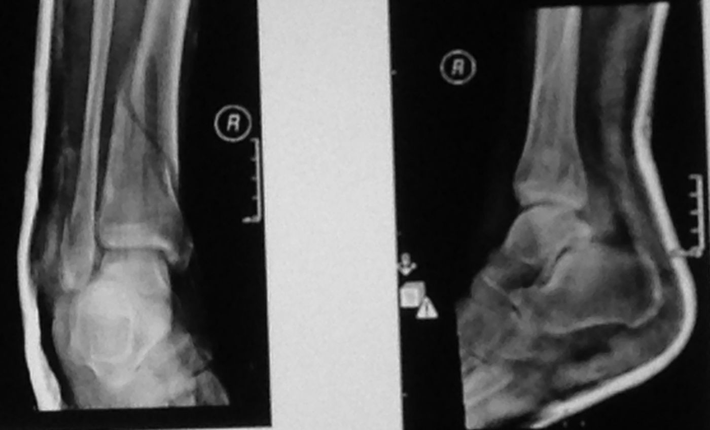
- Anatomical reduction
- Primary bone healing
Q38What are the disadvantages of plating a distal 1/3 tibia fracture?▸
- More dissection, especially at the site of anastomosis between PTA and ATA
- Load bearing construct
- Implant impingement
Q39What are the mechanical and biological advantages of IM nailing?▸
- Mechanical: load sharing implant, more torsional and bending rigidity
- Biology: does not disturb the fracture hematoma; reaming deposits bone graft
- Cons: more anterior knee pain
Q40Which implant is load bearing and which is load sharing?▸
- Plate = load bearing construct
- IM nail = load sharing implant
- Nail has more torsional and bending rigidity
Q41What did the FixDT trial (Costa 2018) show?▸
- IM nail: greater recovery - less disability, better ankle function and health-related QoL at 3 months
- Cost lower with IM nail
- Longer term: similar disability ratings and infection
Q42What did the Injury 2021 Bleeker meta-analysis show?▸
- Nail - more malunion
- Plate - more infection
- Management should be individualized
Q43Should the fibula be plated if the tibia is nailed?▸
- Frodl EFFORT 2021 metaanalysis: no difference in nonunion
- Fixing fibula may reduce secondary valgus/varus malunion but higher risk of wound infection
- Individualized decision
Fact check
Definitive soft tissue coverage should be within 5 days — outdated — BOA/BAPRAS (BOAST 4) standard is definitive closure or coverage within 72 hours of injury if it cannot be performed at the time of debridement — source