Trauma
Femoral shaft fracture - patterns and fixation
Comminuted shaft fracture with butterfly fragment, shaft and neck injury, proximal shaft fracture.
46 questions
6 source pages
2 images
1 fact-check flags
Images appear with the first question taken from each source page — tap a question to open it.
Q1Describe the femoral shaft fracture and degree of comminution on X-ray.▸
- Comminuted femoral shaft fracture with a large butterfly fragment and <50% cortical contact
- No associated femoral neck fracture
- Proximal fragment is abduction and ER (abducted and externally rotated)
- Winquist-Hansen type 3
- XR views required: entire bone + one joint above and below
Q2Describe the Winquist and Hansen classification.▸
- 0: no comminution
- 1: small butterfly fragment
- 2: large butterfly fragment, cortical contact >50%
- 3: large butterfly fragment, cortical contact <50%
- 4: gross comminution
Q3How is bilateral femur fracture managed?▸
- DCO in unstable/selected borderline patients to reduce the second hit (inflammatory response against surgery)
- Hemostasis, revascularisation, decompression, decontamination, stabilisation
- Definitive surgery: IM nail with distal and proximal locking
Q4How do you determine rotation during femoral nailing?▸
- Cortical thickness
- Lesser trochanter and knee X-ray, compare both sides
- Replicate the anteversion of the contralateral side
- Anterior bowing compared with the contralateral side
Q5Describe the AO classification of femoral shaft fractures.▸
- A simple
- B wedge
- C comminuted
Q6How common is a concomitant femoral neck fracture with a femoral shaft fracture, and what is the prognosis?▸
- High energy trauma; 5-10% of femoral shaft fractures have a concomitant femoral neck fracture
- Prognosis of the neck fracture is usually better than isolated NOF (usually undisplaced)
Q7What is the aim and sequence of fixation for ipsilateral femur shaft and neck fractures?▸
- Aim: anatomical reduction + stable fixation, prevent complications
- Dual construct preferred (some studies show high failure rates with a single construct)
- Priority is the neck to prevent AVN; fix the neck first
Q8How is an undisplaced neck fracture managed?▸
- Recognised before nailing: A+P hip screws + centre nail, or hip screw + DHS + long plate, or hip screw x3 + retrograde nail/lateral plate
- Recognised after nailing: miss-a-nail technique
Q9How is a displaced neck fracture managed?▸
- Recognised before nailing: ORIF of NOF with cannulated screw/DHS, then retrograde nailing
- Recognised after nailing: remove sliding hip screw then reduction then fix +/- augment
Q10What are the indications for retrograde nailing?▸
- Reduce time for positioning: bilateral femur fracture
- Reduce further insult to concomitant fractures: acetabular fracture, NOF, spine fracture
- Same incision: floating knee, fracture patella, periprosthetic TKR fracture
Q11What are the contraindications to retrograde nailing?▸
- Open fracture
- Stiff knee
- Patella baja
Q12What factors decide between retrograde and antegrade nailing?▸
- Injury factors: fracture location, entry site condition, associated injury (e.g. concomitant tibial fracture - cannot use traction table), pre-existing proximal implant
- Patient factors: knee ROM, (obesity)
- Cons of retrograde nail: infection will cause septic arthritis
Q13What is the risk of a cephalomedullary nail for combined shaft and neck fractures?▸
- Compared with two separate devices for each fracture, the cephalomedullary nail risks displacing either fracture while introducing the nail
Q14What injuries are associated with femoral shaft fracture?▸
- Hip dislocation, femoral neck fracture, knee fracture
Q15What injuries are shown on this AP X-ray of the right hip and proximal femur?▸
- Femoral shaft fracture: short oblique, medially angulated, laterally translated, no bone contact
- Right hip dislocation (AP view cannot tell anterior vs posterior)
- Small bony fragment inferior to the femoral head, could be femoral head or acetabular fracture
- No femoral neck fracture
Q16What is the initial management and what takes priority?▸
- MOI: RTA/FFH; high energy trauma then ATLS; check open wound and NV status (sciatic nerve)
- If stable: stabilise with Thomas splint, then go for CT scan to see where the bony fragment come from
- Hip dislocation takes priority
- Unstable: DCO; stable: ETC (anatomical reduction, stable fixation, early mobilisation)
Q17Describe damage control orthopaedics in this situation.▸
- Attempt CR +/- OR; insert Schanz screws into proximal femur avoiding the canal, connect to rod
- OR approach depends on the direction of dislocation
- External fixation of the femoral shaft
- Definitive surgery: plate/nail
Q18What are the fracture features and diagnosis on this X-ray of the proximal thigh?📷▸
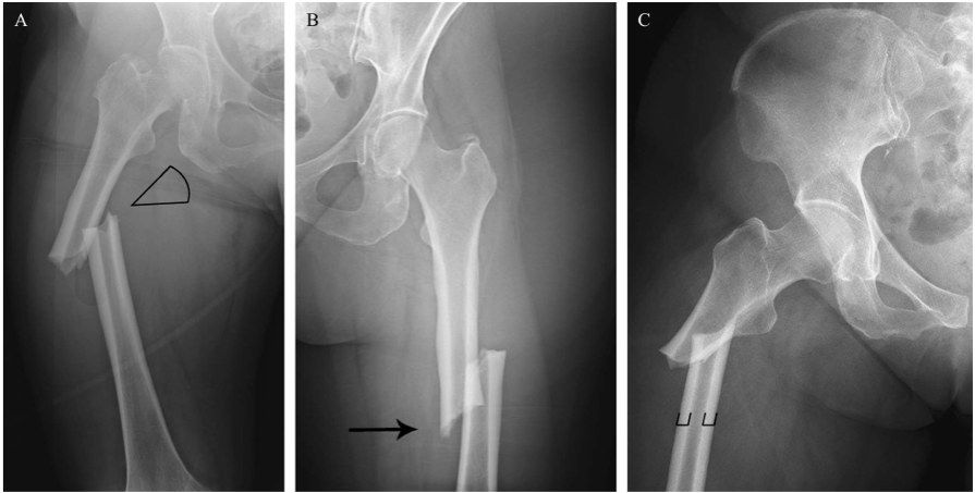
- Transverse fracture over the lateral cortex of the proximal shaft with medial spike, no comminution
- Evidence of beaking over the lateral cortex
- Diagnosis: atypical femoral fracture (AFF)
Q19Describe the 2013 ASBMR case definition of AFF.▸
- MUST be along the femoral diaphysis from distal to the lesser trochanter to proximal to the supracondylar flare
- Plus at least 4 of 5 major features; no minor features required
- Major: minimal/no trauma; origin at lateral cortex, transverse (may become oblique medially)
- Complete fracture may have a medial spike; incomplete involves only the lateral cortex
- Major: beaking (periosteal/endosteal thickening of lateral cortex); not or minimally comminuted
Q20What are the minor features of AFF?▸
- Delayed healing
- Prodromal pain
- Generalised increase in cortical thickness
- Bilateral
Q21What history and management apply to AFF?▸
- History: prodromal symptoms, symptoms on the other side, history of bisphosphonates
- Stop BP and switch to another agent
- Treat fracture side with a long cephalomedullary nail; surveillance of the contralateral side
- Warn about nonunion/malunion (8%)
Q22What is the association between AFF and bisphosphonates?▸
- More common with long-term BP (median 7 years); 1.78/100,000/year with exposure <2 years, rising with 8-9 years exposure
- Risk of AFF declines when BP stopped (70%/yr since last use); RR 2-128, AR <50/100,000 person-years
- Asian 8x risk; oral BP stop after 5 years, IV after 3 years
Q23How is the contralateral limb surveyed?▸
- Clinical + radiological; pain + normal X-ray then rule out referred pain, bone scan
- Pain + beaking then prophylactic nailing
- Beaking then MRI for bone marrow oedema; if MRI positive or stress osteolysis on X-ray then nail (protected weight bearing if patient declines operation)
Q24What is the QEH experience with AFF?▸
- 20% bilateral (UK 7%)
- 30% prodromal pain (UK 40%)
- Nonunion 8%
Q25What did Schilcher 2014 NEJM show about bisphosphonates and AFF?▸
- Risk of atypical femoral fracture during and after bisphosphonate use (2014, New England journal of medicine) by Schilcher on 5300 Swedish men and women
- Risk-benefit ratio for prolonged use >5 years would be inverted
- Preventive effect likely lasts several years after cessation, whereas AFF risk decreases rapidly (70%/yr since last use)
- Unclear benefit for age >80; 2020 NEJM: absolute risk of AFF remains very low vs fracture risk reduction
Q26Describe the distal femoral fracture configuration and articular involvement on X-ray.▸
- Displaced fracture over the metaphyseal region of the femur with a vertical split on AP extending to the articular surface
- Some comminution
- AO complete articular fracture type C
Q27What is the initial assessment and local examination in this distal femoral fracture?▸
- ATLS, primary and secondary survey, rule out fracture in the same limb; AMPLE history
- Locally: skin (impingement, wound), compartment, NV injury (popliteal artery tethered in this region)
- Systemic: secondary survey
Q28What further imaging is required?▸
- XR whole femur to make sure there is no synchronous fracture and to check for proximal implant
- CT scan to delineate fracture pattern, r/o Hoffa fracture (40%)
- XR contralateral side for preop templating
Q29What are the aims of management?▸
- Anatomical reduction of the articular surface
- Stable fixation of the articular block and the shaft for secondary bone healing
- Restore rotation, coronal and sagittal alignment, and length
- Allow early mobilisation; timing ETC
Q30Describe the surgical approaches.▸
- Lateral parapatellar approach with arthrotomy for joint visualisation
- Or lateral approach: Gerdy tubercle to midaxial line, incise ITB, elevate VL anteromedially from the intermuscular septum, ligate perforators (superolateral geniculate artery)
- +/- medial subvastus approach if double plating: plane between sartorius and VMO, then ligate the descending genicular artery
Q31How is the articular block reduced and compressed?▸
- K-wire as joystick, pointed reduction, mini-pelvic clamp; adjuncts: co-linear clamp, cerclage wires
- Reference pins: pin 1 parallel to anterior femoral cortex (axial), pin 2 parallel to joint line (coronal)
- K-wire at junction of anterior 1/4 and posterior 3/4, 2cm from joint line; lag screws for interfragmentary compression
Q32Describe the MIPO plating technique and parameters.▸
- Connect articular block to metaphysis; bolster under the knee to relax gastrocnemius
- Plate span ratio 1:3 comminuted, 1:8 not comminuted; plate screw ratio 1/2
- Working length of plate at least double the fracture length
- Bone graft the void; assess alignment (7.3mm screw parallel to joint line, cable method, Blumensaat line)
Q33What are the cons of non-operative management of a distal femoral fracture?▸
- Stiffness
- Malunion
- Bedbound patient
Q34What other fixation options exist for distal femoral fractures?▸
- LISS plate, angled blade plate, DCS
- Retrograde nail with at least 6cm distal fragment
Q35How is reduction assessed intra-operatively?▸
- Varus/valgus: the 7.3mm screw in the plate parallel to the joint line
- Coronal alignment: cable method
- Rotation by checking lesser trochanter, or comparing cortical thickness
- Sagittal: Blumensaat line-shaft angle
Q36What is the rehabilitation protocol after distal femoral fixation?▸
- Passive ROM
- NWB for 6 weeks
- FWB by 8-10 weeks
- Treat osteoporosis if needed
Q37How do you manage an associated neurovascular injury?▸
- Primary aim is to revascularise the limb
- Early vascular surgeons, vascular shunt, followed by skeletal stabilisation
- +/- prophylactic fasciotomy if warm ischaemic time >6 hrs
Q38What is the evidence for MIPO plating and how does it work?▸
- Baumgaertel Injury 1998 introduced biological plating: indirect reduction and bridge plating superior to direct anatomical fixation for bone healing
- Increase the working length of the construct - evenly distributed forces over a long segment, sharing strain between fragments
Q39How can an elderly patient be allowed to weight bear immediately?▸
- Double plating
- Shortening of the fracture
- IM nail (prerequisite: fracture line >6cm from joint line)
- Megaprosthesis or nail plate construct
Q40Describe the periprosthetic fracture in relation to the TKR components on X-ray.📷▸
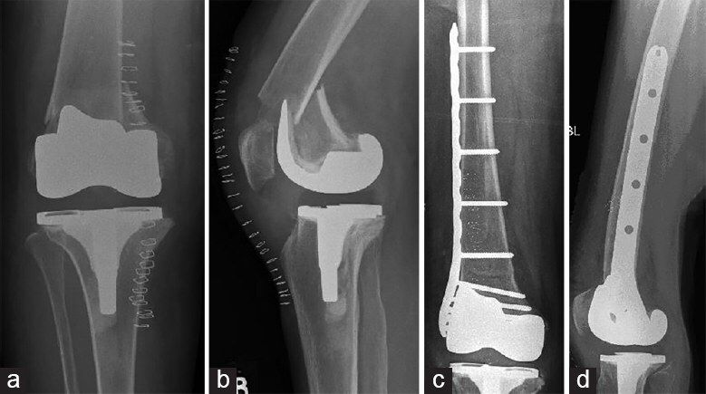
- Supracondylar femur fracture distal to the upper edge of the anterior flange of the femoral component
- Shortening and apex anterior angulation; transverse, not comminuted
- TKR posterior stabilised, no sign of loosening, patella not replaced, no notching
- Osteopenia
- Su type II; Rorabeck type II
Q41What TKR-specific history and examination findings are important?▸
- Premorbid functional status; details of TKR (implant model, timing, posterior sacrificing or retaining)
- Ipsilateral THR; pre-existing pain, stiffness, infection; mechanism of injury
- PE: soft tissue wound/impingement, distal NV status, compartment syndrome
Q42What imaging is required to assess the implant and distal bone stock?▸
- XR of the whole bone
- CT: assess loosening and how much bone stock is left distally
Q43What are the aims of definitive management?▸
- Stable fixation of implant and fracture
- Restore mechanical axis to avoid abnormal stresses on the implant
- Allow early weight bearing
Q44How do you manage a loose implant?▸
- Revision arthroplasty with a long stem to bypass at least 2 cortical diameters (prefer cementless)
- 2/3 zonal fixation
- Address massive bone loss when removing the old TKR; prepare allograft
- Always standby constrain or even megaprosthesis
Q45How do you manage a well-fixed implant?▸
- Locking plate - better restoration of alignment; nail entry/alignment is restricted by the notch; more distal fixation screws
- +/- medial plate for poor bone
- Retrograde nail for posterior cruciate retaining TKR
- Not recommended in ipsilateral THR as a stress riser in the unprotected femoral shaft
Q46What is the immediate management of a periprosthetic distal femoral fracture?▸
- Back slab and analgesics
- Ex fix if the initial soft tissue is compromised
Fact check
AFF incidence is 100 per 100,000/year with 8-9 years of bisphosphonate exposure — imprecise — The source (Dell 2012 JBMR) reports 113.1 per 100,000/year for 8-9.9 years exposure; the 1.78/100,000/year figure for <2 years matches — source