Trauma
Proximal femur fractures - neck and trochanteric
Intracapsular fracture in young and elderly, Watson-Jones approach, nonunion, trochanteric fixation.
89 questions
9 source pages
1 images
1 fact-check flags
Images appear with the first question taken from each source page — tap a question to open it.
Q1Describe the X-ray findings in this young femoral neck fracture.▸
- AP X-ray of the pelvis and bilateral proximal femur
- Over the left side there is a transcervical displaced fracture, with no underlying lytic lesion, +/- medial comminution
- Shenton line broken; bone quality okay
- Garden III, Pauwel III
- Fracture orientation and Pauwel grading are difficult to appreciate
Q2What does Garden III mean in this fracture?▸
- Acetabular and femoral head trabeculae are not aligned
- The capsule is likely intact
Q3What is the initial management of a young femoral neck fracture?▸
- Rule out life-threatening injury; make sure this is an isolated, closed fracture with intact NV
- Targeted history and PE: premorbid status, mechanism and chronicity of the fracture
Q4What is the aim of definitive management in a young femoral neck fracture?▸
- Achieve anatomical reduction and stable fixation to preserve the femoral head and avoid AVN
Q5What is the timing of surgery and the evidence in a young femoral neck fracture?▸
- Papakostidis (Giannoudis group) 2015 meta-analysis: no association between AVN and timing of surgery
- >24hr could increase substantially the odds of nonunion
- Go for reduction and fixation at the next available trauma list by a senior trauma surgeon, under GA
Q6What fixation options are used in a young femoral neck fracture?▸
- DHS + one anti-rotational screw - angular stability + tension band plating for a Pauwel 3 fracture
- Or 3 cannulated 6.5mm hip screws
Q7Describe the Leadbetter technique for reducing a femoral neck fracture.▸
- Hip flexion, adduction and internal rotation to relax the psoas, hamstring and Y ligament
- In-line traction -> book open the fracture
- While maintaining traction and IR: circumduct, abduct and extend the hip
Q8How is reduction assessed clinically and radiographically?▸
- Clinical: heel-palm test - put both legs in IR; good if they stay in IR
- X-ray: Garden's alignment index and Lowell's S lines
- AP view: medial cortex of femoral shaft and central axis of medial trabeculae of the capital fragment should be 160 degrees, accept valgus to 180 degrees
- Lateral neutral (axis of neck and centre of capital fragment), accept 155-180 degrees in both views
Q9What is done if closed reduction fails?▸
- Go for open reduction and internal fixation
- Approach: Watson's Jones
- DHS + anti-rotational screw + capsulotomy
Q10Describe the principle of screw fixation for a femoral neck fracture.▸
- Insert three screws in an inverted triangle configuration
- Avoid multiple drill holes and keep all screws proximal to the lesser trochanter
- Screws perpendicular to the fracture site to subchondral bone
- Parallel screw placement
- All threads pass into the proximal fragment
Q11Describe the screw insertion sequence and why the anterosuperior screw is tightened first.▸
- Use a guide pin to identify neck anteversion
- Insert the anterosuperior pin first, then posterosuperior and inferior
- 6.5mm partial-threaded cannulated screws in the same sequence
- Comminution is usually posteromedial, so tightening the AS screw first gives less varus and retroversion
Q12What is the postoperative care after fixation of a young femoral neck fracture?▸
- TTWB/NWB for 6 weeks
- Treat osteoporosis if present
- Monitor for AVN with XR/MRI
Q13What factors predict AVN after a femoral neck fracture?▸
- Initial fracture displacement
- Quality of fracture reduction
- Loss of reduction post surgery
- Fracture nonunion
Q14Describe the blood supply of the femoral head.▸
- Extracapsular arterial ring: MCFA (posterior), LCFA (anterior)
- Ascending cervical branch (major: posterosuperior)
- Subsynovial intracapsular ring at the subcapital region (major: lateral epiphyseal artery)
- Artery to ligamentum teres
- Intraosseous supply
Q15What are the AVN and nonunion rates by Garden grade?▸
- Garden I/II: AVN <10%, nonunion 5%
- Garden III/IV: AVN >80%, nonunion 30%
Q16What is the controversial evidence regarding timing, capsulotomy and implant choice?▸
- Time to surgery: within or beyond 12 hours (Papakostidis 2015 - no AVN association; >24hr increases nonunion)
- Capsulotomy: theoretically reduces intra-articular pressure
- DHS vs cannulated screws (JBJS 2008): DHS has lower short-term failure rates; backed by Boston group Gardnet 2015 and Singh 2017 for Pauwel II and III
- FEA (Jiang, Injury 2022): supports DHS + BS or FNS
Q17What are the key steps of the Watson-Jones approach to the hip?📷▸
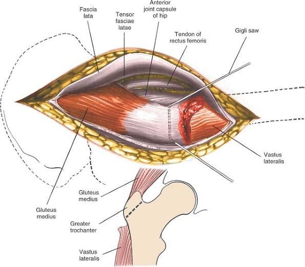
- Anterolateral approach to the hip, no internervous plane
- GA, supine, radiolucent table
- Incision from 2cm inferior and posterior to ASIS to posterior 1/3 of GT down the shaft
- Plane between TF and GM (both supplied by superior gluteal nerve)
- Ligate perforating vessels (superior gluteal arteries) before retraction
- Acetabular-based T capsulotomy
Q18Why is the Watson-Jones approach described as having no true internervous plane?▸
- The interval is between tensor fascia lata (TF) and gluteus medius (GM)
- Both muscles are supplied by the superior gluteal nerve
- Hence it is not a true internervous plane
Q19How is exposure improved proximally and distally in the Watson-Jones approach?▸
- Improve exposure by: proximal - detach the reflected head of rectus femoris
- Distal: release VL anterior fibres or perform trochanteric osteotomy
Q20How do you open reduce a femoral neck fracture through the Watson-Jones approach?▸
- Bone hook at GT + disimpact fracture by traction and ER
- Lever at fracture site (neck always anterior to head)
- LL max IR (maximal internal rotation) of the limb to maintain reduction
Q21What are the advantages and disadvantages of the Watson-Jones approach compared with the Smith-Petersen approach?▸
- Advantages: same wound for fixation; better visualisation of the base of neck/trochanteric region
- Disadvantages: poor visualisation of the head; perforator bleeding
Q22Describe the X-ray findings in this elderly patient with a transcervical femoral neck fracture.▸
- Transcervical displaced fracture of the right femur on AP pelvis
- Osteoporotic bone, no lytic lesion; broken Shenton's line
- No other fractures in the pelvic ring
- Garden IV osteoporotic fracture
Q23What does the focused history and examination include in an elderly hip fracture patient?▸
- Premorbid mental and functional status, PMH, mechanism of injury: 70y fell on level ground, osteoporosis and medications
- Rule out life-threatening conditions, especially HI; make sure it is an isolated injury
- Look for open wound and distal NV deficit
Q24List the BOA six standards of hip fracture care.▸
- Admit to ortho ward within 4hrs
- Surgery within 48hrs if medically fit, during normal working hours
- Pressure sore precautions
- MDT assessment and intervention
- Orthogeriatric combined care within 72hrs
- Secondary prevention of osteoporotic fractures
Q25What is the definitive management of a displaced osteoporotic NOF fracture in a geriatric patient?▸
- Aim: allow weight bearing and mobilisation ASAP and repatriation to place of usual abode
- Surgery: cemented bipolar hemiarthroplasty
- Long run: osteoporosis and fall prevention by the fracture liason service according to the IOF guidelines
- +/- DVT prophylaxis, +/- fascia iliaca block
Q26What is the evidence for THR vs hemiarthroplasty, and cemented vs cementless hemiarthroplasty?▸
- Bhandari 2019 NEJM (HEALTH): THR gave a clinically unimportant improvement in function/QoL over 24months, with more dislocation
- Fernandez 2022 NEJM: cemented hemi = significantly better QoL and lower periprosthetic fracture risk in NOF patients >60yo
- Cemented Exeter stem: 100% survival rate in 17years according to Carrington’s series in JBBS Br 2009; Norwegian registry: lower fracture, infection and revision
- Cemented gives immediate stability, less anterior thigh pain and fewer periprosthetic fractures
Q27What are the risk factors for hip instability after hemiarthroplasty?▸
- Surgeon: posterior approach, unaddressed acetabular defect/GT fracture/glut med tear, failure to restore soft tissue tension, Cement or bone fragment impingement, incorrect stem version
- Improper posterior capsular repair (Kwon meta-analysis: dislocation reduced 10 times if repaired)
- Patient: delirium, Parkinson's, stroke, dementia
Q28What is the evidence for bipolar vs unipolar hemiarthroplasty?▸
- Theoretically bipolar has two bearing surfaces: less dislocation, acetabular erosion and femoral head protrusio
- Australian joint replacement registry 2021: less risk of revision, at least in the young (9.9% vs 6.9%)
- Injury 2019 meta-analysis: greater ROM, less acetabular erosion, lower reoperation; longer operative time; no difference in HHS/mortality
- Cochrane review 2022 Lewis et al: insufficient evidence to determine whether bipolar is superior to unipolar
Q29What is the 1-year mortality after hip fracture and its risk factors?▸
- 25% mortality at 1 year
- Risk factors: male, TOF, ASA 3 or above, OT >48 hrs, age 85 or above
Q30What is the evidence for DHS vs cannulated screw in patients over 50 years old?▸
- FAITH trial: no difference in reoperation rate
- Smokers, displaced or base of neck fractures might do better with a sliding hip screw
Q31What are the minor complications of hemiarthroplasty?▸
- Persistent hip pain
- Poor mobility
- LL muscle wasting
- Superficial infection
Q32What are the major complications of hemiarthroplasty?▸
- Periprosthetic fracture
- Deep infection
- Dislocation
- Aseptic loosening
- Acetabular erosion
Q33How do you comment on screw fixation of a femoral neck fracture on X-ray?▸
- All screw threads passed the fracture site
- All screws reached subchondral bone
- Check the entry site of the screws
- In inverted triangle configuration, screws should be parallel
Q34What does this X-ray show and what is the summary?▸
- Fracture pattern: medial comminuted with a relatively vertical fracture line
- Varus malreduction
- No signs of union; no signs of infection
- Femoral head spherical (signs of AVN); Acetabular side intact
- Summary: NOF fracture with nonunion and varus collapse
Q35List the mechanical and biological reasons for fracture fixation failure.▸
- Mechanical (5): fracture pattern, location, reduction, fixation method, fixation quality
- Biological: patient, fracture, infection
Q36Describe the Leighton classification of femoral neck nonunion and its treatment.▸
- I (<6 weeks): inadequate fixation/malreduction to revision ORIF
- II (3 months): loss of fixation with good initial alignment to valgus osteotomy
- III (fibrous nonunion): free vascularised bone graft (Meyers BG, fibular graft, iliac crest bone graft)
Q37If joint salvage is chosen, what is the revision fixation technique?▸
- Remove implant + valgus osteotomy + revision fixation by angle blade plate + bone graft
- Aim to convert shearing force to compressive force
Q38What investigations are needed in suspected femoral neck nonunion?▸
- WBC, ESR, CRP; +/- hip aspirate if suspicious
- MRI/bone scan to assess AVN
- CT for bone stock
- Compare previous Xray see if there was any mal-reduction or displacement of fracture
Q39What history and examination points are important in failed femoral neck fixation?▸
- Hx: premorbid status, smoker, pre/intra/post-op problems, recurrent injury, current symptoms, limitations, expectations
- PE local: soft tissue, scar, infection signs
- PE systemic: LLD, muscle power, walking status, general fitness for GA
Q40What factors determine the choice of operation for femoral neck nonunion?▸
- Factors to consider: age and premorbid status
- Bone quality and bone stock
- AVN
- Acetabular integrity
- Infection
Q41What are the surgical options: joint salvage versus joint sacrifice?▸
- Salvage: revision fixation - unreliable fixation due to osteoporosis, defeats the goal of early weight bearing
- Joint sacrifice: arthroplasty (hemi vs THR) or Girdlestone
Q42Why are femoral neck fractures difficult to heal?▸
- Biological: synovial fluid, haematoma difficult to form, no cambium layer of periosteum
- Mechanical: high stress, little bone contact
Q43What are the nonunion rates and time definitions after a displaced femoral neck fracture?▸
- 20% nonunion after displaced NOF fracture
- Delayed union: 6 weeks
- Nonunion: 6 months
Q44What is Leighton's classification of femoral neck nonunion?▸
- Leighton's classification (JBJS 2008), based on the cause of nonunion, guides management
- Applies to fractures all with stable fixation
- Type 1: non-anatomical reduction or inadequate fixation
- Type 2: loss of fixation
- Type 3: fibrous nonunion (no displacement, no loss of fixation)
Q45How is each Leighton type managed?▸
- Type 1: remove implant, revise reduction +/- bone graft
- Type 2: change shearing force to compressive force with valgus osteotomy + fixation
- Type 3: drill out nonunion, bone graft
Q46What is Meyer's bone graft and what does it provide?▸
- Quadratus femoris muscle pedicle graft
- Provides mechanical support and brings in blood supply
Q47Describe the X-ray findings at 6 months post fixation.▸
- Failure of hip screw fixation with back out and protrusion of screws
- No healing over the transcervical fracture
- Femoral head collapse suggesting AVN; acetabulum shows no sourcil sign
- Suspected lucency around screw
- Suspected nonunion and AVN
Q48List the reasons for failure of this fixation.▸
- Mechanical: fracture pattern, location, reduction, fixation method, fixation quality
- Biological: patient, fracture, infection
Q49What workup is required?▸
- WBC, ESR, CRP; +/- hip aspirate if suspicious
- MRI/bone scan to see any AVN
- CT for bone stock
- Compare previous X-ray for malreduction or displacement
Q50What factors guide the choice of operation?▸
- Age and premorbid status
- AVN
- Acetabular integrity
- Infection
- Bone quality and bone stock
Q51What reconstruction is chosen and what is the supporting evidence?▸
- Uncemented acetabular cup with cemented PE cup and cemented Exeter stem
- Carrington JBJS Br 2009: 90% 17-year survival for this combination
- Average male life expectancy in HK is 80 so THR is the best option
Q52What history and examination are needed in this failed fixation case?▸
- Hx: premorbid status, smoker, perioperative problems, recurrent injury, current symptoms, limitations, expectations
- PE local: soft tissue, scar, infection signs
- PE systemic: LLD, muscle power, walking status, general fitness for GA
Q53What are the aims and options of the operation?▸
- Aim of operation to restore a functional hip
- Joint salvage vs joint sacrificing
- Salvage: revision fixation - unreliable due to osteoporosis, defeats early weight bearing
- Joint sacrifice: arthroplasty vs Girdlestone vs arthrodesis; hemi vs THR
Q54Describe the right and left hip X-rays.▸
- Right: intertrochanteric fracture, minimally displaced, no features of instability, Kyle 1
- Left: intertrochanteric fracture, unstable - large posteromedial fragment, fracture GT, reversed oblique configuration, Kyle 4
Q55What makes an intertrochanteric fracture unstable?▸
- No medial calcar support
- No lateral buttressing effect (fracture greater trochanter, transverse/reverse oblique fracture)
- Kyle 3 (large posteromedial fragment + GT fracture) and Kyle 4 (extension to subtrochanteric) are unstable
Q56Outline the BOA guideline for initial management of hip fractures.▸
- Admitted to acute ortho ward within 4 hours
- Surgery within 48 hours of admission if medically fit, during normal working hours
- Minimise risk of pressure ulcer
- Routine orthogeriatric medical support from admission
- Bone-protective therapy to prevent further osteoporotic fractures
- Offered MDT assessment and intervention
Q57What are the pre-requisites and technique for a good DHS?▸
- Pre-requisites: no medial calcar comminution, intact lateral buttressing
- Principle: allow controlled collapse across the fracture to enhance healing
- Good reduction to prevent varus; lag screw avoiding superior/posterior cortex; TAD < 25mm; antirotation guide pin
Q58How does a cephalomedullary (PFNA) device stably fix an unstable fracture?▸
- Stability does not rely on lateral buttress or medial calcar
- Efficient load transfer from blade to nail to locking bolt and shaft, not through the medial calcar
- Decreased bending moment as the nail is more medialised than the DHS plate
- Nail can control impaction of fracture by physically blocking the sliding
Q59What are Kaufer's variables affecting construct strength?▸
- Uncontrolled: bone quality, fracture geometry (stable vs unstable pattern)
- Controllable: reduction, choice of implant and mode of application
Q60What focused history and examination are needed in a trochanteric fracture?▸
- Premorbid mental and functional status, PMH, mechanism of injury, osteoporosis and medications
- Rule out life-threatening conditions, especially HI; ensure it is an isolated injury
- Look for open wound and distal NV deficit
Q61How does barrel choice affect DHS sliding and failure?▸
- Short barrel (25mm): longer sliding distance but higher bending moment (increased moment arm)
- Long barrel (38mm): less bending moment, less screw breakage, less friction and better sliding
- Gundle: sliding <1cm gives worse outcome; short barrel if lag screw <85; recommended sliding 25mm
- DHS thread 22mm
Q62How is a pre-existing OA with a trochanteric fracture managed?▸
- CRIF is difficult: the proximal part is stiff
- Fixation +/- second-stage THR if symptomatic
- Or primary THR with fixation of GT/calcar-replacing prosthesis
Q63What is the epidemiology and mortality of trochanteric fractures?▸
- Nonunion/malunion rare <2%
- 20-30% mortality risk in the 1st year (TOF worse than NOF)
- Increased mortality: male, operation >48 hours, age >85, ASA III or above
Q64Describe the Kyle classification of trochanteric fractures.▸
- I - undisplaced
- II - displaced, minimal comminution
- III - greater trochanter fragment
- IV - subtrochanteric extension
Q65Describe the AO classification of trochanteric fractures.▸
- 31A-A1: peritrochanteric simple two part (lateral cortex intact)
- 31A-A2: pertrochanteric with a separate PM fragment (lateral cortex intact)
- 31A-A3: fracture extends through lateral and medial cortices
Q66What is the evidence for cephalomedullary nailing versus DHS?▸
- Nail only shown to be superior to DHS in unstable fractures (Cochrane review 2014)
Q67Describe the fracture, fixation and current status on X-ray.▸
- Fracture TOF with fracture GT fixed with DHS + 1 cerclage wire
- No union, varus deformity
- Cut out of lag screw superiorly, complete sliding of DHS, no implant fracture
- Acetabulum intact, femoral head spherical, no halo/lucency around screws
- Summary: TOF nonunion with implant cut out
Q68What are the reasons for failure in this case?▸
- Mechanical: unstable fracture pattern and location, varus reduction, wrong implant (should have used short barrel), lag screw not in centre-centre position, tip-to-apex
- Biological: infection, osteoporotic patient, fracture
Q69What investigations are required before revision surgery for this nonunion?▸
- Rule out infection: bloods, gallium scan, +/- hip aspirate if suspicious
- Serial XR to see progress of alignment
- CT for acetabular defect and healing status
- MRI for head viability
Q70How would you perform revision fixation?▸
- Remove implant, then revision fixation
- Dimon-Hughston osteotomy: medialized shaft (medialised shaft), reconstruct medial calcar, more valgus configuration
- Converts shearing to compression
Q71What history and examination are needed for this failed fixation?▸
- Hx: premorbid status, smoker, pre/intra/post-op problems, recurrent injury, current symptoms, limitations, expectations
- PE local: soft tissue, scar, infection signs
- PE systemic: LLD, muscle power, walking status, general fitness for GA
Q72What management factors and options apply in this failed TOF fixation?▸
- Factors: age/premorbid, bone quality and bone stock, AVN, acetabulum status, infection
- Joint salvage: revision fixation - unreliable due to osteoporosis, defeats early weight bearing
- Joint sacrifice: arthroplasty vs Girdlestone vs arthrodesis; hemi vs THR
Q73What are the approach and exposure considerations for this revision?▸
- Use the old/incorporate the old incision
- Identify and protect the sciatic nerve; soft tissue release
- Dislocate before removal of the implant
- Careful manipulation of the osteoporotic leg
- Implant removal: correct screwdriver, removal of nail, prepare a broken screw set
Q74How are the hip replacement options chosen in this failed TOF fixation?▸
- Hemi vs THR depending on acetabulum status
- Hybrid/cementless THR with metal-on-PE (joint registry data)
Q75How are the acetabulum and femur reconstructed?▸
- Acetabulum: Acetabular defect (paprovsky) (Paprosky); cementless +/- screw augment, standby cemented
- Femur: Defect management (paprovsky) (Paprosky); cementless calcar replacement vs long diaphyseal stem to bypass screw holes by 2 cortical diameter
- Cemented femur: cement leakage problem; GT reattachment
Q76What is done at reduction if the hip is unstable?▸
- Aim combined anteversion 35
- Femur: increase size of head, increase offset, GT advancement
- Acetabulum: anteversion and opening angle, lipped or constrained liner, MDM
Q77What is the rehabilitation plan after revision?▸
- Rehab plan: Weight bear as tolerated
- Hip precautions
- HO prophylaxis
- Osteoporosis treatment
Q78Describe the displacement and location of this subtrochanteric fracture on X-ray.▸
- Subtrochanteric fracture with displacement, extending into the piriformis fossa and to the lesser trochanter
- Proximal fragment in varus and abducted
- Main distal fragment shortened and adducted
- Lesser trochanter fragment migrated proximally
Q79What is a subtrochanteric fracture and what are the deforming forces?▸
- Area from the lesser trochanter to 5cm distal
- Abduction by gluteus medius and minimus
- Flexion by iliopsoas
- ER by short external rotators
- Adduction and proximal migration of the distal fragment by adductors
Q80Why is a subtrochanteric fracture difficult to heal?▸
- Biologically a watershed area
- Mechanical high stress according to Koch diagram, multiple muscle pull, high cortical-to-cancellous ratio
- Fielding classification: the more distal the fracture, the higher the nonunion rate
Q81What is the management of choice and why?▸
- Long cephalomedullary nail: load-sharing with efficient load transfer
- Good torsional and bending rigidity with high tensile and fatigue strength
- Medialised lever arm decreases bending stress; biologically friendly
- Most recent NICE guidelines recommend IM nail fixation
Q82What difficulties are expected with reduction and fixation?▸
- Difficult reduction from strong muscle pull; reduce on traction table with not too much traction (align distal to proximal with flexion, abduction, ER)
- Assist with Shanz screw (Schanz screw), cerclage wires, colinear clamp; lateral nailing if all fails (especially obese patients)
- More body lateral flexion (entry point); entry point more MEDIAL + POST (medial and posterior) to avoid flexion and coxa vara
Q83Describe the classification systems for subtrochanteric fractures.▸
- Russell-Taylor: 1/2 piriformis fossa intact; A/B loss of medial calcar/lesser trochanter; 1A can use centromedullary nail, others cephalomedullary
- Fielding: type 1 at, type 2 <2.5cm, type 3 >2.5cm from LT (more distal = more nonunion)
- Seinsheimer: 1 undisplaced two-part to 5 subtrochanteric-intertrochanteric extending to GT
Q84What history and examination findings are important in a subtrochanteric fracture?▸
- Premorbid status; history of bisphosphonate/malignancy
- Mechanism of injury, preceding symptoms or prodromal pain
- Rule out HI
- PE: open wound or skin impingement; document distal NV conduction
Q85What imaging is required for a subtrochanteric fracture?▸
- X-ray lateral view, whole femur and one joint below
- Offer operative management for fixation
Q86What is the aim of surgery for a subtrochanteric fracture?▸
- Secondary bone healing with relative stability
- Correct alignment, rotation and length
- Adequate bone contact
Q87If fixed with an IM nail, what is the common malreduced position?▸
- Varus and procurvatum
Q88How do you perform safe reaming for a subtrochanteric nail?▸
- Technique: high speed, slow advancement, serial reaming, irrigation, clear bone debris and suction, vent hole, no tourniquet
- Reamer design: Bixcut with side and forward cutting flutes, sharp flutes
- Anaesthesia: avoid dehydration, maintain BP
Q89When is a plate/angle device used instead of a nail?▸
- For pre-existing deformity or a narrow IM canal
Fact check
Garden III/IV femoral neck fractures have an AVN rate >80% — overstated — Pooled meta-analysis data show AVN after displaced (Garden III-IV) fractures of about 20-33% (Garden IV ~33%, Garden III ~17%); a rate >80% is not supported — source