Trauma
Acetabular fracture and hip dislocation
Acetabular patterns, posterior wall defects, Pipkin fractures, head screw fixation, dislocation reduction.
48 questions
6 source pages
3 images
1 fact-check flags
Images appear with the first question taken from each source page — tap a question to open it.
Q1Describe the X-ray assessment of an acetabular fracture.▸
- Assess IP and II lines, sourcil, teardrop
- Posterior and anterior wall
- Fractures in iliac crest and inferior pubic rami
- Joint subluxation/dislocation
- Associated fracture (femoral neck and head, pelvis)
Q2Outline the immediate management and further imaging of an acetabular fracture.▸
- ATLS primary and secondary survey; rule out other injury, in particular knee, Morel-Lavallee, sciatic and femoral nerve
- +/- Skeletal traction via a distal femur traction pin
- Judet (OO) view - look for the spur sign -> both column
- Fine-cut CT for fracture pattern, loose bodies, marginal impaction, articular step off or gap
- Judet-Letournel classification: no prognostic value, used for surgical exposure
Q3What are the indications for operative fixation of an acetabular fracture?▸
- Location of fracture line - whether it is in the weight-bearing zone (roof arc <45 of Matta on AP, IO and OO views, or subchondral arc measurement by Olson)
- Incongruent joint (>2mm step, marginal impaction, loose body)
- Instability (Moed's method: 20% - posterior wall fragment size % + intraop screening after dislocation)
- Associated fractures (femoral head/neck)
Q4What are the aims and timing of definitive acetabular surgery?▸
- Aim: anatomical reduction of the articular surface, congruent joint, stability for mobilization
- Timing 2-5 days post injury (according to BOAST guidelines); earlier OT gives a higher chance of anatomical reduction
- <2 days - risk of bleeding
- Letournel: >3 weeks - only 50% anatomical reduction
Q5What are the operative details and postoperative care for acetabular fracture fixation?▸
- Single approach to the most displaced fracture +/- an additional approach
- Reduction with joystick, colinear clamp, Farabeuf clamp
- ORIF with 3.5mm reconstruction plate / 3.5mm lag screw / spring plate
- Assess fixation with OOO (joint penetration), IOO (supraacetabular screw within the iliac table), IIO (AP position in the pubic rami)
- Postop HO prophylaxis, NWB for 3 months until healing consolidated, XR + MRI to look for AVN
Q6What are the pros and cons of the lateral versus prone position for acetabular surgery?▸
- Lateral pros: less problematic for the chest, lower infection and revision rates, easier limb manoeuvrability
- Lateral cons: sciatic nerve, difficult manual traction, difficult reduction of the posterior column, impaired access through the greater sciatic notch
- Prone position is better for T-type or transverse fractures
Q7Which approach (ilioinguinal or Kocher-Langenbeck) is chosen for an acetabular fracture?▸
- If choosing one -> depends on which side is more displaced/more unstable
- Both column fracture / anterior column + posterior hemi-transverse -> usually anterior
- T / transverse -> usually posterior approach
- May need a combined approach / staged operation
Q8What are the poor prognostic factors in an acetabular fracture?▸
- Posterosuperior wall (very important for stability/joint congruency)
- Marginal impaction
- Roof arc angle <45 degrees
- Hip dislocation
- Femoral head fracture or impaction
Q9Why may a both-column fracture have a better prognosis?▸
- The whole acetabulum is separated from the axial skeleton
- Secondary congruity: a floating acetabulum - the joint is congruent but its orientation differs from the axial skeleton
Q10How is an elderly acetabular fracture managed with respect to acute THR?▸
- Primary THR - advantages: earlier weight bearing, shorter OT time
- Primary THR disadvantages: risk of aseptic loosening due to poor fixation, risk of dislocation, risk of HO, need for special implants
- ORIF then THR advantage: defined bone stock
- ORIF then THR disadvantages: higher failure rate due to abnormal anatomy, other implants, more scar tissue, HO, soft tissue insufficiency, infection
Q11What is the evidence for combined hip procedures in acetabular fractures?▸
- SICOT 2017: 20-25% of acetabular fractures need revision ORIF or THR, so each case needs an individualised treatment plan
- BJJ 2019 Borg prospective study (minimum 2-year FU): combined hip procedure reduces the need for further surgery compared with ORIF
- Outcome: 78% survival at 10 years
- Poor prognostic factors: male, <50 years old, >80kg, significant acetabular defect
Q12What is Olson's subchondral arc measurement?▸
- Measures the superior 10mm of the acetabulum
- Any fracture line within it will be in the weight-bearing zone
Q13What is the nerve supply of the gluteal muscles and what lies between them?▸
- Gluteus maximus: inferior gluteal nerve
- Gluteus medius and minimus: superior gluteal nerve
- Between maximus and medius lie the superior gluteal artery and nerve
Q14What radiographic features suggest a posterior rather than anterior hip dislocation, and how is the diagnosis confirmed?📷▸
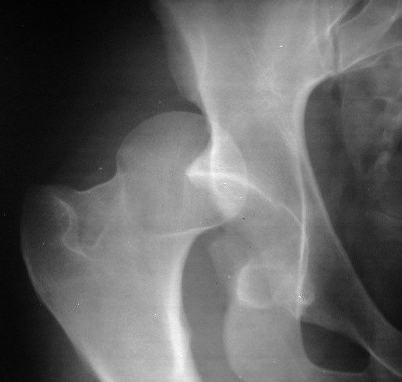
- Femur appears adducted, with the lesser trochanter less prominent
- No fracture over the femoral head and neck
- Likely posterior dislocation, but there is no lateral XR in the trauma setting - confirm with clinical examination
Q15How are hip dislocations classified?▸
- Thompson and Epstein (posterior): 1 small post fragment, 2 large post fragment, 3 comminuted post fragment, 4 acetabular floor, 5 femoral head
- Anterior (Epstein): 1 superior, 2 inferior; A no fracture, B femoral head fracture, C acetabular fracture
Q16Describe the initial ATLS assessment of a hip dislocation.▸
- ATLS, AMPLE history, primary and secondary survey
- Local assessment: wounds, Morelle Lavalle, knee dislocation, sciatic/femoral nerve
- Systemic survey to rule out other injuries
Q17What is the timing and technique of closed reduction for a hip dislocation?▸
- CT if available; if time is needed, proceed with CR and screen for neck fracture under II before reduction
- CR under GA with muscle relaxant within 6 hours
- AVN risk: >6 hours, repeated attempts >2, neck fracture
- Apply traction in line with the deformity regardless of the direction of dislocation
Q18What must be checked after reduction of a hip dislocation?▸
- Stability: hip flex, IR, axial load
- NV status
- +/- skeletal traction if needed
- Post-reduction CT for congruency, iatrogenic fracture, marginal impaction, loose bodies
Q19What is the definitive management of a reduced hip dislocation with a posterior wall fracture?▸
- Traction x 4/52 if unstable but concentrically reduced; PWB for 4 weeks if stable and concentric
- Surgery if instability: posterior wall fragment >20% (dynamic screening is more reliable - Moed, J Orthop Trauma 2009)
- Surgery if incongruent: marginal impaction, articular step >2mm, loose bodies
- Surgery based on fracture location (roof arc angle of Matta or subchondral arc of Olson) and associated fractures
Q20What are the aims, timing and approach of surgery for a dislocated hip with posterior wall fracture?▸
- Aim: anatomical reduction, congruent joint, stability for mobilisation
- Timing 2-5 days post injury (according to BOAST guidelines); earlier OT higher chance of anatomical reduction; <2 days - risk of bleeding
- Letournel: >3 weeks - only 50% anatomical reduction
- Approach: Kocher-Langenbeck
Q21Describe the position, incision and superficial dissection of the Kocher-Langenbeck approach.▸
- Lateral position, hip extended and knee flexed
- Incision from PSIS along the femoral shaft, centred on the GT
- Split fascia lata
- Split gluteus maximus along its avascular plane
Q22How are the short rotators managed in the Kocher-Langenbeck approach?▸
- Cut the short rotators 1.5cm from their GT insertion
- Beware the ascending branch of the medial circumflex artery, which runs anterior to quadratus femoris
- Therefore do NOT incise the quadratus femoris
Q23Which nerves must be identified and protected in the Kocher-Langenbeck approach?▸
- Trace the piriformis to the greater sciatic notch (GSN)
- Trace the internal oblique (internal obturator) to the lesser sciatic notch (LSN)
- Hoffman on the LSN to protect the sciatic nerve
Q24Describe the capsulotomy and exposure adjuncts of the Kocher-Langenbeck approach.▸
- Marginal capsulotomy (acetabular-based), keeping the blood supply to the posterior wall fragment
- Subperiosteal dissection of the greater and lesser sciatic notches to expose the retroacetabular surface
- May insert a Shanz pin into the femoral neck to allow traction and assess for loose bodies
- +/- release the gluteus maximus insertion into the femur to aid retraction and reduce stretch on the sciatic nerve
Q25Describe the trochanteric flip osteotomy used in the Kocher-Langenbeck approach.▸
- Indicated when better visualisation of the joint is needed
- Predrill the GT
- Posterior to anterior oscillating saw digastric cut
- Detach the vastus lateralis and gluteus medius from the underlying attachment
Q26What fixation and postoperative care follow the Kocher-Langenbeck approach?▸
- Reduction with joystick, colinear clamp
- ORIF with 3.5mm reconstruction plate / 3.5mm lag screw / spring plate
- Assess with OOO (joint penetration), IOO (supraacetabular screw within the iliac table), IIO (AP position in the pubic rami)
- Postop HO prophylaxis, NWB for 3 months until healing consolidated, XR + MRI to look for AVN
Q27What are the early complications of the Kocher-Langenbeck approach and hip dislocation?▸
- <2% recurrent dislocation
- 20% sciatic nerve injury
- Femoral nerve injury
- Inferior gluteal artery and nerve injury (from vigorous piriformis retraction)
- Superior gluteal artery and nerve injury (superior retraction of glut med/gluteus medius)
- 1st perforator of the profunda femoris (during gluteus maximus release)
Q28What is the prognosis and management of sciatic nerve injury after hip dislocation?▸
- 50% recovery
- EMG post injury at 3-4 months
- OT if suspected entrapment or no recovery at 1 year
Q29What are the late complications of hip dislocation and acetabular fracture?▸
- Post-traumatic OA (20%)
- AVN (up to 40%)
- Chondrolysis
- Heterotopic ossification
Q30What obstacles may prevent closed reduction of a hip dislocation?▸
- Intracapsular: bony fragment, labrum
- Extracapsular: psoas and rectus femoris in front; gluteus maximus and piriformis at the back
- Open reduction if failed 2 times - via the KL approach
Q31What are the contents of the greater sciatic notch?▸
- Superior and inferior gluteal nerve and vessels
- Piriformis
- Sciatic nerve and posterior cutaneous nerve of thigh
- Pudendal nerve
- Nerves to obturator internus and quadratus femoris
Q32What are the contents of the lesser sciatic notch?▸
- Pudendal nerve
- Internal pudendal artery and vein
- Nerve to obturator internus
- Obturator internus tendon
Q33How is a Pipkin III fracture managed?▸
- No closed reduction (NO CR)
- Young: fix the neck before the head
- Old: hemiarthroplasty or THR
Q34Which approaches are used for Pipkin I, II and III fractures?▸
- Anterior or anterolateral approach
Q35How does fragment size guide treatment of a Pipkin I fracture?▸
- <1cm2: excise the fragment
- >1cm2: fix the fragment
Q36Which approach is used for a Pipkin IV fracture?▸
- Kocher-Langenbeck approach with trochanteric flip osteotomy
- Then fix the fracture
Q37What is the long-term outcome of Pipkin fractures, and how do they relate to the Thompson-Epstein classification?▸
- Around 20% of patients require THA in the first 6 months
- Dislocation of the femur + fracture head = TE5, then use Pipkin
- Both fracture + dislocation = Pipkin 4 + TE5
Q38What approaches are used for open reduction and fixation of femoral head fractures?📷▸
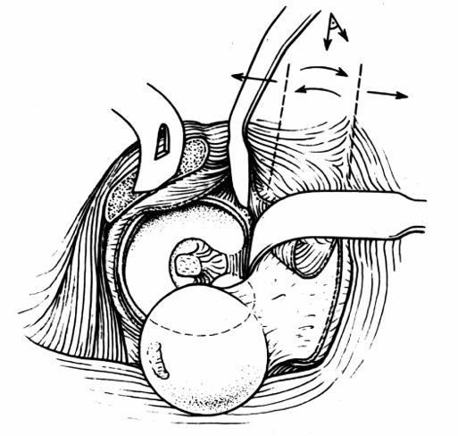
- Depends on the direction of dislocation
- Anterior: Smith-Petersen; posterior; trans-trochanteric
- Surgical hip dislocation - allows 360 degrees direct visualisation of the head
Q39What implant is used for fixation of a femoral head fracture?▸
- the source title states fixation of the femoral head with headless screws
- The approach depends on the direction of dislocation
Q40Describe the Kocher-Langenbeck approach with trochanteric flip.▸
- Subcutaneous fat, TFL and gluteus maximus split
- Trochanteric osteotomy: from the posterosuperior edge of the GT extending distally to the posterior border of the VL ridge; mobilise anteriorly
Q41Describe the Z capsulotomy used in surgical hip dislocation.▸
- First limb along the anterolateral axis of the femoral neck
- Second limb along the distal anterior edge of the capsule around the calcar
- Third limb parallel to the edge of the acetabulum
Q42How is the hip dislocated anteriorly in the surgical hip dislocation approach?▸
- Flexion, adduction and external rotation of the hip
Q43What is the clinical significance of a hip held in flexion, abduction and external rotation?📷▸
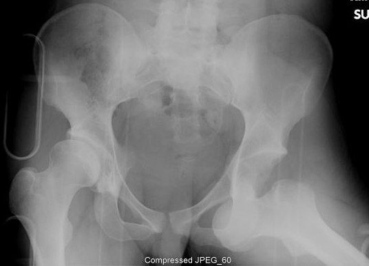
- Clinically this indicates an anterior dislocation
Q44Which Epstein classification applies to this injury?▸
- Epstein II inferior - as the hip is flexed
Q45Describe the Walker modification of Allis reduction for anterior hip dislocation.▸
- Supine; stabilise the pelvis; in-line traction; knee flexed
- Hip external rotation + flexion
- Laterally directed force + abduction
- Stabilise in internal rotation
Q46Describe the injury pattern shown.▸
- Pelviacetabular fracture + bilateral hip dislocation
- Right Thompson-Epstein IV (medial wall)
- Left Thompson-Epstein V
Q47How is anterior hip fracture dislocation classified?▸
- 1 Superior
- 2 Inferior
- Modifiers: A no fracture, B femoral head fracture, C acetabular fracture
Q48In the Thompson-Epstein classification, what do grades IV and V represent?▸
- Type IV: acetabular floor (medial wall)
- Type V: femoral head
Fact check
Acetabular fracture surgery should be performed 2-5 days post injury according to BOAST guidelines — misattributed — The BOAST pelvic fracture standard recommends definitive fixation within 72 hours of stabilisation; a 2-5 day window is not a BOAST recommendation — (medium confidence) — source