Trauma
Pelvic ring fracture - management and approaches
Pelvic disruption, acute resuscitation, definitive fixation, ilioinguinal and Stoppa approaches.
65 questions
7 source pages
3 images
1 fact-check flags
Images appear with the first question taken from each source page — tap a question to open it.
Q1Describe the AP pelvis X-ray findings of gross pelvic disruption.📷▸
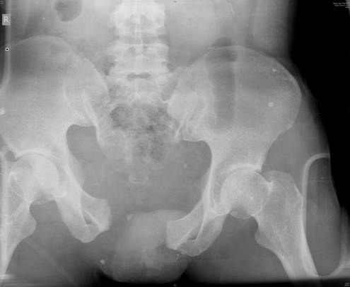
- Widening of the pubic symphysis
- Suspected widening of the right SI joint
- Vertical translation of the hemipelvis
- No fracture over the femoral neck/head
- Remember: assess anterior then posterior; the lines are not as important
Q2How is this injury classified?▸
- Young and Burgess - vertical shear
- Tile type C
Q3What are the three Tile categories of pelvic fracture?▸
- Stable
- Rotationally unstable
- Vertically unstable
Q4What are the three Young-Burgess mechanisms?▸
- Lateral compression
- AP compression
- Vertical shear
Q5Outline the immediate ATLS management of a high-energy pelvic fracture.▸
- High energy -> ATLS protocol + AMPLE -> primary survery ABCDE
- Secure airway and breathing
- C-spine triple immobilisation
- Disability: examine lower limb neurology
- Exposure
Q6How is circulation managed in the acute pelvic fracture?▸
- Pelvic binder application
- Fluid challenge with warmed 2L crystalloid
- Early prescription of unmatched whole blood
- If not responding, activate the massive transfusion protocol
- ROTEM-based substitution of single components
- Time limited permissive hypotension
Q7Describe the pelvic binder application and its goal.▸
- Ask assistance to hold the legs in internal rotation (IR)
- Apply at the level of the greater trochanter (GT)
- Goal: protect the first clot with a tamponade effect
Q8What is the tranexamic acid regimen in pelvic trauma?▸
- IV tranexamic acid 1g over 10 minutes, then 1g over 8 hours
- Given within 3 hours
Q9What scans are ordered after the primary survey?▸
- FAST scan
- XR (trauma series) and CT
- Second survey for life-/limb-threatening injuries
Q10What local associated injuries are sought in the second survey?▸
- Open wound
- Soft tissue: morel Lavelle (15% infection rate)
- Urinary: blood from urethral meatus, distended bladder, floating prostate -> urinary diversion
- GI: blood from rectum -> treat as open fracture + faecal diversion
- GU: blood from vagina
Q11What is a Morel-Lavallee lesion and how is it monitored?▸
- Soft tissue degloving injury in the subcutaneous plane/fascia
- 15% infection rate
- Monitor +/- contrast CT - collection with contrast enhancement at the subcutaneous plane
Q12What is the operative management if the patient remains unstable?▸
- Rule out other sources of bleeding
- DCO operative management (3 in 1) - a local study from a tertiary trauma centre in HK showed 3 in 1 reduces mortality
- Aim: skeletal stabilisation and haemorrhage control -> stabilise and nurture the first clot
- External fixator
- Angiogram + embolization
Q13What are the three sites of retroperitoneal bleeding in pelvic fracture?▸
- One in front of the SIJ
- One in front of the quadrilateral plate
- One behind the pubic rami
Q14What are the definitive management principles for a pelvic ring injury?▸
- AIM: restore integrity of the pelvic ring
- Anterior: plating
- Posterior: SI screw or plating
- If fixation is still tenuous, add an exfix to augment the fixation
Q15What is the New Berlin definition of a polytrauma patient?▸
- AIS >=3 for 2 or more different body regions
- Plus >=1 of: age >=70, GCS <=8, hypotension sBP <90, acidosis BE <=-6, coagulopathy
Q16What are the three peaks of mortality in trauma?▸
- On scene
- Golden hours (bleeding)
- 2-3 weeks (infection, MODS, ARDS)
Q17What are the ISS body regions, AIS grades and ISS threshold?▸
- ISS regions: 1 head and neck, 2 face, 3 chest, 4 abdomen/pelvis, 5 extremities, 6 external
- AIS: mild, moderate, severe non-life-threatening, severe life-threatening, critical, fatal
- ISS >15 = mortality 10%
Q18What is DCO and what are its five components?▸
- Damage control orthopaedics - a staged approach for the polytraumatised patient
- Limits CARS and SIRS by minimising the impact of surgery (the 2nd hit)
- Components: haemorrhage control, decontamination, decompression, temporary skeletal stabilisation, revascularisation
Q19Describe the stages of DCO.▸
- It involves a 3 stage approach
- Stage I: initial life-saving procedure (day 1, +/- day 2-3 for second look)
- Stage II: ICU optimisation to avoid the lethal triad (acidosis, hypothermia, coagulopathy)
- Stage III: definitive treatment once stabilised (day 5-10; stable BP, no inotropes, no hypothermia, normal urine output, no acidosis/coagulopathy)
- Stage IV: immunosuppressed D7-21 - avoid OT
Q20What key physiologic values did Heather Vallier publish for DCO?▸
- pH >7.25
- BE >=-5.5
- Lactate <4
- (J Orthop Surg Res 2015)
Q21How are trauma patients classified, and when is DCO considered in a borderline patient?▸
- Classified stable, borderline, unstable, extremes
- Borderline: ISS >40 or ISS >20 + chest injury
- GCS <=8
- Bilateral femoral fracture, pulmonary contusion, pelvic/abdominal injury
- Hypothermia; head injury with AIS >=3; IL6 above 500pg/dL
Q22What is the more recent evidence about early definitive fracture fixation?▸
- Nahm J Trauma 2011: most polytrauma patients benefit from early definitive fracture fixation
- New concept of early appropriate care (within 36hrs of injury)
- Key is individualised, based on intraoperative response to resuscitation
- Continued reassessment and ability to change from ETO to DCO
Q23Which radiographic lines and walls are assessed on an AP pelvis X-ray?📷▸
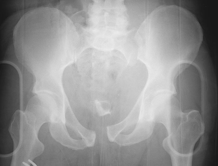
- Ilioischial line
- Iliopecteneal line
- Post/ ant wall
- +/- Shenton's line
Q24What displacement findings indicate vertical/rotational instability?▸
- Obvious displacement
- Vertical/rotational instability
- Widening of the pubic symphysis 2.5mm and SIJ 5mm
Q25What posterior injuries should be looked for on a pelvis X-ray?▸
- Avulsion fracture of L5 transverse process, ischial tuberosity, ischial spine
- Sarcal foramen + fracture
Q26What are the indications and contraindications for a pelvic external fixator?▸
- Indications: mechanically unstable ring (VS, APC2-3, LC2-3) and haemodynamically unstable patient
- Contraindications: iliac fracture involving tracking of pins; acetabular fracture
Q27What is the principle of pelvic external fixation?▸
- Decrease pelvic volume
- Stabilise the bleeding bone surface + venous plexus bleeding (tamponade effect) -> form the first clot
Q28What is the preoperative setup for pelvic external fixation?▸
- 2 surgeons
- Iliac crest draped with the groin well covered
- Remove the pelvic binder at the last minute
- Radiolucent table + X-ray
Q29Describe the ASIS frame for pelvic external fixation: advantages and disadvantages.▸
- Advantages: suitable for most patterns, fast and relatively easy, minimum X-ray
- Disadvantage: not for AP dissociation
Q30Describe the technique of ASIS frame pin placement.▸
- Start 2cm posterior to ASIS (thickest gluteal pillar, avoid LFCN)
- Skin incision more medial than anticipated as the pelvis will close up
- Stab wound, then 3 x 5.0mm pins
- Entry point on the iliac crest: more medial for older, middle for young
- Drill trajectory 30-45 degrees towards the ipsilateral hip
- Confirm with the OOO view (45/45)
Q31Describe the AIIS frame for pelvic external fixation: advantages and disadvantages.▸
- Advantages: no limitations (usable in AP dissociation), less bulky construct, facilitates nursing care
- Disadvantages: needs XR and a good OOO view; increased time and technique
Q32Describe the technique of AIIS frame placement.▸
- Use a good OOO view (tear drop sign at the acetabular roof) for the entry site
- Stab wound 2-3cm medial and below ASIS
- Blunt dissection (watch out for the LFCN)
- Trajectory AIIS to PSIS
- Confirm trajectory with the IOO view (20/35)
- Rod connection to form L and R modules -> reduction by using the modules as handles
Q33What are the indications and contraindications for a C clamp?▸
- Indications: posterior instability not controlled by an anterior frame; fracture of ilium and acetabulum; refractory haemodynamic instability
- Contraindications: fracture within the ilium (transiliac fracture dislocation); comminuted sacral fracture with risk of compression of the sacral nerve plexus (relative)
Q34How is a C clamp applied?▸
- 3 surgeons: hold device, reduction, tightening; preassemble at the side; involved limb draped free for manipulation
- Landmarks: line joining ASIS and PSIS, and another along the femoral shaft
- Push clamps along the transverse rail until bilateral bone contact, while applying traction and IR to the affected leg
- Bony landmark: intersection between anterior and posterior iliac wing
- Hammer the nail 1cm into bone, then push the side arms towards each other to tighten
Q35What are the indications, positioning and incision for the ilioinguinal approach?📷▸
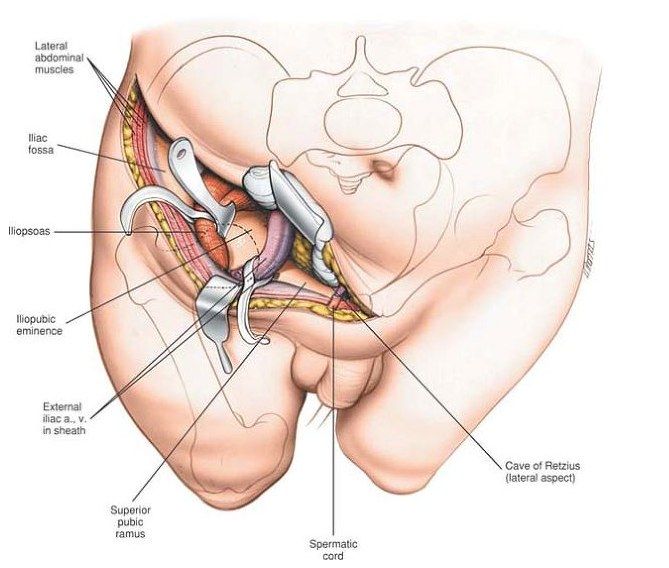
- Anterior approach to the pelvis and acetabulum
- Supine on a radiolucent table, GA, Foley
- Flex the hip to relax the psoas and NV bundle
- Incision from 5cm above ASIS to 1cm above pubic symphysis
Q36How is the internal iliac fossa exposed in the ilioinguinal approach?▸
- Skin + fascia from lateral to medial
- Release the external oblique insertion from the iliac crest
- Subperiosteal dissection to mobilise the iliacus muscle
- Pack the iliac fossa with gauze -> lateral window
Q37How is the inguinal canal unroofed in the ilioinguinal approach?▸
- Incise the external oblique aponeurosis from ASIS to the external inguinal ring (beware of the LFCN)
- This unroofs the inguinal canal, exposing the inguinal ligament
- Mobilise the spermatic cord/round ligament medially and sling with a Penrose drain
- Sharply incise the inguinal ligament, leaving a cuff of IO and transversus abdominis for later repair (beware of the ilioinguinal nerve)
Q38Describe the medial deep dissection of the ilioinguinal approach.▸
- Divide the rectus sheath and rectus 1cm proximal to its insertion
- Develop the plane between the symphysis pubis and bladder (Cave of Retzius)
Q39How is the iliopectineal fascia delineated in the ilioinguinal approach?▸
- Retract the femoral vessels (lacuna vasorum) medially
- Retract the femoral nerve and iliopsoas (lacuna musculorum) laterally
- Divide distally down to the pectineal eminence
Q40What are the three slings used in the ilioinguinal approach and what do they contain?▸
- Lateral sling: iliopsoas and femoral nerve
- Middle sling: femoral vessels
- Medial sling: spermatic cord
Q41What are the three windows of the ilioinguinal approach and what does each expose?▸
- Lateral window (lateral to the lateral sling): entire iliac fossa, SIJ, sacral ala, superior iliopectineal eminence
- Middle window (between middle and lateral slings): pelvic brim, quadrilateral surface
- Medial window (between medial and middle slings): pubic ramus, pubic symphysis, retropubic space of Retzius
Q42How can visualisation through the medial window be improved?▸
- The surgeon stands on the opposite side of the table
- With tilting of the table
Q43What is the corona mortis and how is it managed?▸
- Ligate retropubic anastomosis - obturator artery from the external iliac (inferior epigastric branch)
- Ligated during the ilioinguinal approach while developing the space of Retzius
Q44What are the dangers and complications of the ilioinguinal approach?▸
- Femoral nerve; femoral and external iliac arteries
- Lymphatics; LCFN; inferior epigastric artery; spermatic cord
- Heterotopic ossification; obturator nerve; ilioinguinal nerve
Q45What are the indications and access of the modified Stoppa approach?▸
- Indication: acetabular and pelvic ring fractures
- Access: pubic body, superior pubic ramus, pubic root, anterior column, mid posterior column, quadrilateral plate, anterior SIJ
Q46What is the setup and incision for the modified Stoppa approach?▸
- Radiolucent table, supine, Foley, flex hips and knees
- Operating surgeon on the opposite side
- Midline transverse incision 2cm above the pubic symphysis (laterally beware the external inguinal ring)
Q47Describe the superficial dissection of the modified Stoppa approach.▸
- Incision of subcutaneous tissue and rectus fascia in line
- Rectus abdominis fascia split along the linea alba
- Pyramidalis muscle released
- Retract the rectus abdominis muscle laterally
Q48Describe the deep dissection of the modified Stoppa approach.▸
- Transversalis fascia opened sup to PS
- Enter the space of Retzius (between pubic symphysis and bladder)
- Hohman retraction over the pubic tubercle to retract the rectus abdominis
- Subperiosteal dissection along the pubic ramus, then continue retroramus dissection to the quadrilateral surface
- Release the iliopectineal fascia from the pelvic brim
Q49What is the corona mortis and how is it managed in the modified Stoppa approach?▸
- Identified during subperiosteal dissection along the pubic ramus
- Ligated: 2 veins and 1 artery
Q50Describe the X-ray findings in this pelvic disruption.▸
- Widening of the pubic symphysis ~2cm
- Fracture of the right inferior pubic ramus
- Widening of the left SIJ with vertical displacement of the left hemipelvis
- Fracture of the left L5 transverse process
Q51How would you classify this fracture?▸
- Vertically and rotationally unstable fracture
Q52Which ligaments provide vertical and rotational stability of the pelvic ring?▸
- Vertical stability: iliolumbar, sacrotuberous, posterior SI ligaments
- Rotational stability: pubic symphysis, sacrospinous (suspect pelvic floor injury), anterior SI ligaments
Q53Describe Xray findings in a suspected pelvic fracture.▸
- Pubic symphysis, rami (transverse -> LC; vertical -> open book), iliac wing, SIJ, sacrum
- Avulsion fracture: L5 transverse process, STL, SSL
- Look for an associated hip fracture
- This is a rotational/vertical unstable fracture
Q54What are the ATLS priorities in the acute management of a pelvic fracture?▸
- ATLS - resuscitate according to ABC
- Rule out other life-threatening chest/abdomen trauma by clinical examination, Xray and FAST
Q55What is the role of the pelvic binder and what are its indications?▸
- Pelvic binder centre at the GT with LL IR
- Indication: blunt trauma, high energy, shock
- Protect the first clot by tamponade effect
Q56If haemodynamically unstable, how is the pelvic fracture resuscitated?▸
- Give 2L crystalloid and see response
- Take blood for cross-match
- If still unstable give O-negative blood and start massive transfusion protocol -> DCO
Q57How does FAST alter the acute management sequence?▸
- FAST +ve (intraperitoneal bleeding): laparotomy first
- FAST equivocal (60% of bleeding due to pelvis) or -ve: ex fix first
Q58What is the role of embolization in pelvic trauma?▸
- Considered after pelvic packing if still not stable
- Stable patient -> highly selective embolization
- Unstable patient -> non-selective embolization of the internal iliac
- If no extravasation, bleeding may be masked by shock/vasospasm
- Material: gel foam / metallic coil
Q59What anterior frame is used acutely and how are pins placed?▸
- Anterior frame iliac crest construct if there is no iliac fracture
- +/- C clamp
- Pins start 2cm posterior to ASIS
- Use the OOO Xray view and orient to ASIS-PSIS and the femoral shaft
Q60What is pelvic packing and how is it performed?▸
- Performed via the Stoppa approach (2cm above PS, between transversus abdominis and bladder)
- Pack anterior to the SIJ, the quadrilateral plate, and posterior to the PS
- Performed if the patient is still not stable before embolization
Q61How are PR and GU bleeding managed in pelvic trauma?▸
- PR/GU bleeding: +/- diversion + antibiotic
- Morel-Lavallee: subcutaneous/fascia plane with contrast enhancement; drainage in 2nd look OT - there is a risk of injury to the inferior epigastric artery/obturator artery; no drainage in D0
Q62What trauma patterns must be bewared of in pelvic fractures?▸
- Tilt fracture (lateral compression): starts benign-looking, then severe displacement, LLD, pain in sexual intercourse, difficult childbirth
- Pelvic acetabular fracture
Q63What is the load transmission of the posterior and anterior pelvic complexes?▸
- Posterior complex: 60% of body weight
- Anterior complex: 40% of body weight
Q64Why should both the anterior and posterior complexes be fixed?▸
- Fix both for early mobilization
- If treating the posterior alone: PWB
Q65What must be considered when there is both column involvement?▸
- Ddx of both column involvement (4) - the four differentials are listed on the source but not expanded in the notes
- May need a combined approach
- If just one column, choose the more displaced one
Fact check
Pelvic ring instability is indicated by widening of the pubic symphysis of 2.5mm and SIJ of 5mm — wrong unit — Classic teaching defines anterior instability by symphysis diastasis >2.5 cm (not mm); SI joint widening >5 mm is a sign of posterior injury, though recent literature questions the exact 2.5 cm cut-off — source