Trauma
Fatigue, fragility and pathological fractures
Fatigue and fragility fractures, osteoporotic bone, pathological subtrochanteric fracture.
35 questions
3 source pages
1 images
Images appear with the first question taken from each source page — tap a question to open it.
Q1What is a stress fracture?▸
- A fracture of bone at stresses below the ultimate tensile strength
Q2Differentiate an insufficiency fracture from a fatigue fracture.▸
- Insufficiency fracture: physiological stress on a weakened bone (normal stress, abnormal bone)
- Fatigue fracture: repetitive loading below the ultimate tensile stress on normal bone (abnormal stress, normal bone)
Q3Describe the pathogenesis of stress fractures.▸
- Bone formation lags behind bone resorption
- Ultimate tensile strength reduced, rendering the bone susceptible to microfractures
- This propagates to a stress fracture
Q4Which femoral neck stress fractures are more serious and how are they treated?▸
- Tension fractures are more serious
- They may require operation
Q5What is the concern with a femoral shaft stress fracture?▸
- It responds to protected weight bearing
- It can progress to a complete fracture if unrecognised
Q6What is notable about pelvic stress fractures?▸
- They are rare
- They involve the sacrum and pelvis
Q7What is the presentation of a stress fracture?▸
- History of overuse
- Insidious onset of pain
- Localised swelling and tenderness
Q8What is the fulcrum test used for?▸
- It is used for a femoral shaft stress fracture
Q9What are the X-ray findings and timing in a stress fracture?▸
- XR is normal in the first 2-3 weeks
- Later: periosteal reaction, thin fracture line, cortical lucency
Q10What is the role of MRI in stress fractures?▸
- MRI (STIR sequence) is the most specific test but is not specific
Q11What are the bone scan findings in an acute stress fracture, a healing fracture and a soft tissue injury?▸
- Bone scan is highly sensitive but non-specific
- Acute stress fracture: linear area of increased uptake at all 3 phases
- Healing fracture: back to normal at phase I, II and III sequentially
- Soft tissue injury: uptake in the first 2 phases
Q12What is the general management of a stress fracture?▸
- Rule out anorexia nervosa or metabolic disease
- Nonoperative: protected weight bearing, rest, cross training, analgesics, therapeutic modalities
Q13What is risk stratification in stress fracture management?▸
- Low-risk fracture
- High-risk fracture
Q14Which is the most common stress fracture and where does the compression type begin?▸
- Tibial stress fracture - the most common
- Compression fracture begins at the posteromedial (PM) cortex
- Transverse in orientation, common in distance runners
- Treated nonoperatively with pneumatic bracing
Q15Describe the tibial tension stress fracture.▸
- Anterior cortex of the middle 1/3 of the tibia
- XR features are subtle but better seen on the lateral view - a V-shaped defect at the anterior cortex
- Common in repetitive jumping and heaping activities
Q16Why do tibial tension fractures predispose to complete fracture, delayed union or nonunion?▸
- The tension side effect (Wolff's law)
- Hypovascularity of the anterior cortex
Q17How are tibial tension stress fractures treated?▸
- Initial: trial of rest +/- immobilisation
- Once a wide fissure develops, operative treatment is needed
- Delayed/nonunion: excision and bone grafting, or IMN
Q18How is a femoral neck stress fracture managed?▸
- Compression type -> nonoperative
- Tension type -> operative
Q19Where do 5th metatarsal stress fractures occur and how are they managed?▸
- At the metadiaphyseal junction
- Sx+ / XR- -> NWB + brace
- Sx+ / XR+ -> low demand: NWB; high demand: IM screw
- Delayed union -> intramedullary screw fixation
Q20Describe the X-ray findings in this osteopetrotic subtrochanteric fracture.▸
- Polyostotic sclerotic bone
- Bone in bone pelvis
- Pathological fracture with lateral beaking and medial spike, non-comminuted, subtrochanteric, short oblique
- Suspect underlying osteopetrosis
Q21What is the differential diagnosis of this sclerotic fracture?▸
- Metastasis
- Hypoparathyroidism
- Paget's disease
- Pyknodysostosis
Q22What other X-rays should be requested and what do they show in osteopetrosis?▸
- Spine: rugger jersey appearance
- Hand: bone in bone
- Lower limb: Erlenmeyer flask appearance
Q23What is pyknodysostosis?▸
- Autosomal recessive trait with mutation in cathepsin K
- Generalized osteosclerosis like osteopetrosis but with preservation of the medullary canal of long bones
Q24What is the pathophysiology of osteopetrosis?▸
- Defect in osteoclastic resorption due to defective carbonic anhydrase / chloride channel
- Osteoclasts also lack a normal ruffled border and clear zone
Q25How do patients with osteopetrosis present?▸
- Fracture (cartilage remains in the diaphysis -> inferior mechanical properties)
- Deformity from repeated fractures
- Infection (osteomyelitis)
- Pancytopenia
- Cranial nerve palsy from overgrowth of skull foramina
- Osteoarthritis
Q26What are the types of osteopetrosis?▸
- AD tarda = Albers-Schonberg disease: mild anaemia, pathological fractures, premature OA, increase serum acid phosphatase
- AR: patients die in infancy
- Intermediate type
Q27What investigations are performed in osteopetrosis?▸
- Biopsy: empty lacunae and plugged-up haversian system with necrotic cartilage
- Enzyme studies: TRAP and Creatinine kinase BB isoenzyme
Q28What medical treatment is used in osteopetrosis?▸
- Bone marrow transplant
- High-dose vitamin D with a low-calcium diet
- Inferon 1 gamma beta
Q29What are the special considerations when fixing a fracture in osteopetrosis?▸
- Freshen the fracture edges
- Fix with plating (tension band +/- medial plating)
- Sharp reaming with irrigation and a prepared burr
- Rigid fixation and bone graft
- Warn of a high failure rate
Q30What are the special considerations for replacement arthroplasty in osteopetrosis?▸
- Use cementless implants - there is no interdigitation for cement
- Use a short stemmed implant
- The Acetabulum need multiple screws
Q31Describe the Rommens classification of pelvic fragility fractures.📷▸
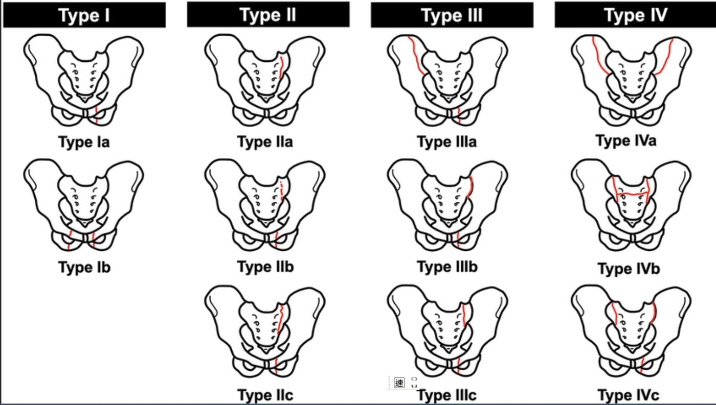
- 1: anterior only
- 2: undisplaced + posterior
- 3: displaced posterior
- 4: bilateral posterior
Q32What is the management principle and recommendation by Rommens type?▸
- Regain walking ability ASAP while balancing the high operation risk
- Type 1: non-operative
- Type 2: early weight bear; if OK non-op, if not tolerated then operation
- Types 3/4: operative
Q33What are the problems of non-operative management, and those specific to pelvic fragility fractures?▸
- Non-operative: non/malunion, fracture progression
- PFF-specific: poor bone, higher rate of loss of fixation
- Measures: cemented screw, interdigitation of fixation, robust fixation
Q34What is the evidence on fracture progression and anterior fixation in FFP?▸
- Rommens JBJS 2019: fracture progression rate 14%
- Herteleer Eur J Trauma Emerg Surg 2022: no correlation between anterior fixation and overall fixation failure (no consensus)
Q35When should pelvic fragility fractures be operated on?▸
- SR by Daniel BMC MSK Disord 2021: fixation considered for patients failing a brief period of non-operative management
- Fixation should be robust enough to allow immediate weight-bearing but minimise operative morbidity and post-op complications