Foot and Ankle
Hallux valgus - clinical and radiographic assessment
Hallux valgus severity on clinical and X-ray assessment including Harris axial view
41 questions
5 source pages
2 images
Images appear with the first question taken from each source page — tap a question to open it.
Q1What are the causes of ankle arthritis?▸
- Post-traumatic (70%): altered load following injury; 2/3 fracture, 1/3 sports-related ligamentous injury (Iowa study)
- Primary OA
- Inflammatory arthritis (RA, gout)
- Neuropathic
- Post-infection
Q2What is the Takakura classification of ankle osteoarthritis?▸
- I: early sclerosis
- II: medial joint space narrowing, no subchondral bone contact
- III: subchondral bone contact
- IIIA: medial malleolus joint space obliteration; IIIB: talar dome roof obliteration with bone contact
- IV: all joint space with complete bone contact
Q3What are the prerequisites for a joint-preserving supramalleolar osteotomy?▸
- Medially focused ankle arthritis (stage 2/3a)
- Nearly normal ROM
- Minimal talar tilting
- Realignment targets: TAS(tibial anterior surface angle)=LDTA (lateral distal tibial angle)=89, talar tilt <10, TLS(tibial lateral surface angle)=ADTA(anterior distal tibial angle)=80
Q4What are the advantages and disadvantages of ankle fusion?▸
- Advantages: more stable and durable than replacement, corrects deformity, less infection
- Disadvantages: longer rehab (3-6 months), adjacent joint degeneration, motion loss, stress fracture, require shoe modification if active
- Complications: adjacent joint arthritis 50% in 10 year, non union 20%
Q5What is the optimal position for ankle fusion?▸
- Neutral dorsiflexion
- 5 deg external rotation (similar to the other side)
- 5 deg hindfoot valgus
- Talus translated 5mm posteriorly (anterior edge of talus same level as anterior edge of tibia - normalize gait and decrease stress at knee)
Q6What are the contraindications to total ankle replacement?▸
- Young and active patients
- Poor bone stock or talar AVN
- Infection
- Poor ligamentous stability or absent malleoli
- Significant malalignment
Q7Describe the X-ray findings on this section.▸
- Osteoarthritic change at the tibiotalar joint: decreased joint space, subchondral sclerosis
- Evidence of healed tibia and fibula fracture; syndesmosis appears intact
- Diagnosis: post-traumatic ankle arthritis
Q8What history, examination and imaging workup is needed?▸
- Hx: risk factors, pain, functional limitations
- PE: skin status, tenderness, ROM, deformity, adjacent joints, overall alignment
- XR: LL scannogram, foot and ankle for subtalar/other joint degeneration, Saltzman view
- MRI if early stage
Q9What patient and disease factors guide management?▸
- Patient: age, functional status, rehab potential, expectations
- Disease: cause (post-traumatic, inflammatory - may be bilateral/osteopenic)
- Adjacent joint OA/bilateral disease, extra-articular malalignment, bone stock
- Joint congruency and correctability, OA stage
Q10What is the overall management principle and what conservative options exist?▸
- Principle: resume stability, correct alignment and preserve cartilage if possible
- Conservative: physio, NSAID, rocker bottom sole, AFO to simulate fusion, off-loading brace
Q11What joint-preserving options exist for ankle arthritis?▸
- Realignment: supramalleolar osteotomy +/- soft tissue
- Joint resurfacing: MACI, stem cells
- Ankle scope: synovectomy, cheilectomy
- Distraction diastasis: Exfix for 3/12 (3 months), stimulates PG production
Q12What are the contraindications to ankle fusion?▸
- Bilateral disease
- Adjacent joint degeneration
Q13What techniques and fixation are used for ankle fusion?▸
- Arthroscopic arthrodesis in minimal deformity
- Anterior +/- transfibular approach (fibula can be used as onlay graft)
- Fixation: plate/screw, Blair's fusion, or retrograde hindfoot nail if subtalar degeneration
Q14Which features do you comment on in a total ankle replacement X-ray?▸
- Cement, bearing, syndesmosis fixation, alignment, sizing
- Bone cut (just below the syndesmosis)
Q15What are the specific complications and commonly used implants in TAR?▸
- Specific complications: malleolar impingement, medial malleolus fracture
- Common implants: STAR (no tibiofibular fusion) and Agility
Q16What is the survival of the STAR prosthesis and what trial is ongoing?▸
- STAR 70.7% survival 10 yrs; 80% at 5 years; 50% at 15 years
- TARVA trial ongoing
Q17What are the properties of ankle cartilage and why is ankle arthritis uncommon?▸
- Thinnest cartilage, smaller contact area, stiffer
- 1 mm shift of a fracture decreases contact surface area by 42%; medial instability by 15%
- Uncommon because the joint is highly congruent/stable with rolling (not rolling + shear)
- Cartilage is well nourished, with resistance to inflammatory mediators like MMP, IL1 --> stiffness does not decrease with age
Q18Describe the three generations of total ankle replacement.▸
- 1st: two components, highly constrained, cemented tibia and talus, high incidence of loosening
- 2nd: two components, polyethylene bearing, more conservative bone cuts, cementless
- 3rd: three components, independent PE mobile bearing, cementless, More rely on ligamentous stability
Q19What is used if both the ankle and subtalar joints have OA?▸
- Hindfoot arthrodesis nail
Q20Describe the deformity shown in the clinical photo.📷▸
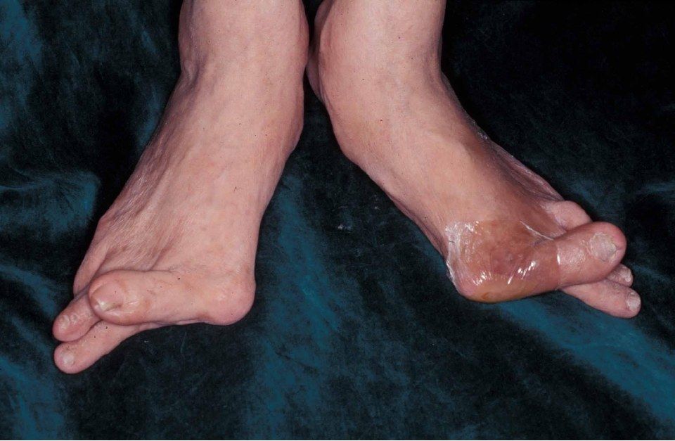
- Severe bilateral hallux valgus: big toe deviated laterally, 1st MT medially
- Hallux also in a pronated position
- Lesser toe deformity is present
- Bunion with no callosity, no scars
Q21What would you examine in this patient?▸
- Standing foot arch and hindfoot alignment; sole for callosity
- Single and double heel raise
- Palpate bunion, 2nd MT plantar and 5th MT head; check correctability; Check TMTJ hypermobility
- NV exam, Silverskiold test, gait (loss of third ankle rocker)
- Look for RA features and generalized ligamentous laxity
Q22What X-ray parameters do you assess in hallux valgus?▸
- DP foot: HVA, IMA, HVI, DMAA, 1st MTPJ congruence, sesamoid station, MTPJ/TMTJ arthritis, Maestro's cascade
- Lateral: calcaneal pitch and Meary angle
Q23What determines the choice of surgery in hallux valgus?▸
- 1. severity, 2. congruence, 3. TMTJ laxity, 4. 1st MTPJ arthritis
- This case needs a combined soft tissue and bony procedure: modified McBride (DSTP) + 1st MT realignment osteotomy (scarf osteotomy)
Q24Compare chevron and scarf osteotomies for hallux valgus.▸
- Chevron: no more than 1/3 shift, 1 mm ~ 1 degree of IMA correction, not for severe deformity
- Scarf: versatile, corrects deformity in all three planes, stable allowing early weight bearing
- Scarf complications: troughing, nonunion, hallux varus
Q25Explain the pathophysiology of hallux valgus.▸
- Defined as lateral deviation of the great toe with medial deviation of the 1st MT
- Overall: destabilization of 1st MTPJ
- Medial capsular attenuation; the 1st PP deviates laterally while the 1st MT drifts medially off the sesamoids
- Plantar-lateral migration of abductor hallucis plantarflexes and pronates the phalanx
- EHL and FHL subluxate laterally and become deforming forces; secondary contracture of the lateral capsule, adductor hallucis and intermetatarsal ligament
- The 1st ray defunctions, the 2nd MT head takes body weight and the plantar plate eventually become attenuated
- Sagittal imbalance causes claw toe from EDL overpull
Q26What history would you take in hallux valgus?▸
- RA, family history
- Pain, walking tolerance
- Expectations
Q27What conservative management is offered for hallux valgus?▸
- Wide toe box, advise against high heels
- NSAIDs
- Toe spreader or spacer
Q28Describe the modified McBride procedure.▸
- Lateral: release adductor hallucis from the sesamoid
- Lateral: release the intermetatarsal ligament (watch the common digital nerve), capsulotomy, release the suspensory sesamoid-metatarsal ligament
- Medial: capsulorrhaphy
- Medial: bunionectomy (cut 2 mm medial to the sulcus)
Q29Describe the scarf osteotomy technique and aftercare.▸
- Medial incision; protect the dorsal medial cutaneous branch of the superficial peroneal nerve and the medial cutaneous branch of the hallux (medial plantar nerve)
- Z-shaped osteotomy: longitudinal cut parallel to the sole, transverse cut perpendicular to the 2nd MT; distal limb points dorsal, proximal limb plantar
- Translate the distal fragment laterally, fix with 2 x 2 mm headless compression screws
- Rehab: heel walking for 6 weeks
- Complications: troughing, nonunion, hallux varus
Q30What are the risk factors for hallux valgus?▸
- Multifactorial: female, genetic predisposition
- Extrinsic: shoewear
- Intrinsic systemic: RA, ligamentous laxity
- Intrinsic local: flat foot, amputated 2nd toe
Q31What are the deforming forces in hallux valgus?▸
- Dynamic: adductor hallucis - pronation and adduction of the proximal phalanx
- Dynamic: abductor hallucis - plantarflexion and pronation of the proximal phalanx
- Dynamic: EHL and FHL subluxed laterally in relation to the MTPJ
- Static restraint loss: plantar plate (sesamoid pronation), lateral capsule contracture, intermetatarsal ligament contracture, crist erosion
Q32Describe the X-ray findings and their significance.📷▸
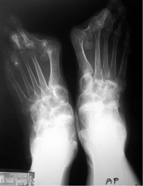
- Severe bilateral hallux valgus deformity
- OA changes at the 1st MTPJ
- Rule out an underlying systemic cause
- Operative management if symptomatic
Q33What are the indications for 1st MTPJ fusion?▸
- OA
- RA
- Spasticity
- Fusion corrects the IMA with realignment of the hallux
Q34What are the radiographic findings?▸
- Bilateral hallux valgus
- Eyeballing HVA >40 deg, IMA >13 deg
- DMAA <10 deg
- No 1st MTPJ arthritic changes
- Clinically need to check 1st TMTJ laxity
Q35What is the operative plan?▸
- Proximal metatarsal osteotomy
- Distal soft tissue release
- Medial eminence resection and capsular repair
Q36What are the complications of hallux valgus surgery?▸
- Overcorrection causing hallux varus
- Undercorrection (OT factor, patient factor)
- Malunion or nonunion
- AVN (medial artery to metatarsal neck), recurrence
- Troughing leading to elevation or rotation of the 1st MT
Q37How do MTPJ, TMTJ, severity, DMAA and HVI guide the operation?▸
- 1st MTPJ pathological: fuse
- 1st TMTJ lax: fuse
- Severe HVA/IMA: proximal osteotomy; mild: distal
- Abnormal DMAA: proximal + distal medial closing wedge; both severe: biplanar closing wedge
- HVI >10: Akin
Q38What conservative management would you try first?▸
- Shoewear advice
- Orthosis
- Operate if symptomatic despite conservative measures
Q39How is the Harris axial view taken and what does it show?▸
- Foot in dorsiflexion with the beam angled 45 deg cephalad
- Visualizes the joint surface, loss of height, increasing width and angulation of the tuberosity fragment (usually varus)
Q40How is the Canale view taken?▸
- Ankle in maximal equinus
- Foot on the cassette pronated 15 deg
- X-ray beam 15 deg cephalad
Q41How is the Broden view taken?▸
- Patient supine, cassette under the leg and ankle
- Foot in neutral flexion with the leg internally rotated 15 deg (mortise)
- X-ray centred over the lateral malleolus
- Beam directed toward the head at 40, 30, 20 and 10 deg