Foot and Ankle
Lateral ankle ligament and tendon injuries
Lateral ligament insufficiency on stress views, talar tubercle avulsion and peroneal subluxation
29 questions
3 source pages
2 images
1 fact-check flags
Images appear with the first question taken from each source page — tap a question to open it.
Q1What types of ankle sprain do you know of?▸
- Low (90%) or high
- Eversion or inversion
Q2Which lateral ankle ligaments are injured and by what mechanism?▸
- ATFL is commonest (plantarflexion + inversion)
- CFL is 2nd most common (dorsiflexion + inversion)
- PTFL is the strongest
Q3How do you distinguish low from high ankle sprain on examination?▸
- Low: anterior drawer (dorsiflexion tests CFL, plantarflexion ATFL), talar tilt indicates CFL torn
- High: syndesmosis tenderness, squeeze test, cotton test, Frick test (external rotation), cross leg test
Q4When would you X-ray a patient with an ankle sprain?▸
- Follow the Ottawa rules
- Pain within 6 cm of the malleolar tip or unable to bear weight: X-ray ankle
- Pain over the 5th MT base or navicular or unable to bear weight: X-ray foot
Q5What is the Chorley classification of low ankle sprain?▸
- I: partial ATFL
- II: complete ATFL +/- partial CFL (anterior drawer I)
- III: complete ATFL + CFL +/- capsule +/- PTFL (anterior drawer II, talar tilt)
Q6What is the treatment for a low ankle sprain?▸
- Conservative: RICE, physio, functional training, peroneal and proprioception training
- 20% develop symptoms of chronic ankle instability
- Surgery if bony avulsion, both medial and lateral ligament rupture, or talar tilt in athlete > 15deg
- Recurrent instability: anatomic repair (Brostrom +/- Gould modification), anatomic reconstruction, or non-anatomic (Chrisman Snook, Watson Jones, Evans)
Q7What does the stress view X-ray show?▸
- Significant talar tilt suggesting lateral ankle ligamentous disruption
Q8What history and examination features determine functional vs mechanical instability?▸
- History: age, body build, sport, occupation
- History of hypermobility, mechanism of injury, recurrence
- Examination aims to determine functional vs mechanical instability and high vs low sprain
Q9What concomitant pathologies must be ruled out in an ankle sprain?▸
- Tendon: peroneal split tear or subluxing tendons
- Bone: fractures of the anterior calcaneal process, lateral or posterior talar process, 5th MT base
- Cartilage/joint: OCD, loose bodies
- Predisposing factor: cavovarus
Q10What X-ray views and MRI are used in ankle sprain?▸
- Ankle AP, lateral, mortise weight bearing, Saltzman view (syndesmosis and OCD)
- Foot DP, oblique, weight-bearing lateral
- Stress view with ER/ varus: talar tilt 5 deg+ (or absolute 10) abnormal; anterior drawer same
- Scannogram; MRI for OCD, cartilage status, degree of tear and tissue quality
Q11How is an acute high ankle sprain (syndesmosis injury) treated?▸
- No diastasis: immobilize 1-3 weeks, then functional rehab + strengthening
- Diastasis: obtain MRI
- Isolated AITFL: immobilization + NWB 4-6 weeks
- AITFL + others: 2 syndesmosis screws, then NWB 6-12 weeks
Q12How is chronic syndesmosis instability treated?▸
- Debridement + 2 syndesmosis screws or suture button
- Reconstruction by peroneus longus: half PL harvested from the LM tip
- Free end secured by a 3.5 mm screw + washer, protected with a syndesmosis screw
Q13What are the surgical options for recurrent ankle sprain with mechanical instability?▸
- Anatomic repair with endogenous tissue when adequate (Brostrom +/- Gould modification: anatomical shortening and reinsertion of ATFL + CFL with inferior extensor retinaculum and fibular periosteum); fewer complications, quicker rehab
- Anatomic reconstruction: semiT or plantaris autograft or synthetic graft replicating ATFL/CFL anatomy
- Non-anatomic checkrein: Chrisman Snook, Watson Jones, Evans
- Consider diagnostic arthroscopy: intraarticular pathology amenable to arthroscopy was found in 83% of Brostrom repairs
Q14What is the evidence for lateral ligament augmentation?▸
- AJSM 2019 RCT: primary repair + LARS gave better FAOS at 5-year follow-up and high Tegner scores vs MBG
- Foot and Ankle Surg 2021 meta-analysis: no difference
Q15What is a Shepherd fracture?📷▸
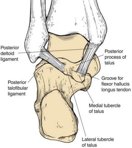
- Fracture of the posterolateral talar tubercle
- Contrast with a Cedell fracture = posteromedial talar tubercle
Q16What is a Cedell fracture and what structure lies between the two tubercles?▸
- Posteromedial talar tubercle fracture
- The FHL tendon runs between them
Q17Describe the Oden classification of superior peroneal retinaculum (SPR) injury.📷▸
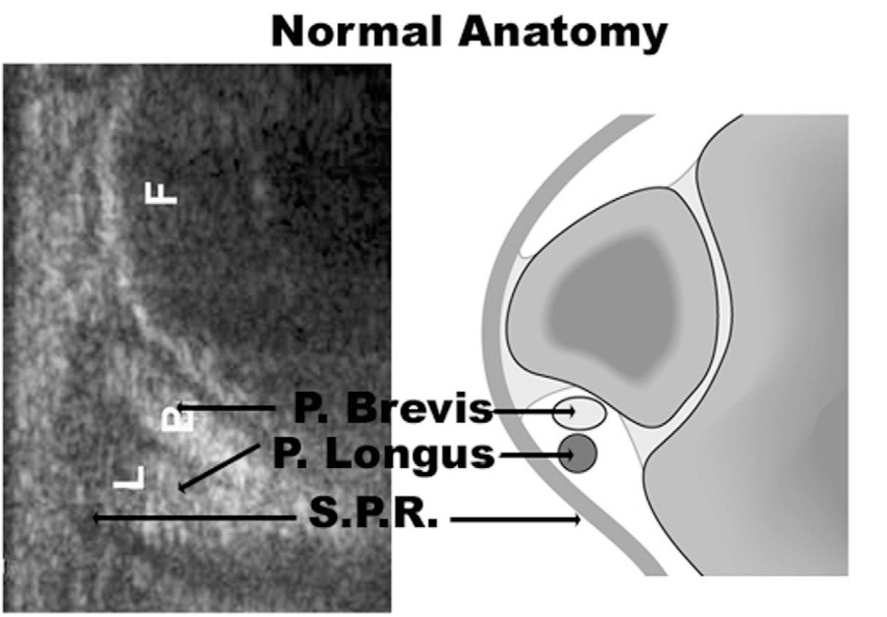
- Grade 1: SPR elevated off the fibula, tendons lie between bone and periosteum (most common, >50%)
- Grade 2: fibrocartilaginous ridge avulses with the retinaculum; tendons displaced beneath the ridge
- Grade 3: avulsion of a small cortical osseous fragment from the fibular insertion; tendons dislocate beneath the fragment
- Grade 4 (rarest): complete avulsion/rupture of the SPR; tendons lie external and superficial to the retinaculum
Q18Which Oden grade is most common and what is its pathoanatomy?▸
- Grade 1, >50% of cases
- SPR elevated off the fibula
- Tendons come to lie between the bone and the periosteum
Q19What is the pathoanatomy of an Oden grade 2 lesion?▸
- The fibrocartilaginous ridge behind the lateral insertion of the SPR avulses together with the retinaculum
- The peroneal tendons are displaced beneath the ridge
Q20What is the pathoanatomy of an Oden grade 3 lesion?▸
- Avulsion of a small cortical osseous fragment from the fibular insertion
- The tendons dislocate beneath the fibular fragment
Q21What is the pathoanatomy of an Oden grade 4 lesion?▸
- The rarest type of lesion
- Complete avulsion or rupture of the superior peroneal retinaculum
- Tendons lie external and superficial to the retinaculum
Q22What other classifications are used for peroneal tendon pathology?▸
- Raikin for intrasubstance subluxation
- Redfern for peroneal tendon tear (both okay/partial tear, one torn, both torn; A not retracted, B retracted)
Q23Describe the anatomy of the peroneal tendon groove.▸
- Peroneal tendon sheath splits at the level of the peroneal tubercle
- Groove over the posterolateral aspect of the fibula
- Bounded by the superior peroneal retinaculum, fibrocartilaginous ridge and Talus/ calcaneum
Q24How do you assess suspected peroneal tendon subluxation?▸
- History: timing, recurrence
- Apprehension test (resisted dorsiflexion and eversion, or plantarflexion + inversion to dorsiflexion + eversion)
- Check for voluntary subluxation and any hindfoot varus
Q25How is the apprehension test for peroneal tendon subluxation performed?▸
- Resisted dorsiflexion and eversion, or
- Move the ankle from plantarflexion + inversion to dorsiflexion + eversion
Q26What imaging is used for peroneal tendon subluxation?▸
- XR: look for the fleck sign
- Dynamic USG
- CT
- MRI
Q27How is acute peroneal tendon subluxation managed nonoperatively?▸
- Short leg cast and protected weight bearing for 6 weeks
Q28What is the operative management of acute peroneal tendon subluxation?▸
- Acute repair of the superior peroneal retinaculum
- Deepening of the fibular groove
Q29How is chronic or recurrent peroneal tendon dislocation managed?▸
- Groove deepening with soft tissue transfer and/or osteotomy
Fact check
Ankle X-ray is indicated if pain is within 6 cm to the tip of the malleolus — imprecise phrasing of the Ottawa ankle rules — Rule = pain in the malleolar zone AND bone tenderness along the distal 6 cm of the posterior edge/tip of either malleolus, or inability to bear weight 4 steps — source