Foot and Ankle
Diabetic foot ulceration and offloading
Plantar forefoot ulceration, Symes amputation levels and pressure-relieving shoe wear
21 questions
3 source pages
1 images
1 fact-check flags
Images appear with the first question taken from each source page — tap a question to open it.
Q1Describe the ulcer shown on the plantar aspect of the foot.📷▸
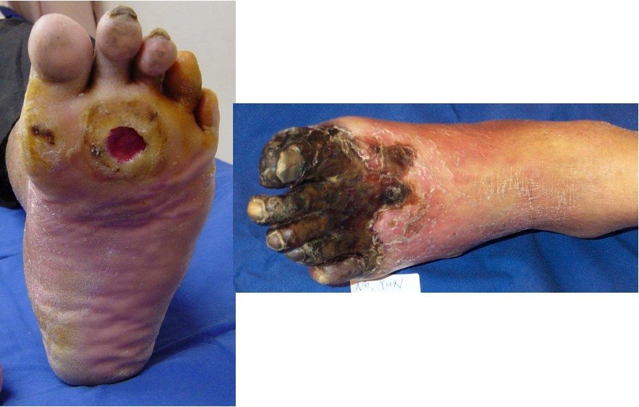
- Erythematous base with punched out edges
- No exposed tendon or bone, no pus
- Surrounded by callosities
- Associated hallux deformity with the 2nd and 3rd toes in clawed position
- Site and deformities suggest a DM neuropathic ulcer
Q2What is the Brodsky depth-ischemia classification of diabetic foot ulcers?▸
- Depth: 0 at risk, 1 superficial ulcer, 2 deep ulcer (tendon or joint exposed), 3 extensive ulcer or abscess
- Ischemia: A not ischemic, B ischemia without gangrene, C partial forefoot gangrene, D complete gangrene
- 4+4 system combining depth and ischemia
Q3What neurological and vascular examination would you perform?▸
- Proprioception
- Vibration with 128 Hz tuning fork
- Semmes Weinstein 5.07 for protective sensation
- Palpate pulses, ABI
- Check muscle power PL, PB, TA, PT; Silverskiold test; shoewear
Q4How do you differentiate a neuropathic from an arterial ulcer?▸
- Neuropathic: plantar, not painful, foot deformity, pulse likely present, punched out lesion
- Arterial: tip of foot or edges, painful, atrophic changes, absent pulse, fibrotic base, no surrounding callosity
Q5How would you manage this patient with a neuropathic ulcer and absent pulses?▸
- Multidisciplinary: medical, vascular, P&O, ortho; patient education
- Goal: ulcer free, functional, plantigrade foot fitting a brace or shoe, with minimal complications and hospital stay
- Systemically control/correct risk factors eg DM, PVD; improve nutrition
- 1. unload prominent area: total contact casting (increases contact area, transmits load proximally, typically 6 months)
- Prevent infection: daily dressing (Medihoney), debridement or amputation
Q6When and at what level would you amputate?▸
- Indications: dangerous (infected), dead (gangrene), damn nuisance
- Level balances healing potential against energy expenditure
- Longer residual limb is more energy conserving; the exception is Symes
- Order: Transmetatarsal > Symes > Lisfranc > Chopart > transtibial
- Lisfranc and Chopart need TA lengthening
Q7What blood tests and X-ray would you request for this foot ulcer?▸
- WCC (>1.5), CRP, ESR, albumin (lecturer quotes >30 g/dL; see factcheck for unit correction)
- XR to look for fragmentation signifying Charcot arthropathy or osteomyelitis
Q8Which laboratory tests assess healing potential?▸
- Serum albumin >3.0 g/dL
- Total lymphocyte count >1,500/mm3
Q9What further vascular investigations would you perform?▸
- ABI >0.45
- Ischemic index (Doppler pressure at intended level / brachial sBP) >0.5
- Absolute toe pressure: minimum for healing 40 mmHg, normal 100 mmHg
- Transcutaneous O2 pressure of toe >40 mmHg
- In this patient the pulse is not palpable; DM patients can have mixed cause of ulcer
Q10What is the pathophysiology of the diabetic foot ulcer?▸
- Neurology (most important), vasculopathy, or mixed
- Sensory: loss of protective sensation; 90% who fail to sense 10 g with the 5.07 Semmes-Weinstein monofilament for 1 s; 128 Hz vibration at the base of the big toe nail
- Motor: muscle imbalance causing deformity (most common common peroneal nerve)
- Autonomic: drying of skin from glandular dysfunction
- Angiopathy present in 60%
- Net effect: mechanical stress on skin that is more prone to injury due to drying, and lacks protective sensation
Q11How would you risk-stratify a diabetic foot?▸
- By sensation, pulse, deformity and ulcer
- Low if all normal; medium if one impaired; high if >1 impaired or previous ulcer; active ulcer
- High risk: screening every 3 months; immediate attention if Charcot
Q12What is the principle of total contact casting?▸
- Increase contact surface area and transmit vertical load proximally
- Cast in plantigrade to prevent weight bearing and limit toe movement
- Typically cast for 6 months
- Also corrects deformity
Q13What local wound care is used?▸
- Daily dressing; wound nurse uses Medihoney - hygroscopic to dehydrate the wound
- Contains glucose oxidase producing small amounts of H2O2 and lowers pH to speed healing
- Debridement or amputation
Q14What long-term surveillance does an at-risk foot need?▸
- Screen the at-risk foot: NV status, deformity, footwear, ulcers
- Refer medical for home glucose monitoring control
- Refer P&O for unloading orthosis over bony prominences; refer podiatry
Q15What is a Symes amputation?▸
- Amputation at the level of the talotibial joint
- End-bearing amputation
- More energy saving than midfoot amputation
- Requires a patent posterior tibial artery to keep the heel pad viable
Q16Describe the technique of a Symes amputation.▸
- Fish mouth incision starting 1 cm distal and anterior to both malleoli, one along anterior ankle joint and one at 90 deg along the CCJ
- Ligate posterior tibial artery as distal as possible
- Expose the ankle joint
- Resect bone 0.5 cm above the joint line
- Drill holes over anterior tibia and fibula and attach the heel pad
Q17What are the common complications of a Symes amputation?▸
- Posterior migration of the heel pad
- Skin slough from overly vigorous trimming or circulation compromise over the heel pad
Q18What are the three key features of proper shoe wear?▸
- Rotational rigidity (protects the flatfoot)
- Heel counter stability
- Flexion stability (protects the forefoot)
Q19What does rotational rigidity in a shoe protect?▸
- It protects the flatfoot
Q20What does heel counter stability provide?▸
- Heel counter stability is one of the three key features of proper shoe wear
- The notes do not specify the structure it protects
Q21What does flexion stability in a shoe protect?▸
- It protects the forefoot
Fact check
Serum albumin threshold for healing potential is Alb >30g/dL — wrong units; 30 g/dL is physiologically impossible — Threshold is serum albumin >3.0 g/dL (= 30 g/L); total lymphocyte count >1,500/mm3 (>1.5 x10^9/L)