Foot and Ankle
Achilles tendon rupture and enthesopathy
Achilles rupture diagnosis and management with insertional spur changes on radiograph
25 questions
3 source pages
1 images
1 fact-check flags
Images appear with the first question taken from each source page — tap a question to open it.
Q1Describe the X-ray findings in a Charcot foot.📷▸
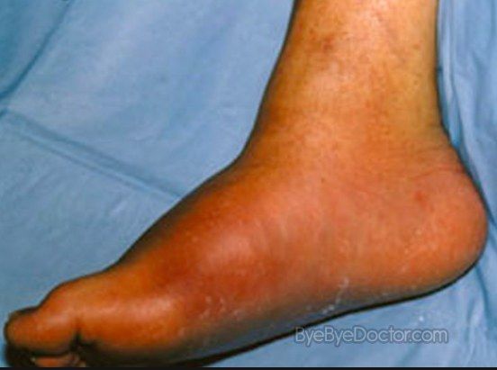
- Destruction of the Chopart joint with subluxation and deformity
- Increased density with bone debris
- +/- dislocation, +/- soft tissue swelling
- Ddx: infection, post-trauma, Charcot (DM, syringomyelia, syphilis, leprosy)
- The 5 Ds: destruction, debris, dislocation, deformity, density
Q2What is a Charcot joint and what is its pathophysiology?▸
- Neuropathic arthropathy: progressive, non-infectious destructive bone and joint disorder with sensory neuropathy
- Neurotraumatic: loss of protective sensation and repeated microtrauma
- Neurovascular: abnormal autonomic vessel tone, increased blood flow causing bone resorption
- Causes: DM, Hansen’s disease (Leprosy), syphilis, syringomyelia, spina bifida
Q3What examinations differentiate infection from Charcot in the foot?▸
- Look for sinus, ulcer, deformities
- Heel raise test differentiates infection from Charcot (OM has less deformity)
- Neuro exam: 128Hz tuning fork, 10g monofilament, proprioception
- Palpate pulses; toe pressures, ABI, transcutaneous O2 for healing potential
Q4What are the Eichenholtz and Brodsky classifications of Charcot?▸
- Eichenholtz: stage 0 (joint oedema), fragmentation, coalescence, consolidation
- Brodsky I: 60% TMTJ
- Brodsky II: Chopart/subtalar
- Brodsky III: 20% ankle/calcaneal tuberosity, IV mixed, V forefoot
Q5What are the principles, applications and contraindications of total contact casting?▸
- Immobilise immediately; admit for infection, dislocation or debridement
- Principles: increase contact surface area, evenly distribute forces, control oedema
- Well padded, limit toe movement, change every 2-4 weeks, allow WBAT
- Duration: forefoot 3-6 months, midfoot 6-12 months, hindfoot 12-24 months; 70-80% wound healing
- Contraindications: passed coalescence, severe deformity, uncontrolled oedema, active infection, severe obesity
Q6What are the surgical options and long-term aims in Charcot foot?▸
- Aim: take patient through Eichenholtz stages while minimising the deformity and subsequent skin breakdown and infection; stable plantigrade shoeable foot
- Patient education!; multidisciplinary input (endocrinologist, vascular surgeons, P&O, occu physio); Systemic: control DM (better outcomes in surgical and non-surgical treatment), optimise nutrition
- Some centres may give bisphosphonates, but evidence is not strong; ongoing RCTs investigating denosumab in treatment of Charcot
- Acute surgery indications: fracture, dislocation or impending skin breakdown - exostectomy + TAL
- Superconstruct: fusion beyond zone of injury, shorten to reduce, strongest tolerated device
- Long term: prevention, identify foot at risk, early podiatry referral
Q7What is the history in a suspected Charcot foot?▸
- Ask about DM, trauma, other peripheral neuropathies
- 50% of patients have pain - dorsal columns are affected but the spinothalamic tract remains intact
- Assess functional limitations
Q8What investigations are performed to differentiate Charcot from infection?▸
- Bloods
- WBC scan
- Biopsy
- Infection: C/ST +ve
- Charcot: bone and cartilage debris within synovium
Q9What is the mid-term management of Charcot foot?▸
- Midterm management: CROW boot after contact casting: bivalved AFO with full foot enclosure, hard outer shelf to prevent deformity, rigid rocker bottom base, custom foam padding
- Double rocker shoe modifications reduce ulceration risk at the plantar apex of the deformity
Q10When is surgery indicated acutely in Charcot foot and what are the difficulties?▸
- Fracture, dislocation or impending skin breakdown
- Usually exostectomy + TAL; deformity correction with rigid fixation and immobilisation is risky; final option is amputation
- Difficulties: bleeding, wound infection, failed fixation
Q11What is a superconstruct in Charcot foot surgery?▸
- Fusion extends beyond the zone of injury
- Bone resection shortens the extremity to allow adequate reduction
- Use the strongest device tolerated by the soft tissue envelope
- Applied in a position that maximises mechanical function
Q12What is the long-term management of Charcot foot?▸
- Aim at prevention
- Identify the foot at risk
- Early referral and frequent podiatry assessment
Q13Describe the clinical findings of an Achilles tendon rupture.▸
- Ankle swelling with absence of the normal Achilles tendon contour
- Palpate the gap (size of gap, distance between insertion and gap)
- Thompson's test positive
Q14What risk factors and mechanisms do you ask about in Achilles rupture?▸
- Mechanism: eccentric loading of gastrosoleus in middle-aged patients
- Hx: AS, steroid, fluroquinolones, flat foot, gout, hyperlipidemia, chronic renal disease
- Chronicity of injury and pre-existing pain
- Poor wound healing risks: PVD, smoking, diabetes
Q15What investigations are performed for Achilles rupture?▸
- USG to delineate site and size, complete versus partial rupture
- X-ray: calcinosis suggests pre-existing tendinosis - may need intraop debridement and preparation for tendon gap
- Kager's triangle is distorted when the tendon is ruptured
Q16How is an acute Achilles rupture treated and what is the evidence?▸
- Treatment is individualised: age, functional demand, tendon retraction, timing
- Repair aims for intrinsic healing + aggressive rehab
- JBJS 2001 meta-analysis: less rerupture with surgery (2% vs 20%)
- Accelerated functional rehab concept: Willits RCT (JBJS 2010) - no significant difference in rerupture rate or power
- Later confirmed with (Soroceanu JBJS 2012 metanalysis)
- NEJM 2022 Myhrvold Norway multicentre RCT MIS vs open vs nonop: Rupture rate: conservative 6.2% vs 0.6%; Infection MIS 5.2% vs open 2.8% vs 0.6% nonop; Overall surgery was not associated with better outcomes
- Should be shared decision making, weighing avoiding surgery against higher rerupture risk
Q17How is a delayed presentation with a gap managed?▸
- Aim to reconstruct tension and length - shorten first, then retension
- Gap <3cm: direct repair
- Gap 3-5cm: turn-down flap, VY plasty +/- augmentation (Lindholm, plantaris)
- Gap >5cm: FHL transfer (dispensable, good vascularity, in phase with gastroc)
- FHL released at the Knot of Henry, retrieved to repair site +/- bone tunnel at calcaneal tuberosity
Q18What are the complications and rehabilitation after Achilles repair?▸
- Complications: skin necrosis 2%, rerupture 2%, deep infection 1%, sural nerve injury (especially percutaneous)
- Accelerated rehab: 0-2wk CAM NWB 2cm raise; 2-6wk protected weight bear, 1cm raise, active PF/DF to neutral; 6-8wk WBAT, no heel raise, gradual DF ex, OKC/CKC ex; 8-12wk strengthening + proprioception; >12wk plyometrics
- Return to athletic activity at 4-6 months
Q19What is the non-operative treatment for an old, low-demand patient with Achilles rupture?▸
- Conservative with an accelerated rehab program
- UKSTAR trial Lancet 2020: cast vs functional bracing - no difference in functional outcomes
Q20What is the essence of accelerated rehabilitation after Achilles repair?▸
- Early weight bearing and early mobilisation
- allow collagen fibril crosslinking, also healing tendons undergo intrinsic healing, increasing load to failure
Q21What anatomy must be known for an FHL transfer?▸
- FHL crosses superficial to FDL at the Knot of Henry
- The Knot of Henry lies below the bony prominence of the navicular
Q22Describe the anatomy of the Achilles tendon.▸
- Collagen accounts for 70% of dry weight, of which 95% is type I, with a very small amount of elastin
- Fibres rotate 90 degrees toward insertion into the calcaneal tuberosity (gastrocnemius laterally, soleus medially)
- Watershed blood supply at 5-6cm from insertion
Q23Describe the Cook and Purdam model of Achilles degeneration.▸
- Reactive: myofibroblast proliferation and collagen synthesis in response to repeated mechanical stress
- Reparative: inflammation of the paratenon as an attempt at soft tissue repair
- Degenerative: vascular constriction leading to ischaemia
Q24What is the diagnosis from this X-ray?▸
- Bony spur over the posterosuperior aspect of the calcaneal tuberosity
- Calcification over the insertion of the Achilles tendon
- Diagnosis: Haglund deformity with insertional Achilles tendinopathy
Q25What is Haglund syndrome?▸
- Triad of retrocalcaneal pain
- Insertional tendinopathy
- Haglund deformity
Fact check
NEJM 2022 trial infection rates: minimally invasive surgery 5.2%, open 2.8%, nonoperative 0.6% — misattributed — Those percentages are sensory nerve injury rates; deep infection was 1.1% nonoperative, 1.1% open and 1.7% minimally invasive surgery — source