Foot and Ankle
Hindfoot and knee malalignment
Hindfoot calcaneus alignment and limb deformity patterns including genu valgum and recurvatum
29 questions
2 source pages
1 images
Images appear with the first question taken from each source page — tap a question to open it.
Q1Describe the deformity in this spina bifida patient.📷▸
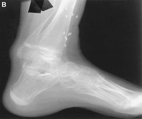
- Hindfoot calcaneus (not varus)
- Midfoot cavus with 1st ray plantarflexion
- Claw toes
- Scar at heel from previous TA lengthening
- Calf muscle wasting
Q2How does a calcaneus foot present and how is it managed?▸
- History of instability and pain, previous TA lengthening
- Neuromuscular causes: spina bifida and polio
- Gait: slapping (lack of gastrocnemius eccentric contraction); heel ulcer
- XR: pistol grip deformity
- Mx: non-operative; tibialis anterior to TA transfer; dome calcaneal osteotomy with posterior tuberosity proximalisation
Q3What is spina bifida?▸
- Neural tube defect
- Failure of neural tube closure at 3 weeks
Q4What are the risk factors for spina bifida?▸
- Folate deficiency
- Maternal diabetes
- Valproic acid
Q5What prenatal screening is done for spina bifida?▸
- Maternal AFP level (high = bad)
- Amniotic fluid AFP
- Structural scan
Q6What is the postnatal treatment of spina bifida and what must you beware of?▸
- Neurosurgery: untethering of cord, drainage of syrinx, screen for Arnold-Chiari syndrome
- Beware malignant hyperthermia
- Beware latex allergy
- Beware neurogenic bladder
Q7Classify spina bifida.▸
- Occulta: vertebral arch failure
- Meningocele: meninges protrusion, no neural element
- Myelomeningocele: meninges and neural elements protrusion
- Rachischisis: neural elements protrusion with no coverings
Q8What are the features of a pre-L3 spina bifida lesion?▸
- Hip FABER contracture
- Knee flexion contracture (FFC)
- Feet equinovarus + claw toe
- Non-ambulatory
Q9What are the features of an L4 (L5 defect) spina bifida lesion?▸
- Hip: full quadriceps, Trendelenburg gait
- Knee: recurvatum (unopposed knee extension)
- Feet: calcaneus
- Limited community ambulator
Q10What are the features of an L5 (S1 defect) spina bifida lesion?▸
- High normal
- Knee normal
- Feet: calcaneus
- Community ambulator
Q11What are the features of S1 or below spina bifida lesions?▸
- Only claw toe
- Near normal
Q12Why must VP shunt function be checked before spine surgery in spina bifida?▸
- Check VP shunt function before spine surgery
- Otherwise it may result in death
Q13Describe the limb deformity in this polio patient.▸
- Right lower limb: hip extension, knee extension +/- recurvatum
- Ankle plantarflexed, tibial external rotation, genu valgum
- Contralateral hip and knee flexion compensating for LLD
- Pelvic obliquity; no claw toes
Q14What do you assess in a polio patient with deformity?▸
- Check shoewear
- Gait: Trendelenburg, short limb, recurvatum, hand-knee gait
- Sitting: inspect for neuropathic ulcers and callosity
- Contractures: Silverskiold (Achilles), knee FFC (recurvatum), hip FABER (Ober)
- Power of antigravity muscles: ankle plantarflexion, knee extension, hip extension
Q15What is the current disability history in this polio patient?▸
- Pain from ulcers, callosity, stress fracture
- Instability
Q16How does quadriceps weakness cause knee flexion contracture and how is it managed?▸
- Knee collapse and unopposed hamstrings perpetuate flexed knee posture
- Ankle PF + hip flexion bring CG forward to lock knee; FFC becomes contracture over time
- Hand-knee gait; first line hinged KAFO
- Flexible: biceps femoris to quadriceps transfer (needs normal hip extension/ankle PF, no recurvatum)
- Rigid: <15 degrees serial casting, 15-30 degrees hamstring/ITB/posterior capsule release, 30-70 degrees distal femoral extension osteotomy, >70 degrees staged release then osteotomy
Q17How is knee recurvatum managed in polio?▸
- <15 degrees: observe
- Solid AFO
- Triple tenodesis (gracilis, hamstrings, ITB)
- Posterior capsular advancement; gracilis/semitendinosus or biceps femoris/tibialis tenodesis
Q18How does exaggeration of the passive stance phase cause knee recurvatum in polio?▸
- By exaggeration of passive stance phase: knee recurvatum assists in locking of the knee (ITB becomes extensor, CG falls in front of knee)
- Posterior knee capsules and hamstrings act as restraints, counterbalanced by hip extension and ankle PF
- Over time the knee loses its posterior constraint causing instability; pain occurs over the anterior knee from excessive hyperextension (perpetuated by fixed equinus)
Q19What are the aims of management in polio deformity?▸
- Maximise gait efficiency
- Prevent deformity with growth
- Prevent upper limb contractures and scoliosis
Q20How does gluteal weakness present in polio and how is it managed?▸
- Gluteus medius/minimus weakness -> Trendelenburg gait
- Gluteus maximus weakness -> back lurch
- Abductors: external oblique transfer
- Glut max (iliopsoas to GT transfer)
Q21What is the FABER contracture in polio and what problems does it cause?▸
- Frog posture: hamstring, hip flexors, TFL spasm -> ITB contracture
- Secondary pelvic obliquity
- Leads to scoliosis and increase contralateral hip dislocation
Q22How is FABER contracture managed in polio?▸
- Mild: fasciotomy
- Moderate: Ober release
- Ober release: ASIS - sartorius + TFL; AIIS - rectus femoris; GT - anterior fibres of glut med/min; LT - iliopsoas
- Severe: Campbell procedure
- Campbell: outer table glut med/min + TFL; inner table iliacus; AIIS rectus femoris; ASIS distalisation of sartorius
Q23What is poliomyelitis?▸
- Non progressive motor paralysis secondary to polio virus infection at anterior horn cells
- Enterovirus, faecal-oral route
Q24What is the pattern of poliomyelitis infection?▸
- 80% asymptomatic
- 10% flu-like
- 10% neurological symptoms, of which 2% paralysis
Q25What are the phases of poliomyelitis?▸
- Acute: flu-like symptoms, flaccid paralysis
- Recovery: no more muscle ache, some recovery of power
- Residual: >2 years, deformity
Q26What is post-polio syndrome?▸
- New onset weakness after a prolonged period of recovery
- Occurs in 50% of patients
Q27What are the diagnostic criteria for post-polio syndrome?▸
- History of paralytic poliomyelitis by clinical/NCT
- Complete recovery >15 years with neurological and functional stability
- New onset weakness
- New onset muscle/joint pain, deformity, cold intolerance
- Exclude other causes
Q28What is the pathophysiology of post-polio syndrome?▸
- Immunological/viral reactivation
- Aging/degeneration of neurons
Q29What are the 5 potential problems after the residual paralysis stage?▸
- Isolated muscle weakness - splintage, passive mobilisation
- Deformity
- Shortening (LLD)
- Flail joint - may need arthrodesis or splintage
- Vascular dysfunction