Foot and Ankle
Pes cavus - deformity and causes
Cavus arch patterns with neurological causes such as Charcot-Marie-Tooth and Friedreich ataxia
45 questions
4 source pages
3 images
Images appear with the first question taken from each source page — tap a question to open it.
Q1Describe the clinical photo of an adult acquired flatfoot.📷▸
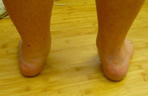
- Adult with forefoot abduction
- Decreased medial foot arch (midfoot supinated in relation to hindfoot)
- Hindfoot valgus
- Medial prominence
Q2What is the differential diagnosis of acquired flatfoot (bilateral vs unilateral)?▸
- Bilateral/any: PTTD, tarsal coalition, congenital vertical talus
- Unilateral: post-traumatic (Lisfranc, spring ligament injury), degenerative, Charcot
- Bilateral systemic: rheumatoid arthritis, neuromuscular (spina bifida), cerebral palsy
- Generalised ligamentous laxity (Marfan, Ehlers-Danlos)
Q3What are the possible sources of pain in acquired flatfoot?▸
- Medial: PTT, deltoid ligament, tarsal tunnel
- Lateral: subfibular impingement
- Heel: plantar aponeurosis, retrocalcaneal bursitis
- Diffuse: ankle, subtalar joint
- Proximal: knee, back; distal: bunion, metatarsal heads
Q4How do you clinically assess a flatfoot?▸
- Determine whether the flatfoot and hindfoot valgus are flexible or rigid, and identify cause and complications (arthritis)
- Inspection: double heel raise and single leg heel raise (PT power, flexibility)
- Too many toes sign, navicular bulging signifying accessory navicular; look for scars (post trauma)
- Palpate sinus tarsi, deltoid ligament, PTT; test PT, FDL/ FHL power (transfer)
- Subtalar ROM and residual forefoot supination with correction; Silverskiold test
- Beighton score, check hands for RA, check spine if bilateral; assess gait
- X-ray: weight-bearing DP (Simmons angle, talocalcaneal angle, talus uncoverage) and lateral (Meary angle, calcaneal pitch, foot collapse at TN/NC/TTJ), No feature of previous fracture/ fragmentation, Saltzman view for hindfoot alignment, XR ankle mortise, Scannogram for LL alignment, MRI for PTT
- Bloods to rule out RA
Q5How is the foot arch maintained and what is the pathophysiology of PTTD?▸
- Medial longitudinal arch static support: bony (calcaneum, talus, navicular, medial 3 rays) and soft tissue (spring ligament, plantar aponeurosis, deltoid ligament)
- Dynamic support: PTT, peroneus longus and intrinsics
- Lateral arch: calcaneum, cuboid, lateral 2 rays with long and short plantar ligaments, bifurcate ligament
- Transverse arch: keystone is 2nd MT and middle cuneiform, supported by the Lisfranc ligament
- Pathophysiology: PTT tenosynovitis causes loss of dynamic support, failure to lock the transverse tarsal joint --> further cause static stabilizer to fail
- Result: loss of one foot of the tripod, hindfoot valgus, MF/FF compensatory supination to maintain the tripod; PTT has a watershed blood supply 2-6cm proximal to insertion
Q6What is the aim of management in adult acquired flatfoot?▸
- Stable, plantigrade, painless foot
- Rebalance the foot through bone and/or soft tissue to restore a balanced tripod
- Treat according to the patient's symptoms
Q7What is the anatomy and action of the posterior tibialis tendon (PTT)?▸
- Origin: posterior fibula, tibia and interosseous membrane
- Inserts: navicular tuberosity, 1st – 3rd cuneiform, cuboid, 2-4 metatarsals, sustentaculum tali
- Invertor of the subtalar joint
- Plantarflexion of the ankle joint
- Adductor of the forefoot
- Watershed blood supply 2-6cm proximal to insertion
Q8What is the Johnson classification of PTTD?▸
- 1: PTT power intact, no flatfoot
- 2: A flexible flatfoot, B abducted forefoot, C forefoot varus
- 3: Subtalar joint arthritis
- 4: Deltoid ligament insufficiency
Q9How is stage 1 PTTD managed?▸
- Physio for TA stretching
- Orthosis: UCBL, moulded AFO, medial heel wedge, longitudinal arch support
- Tenosynovectomy
Q10How is stage 2 PTTD managed?▸
- Combination of bony + soft tissue procedure, tailored to the individual’s deformities and muscle imbalance
- Soft tissue: FHL or FDL tendon transfer; TA lengthening also protects the transfer; +/- spring ligament reconstruction
- Coronal plane bone: calcaneal wedge osteotomy (85% pain relief/improved function at mean 15y, Chadwick BJJ 2015)
- Sagittal plane bone: 1st TMTJ fusion or Cotton medial cuneiform opening wedge osteotomy for forefoot varus - restores tripod
- Transverse plane bone: lateral column lengthening for forefoot abduction - protect FDL transfer
Q11Which tendon transfer is preferred for PTTD - FHL or FDL?▸
- FHL is stronger
- FDL gives better functional outcomes because FHL is harder to mobilise
Q12How are stage 3 and stage 4 PTTD managed?▸
- Stage 3: subtalar fusion +/- triple or double (TN + subtalar) fusion
- Double fusion (TN + subtalar) has similar functional scores and shorter OT time (Int Orthop 2021 Fadle)
- Stage 4: deltoid ligament reconstruction +/- ankle replacement or fusion
Q13What is the FAI 2020 consensus statement on adult acquired flatfoot?▸
- Rename the condition Progressive Collapsing Foot Deformity (PCFD)
- New classification to stage the deformity
- A: hindfoot valgus deformity
- B: midfoot/forefoot abduction deformity
- C: forefoot varus/medial column instability
- D: peritalar subluxation/dislocation
- E: ankle instability
Q14What is the role of arthroereisis in flatfoot?▸
- Subtalar arthroereisis induces a triplanar modification of the foot limiting pronation
- EFFORT review 2017 and AAOS 2021: no long-term studies, only poor quality evidence supporting its use
Q15What are the positions of fusion in a triple arthrodesis?▸
- Subtalar joint 5 degrees valgus (TA is not a deforming force)
- TN joint: adequate coverage of the talar head; restore hindfoot-forefoot relationship so the 1st and 5th MT heads both touch the ground
- Restore the height of the medial arch
- CC joint: neutral (in situ)
Q16Describe the clinical features of a cavovarus foot.📷▸
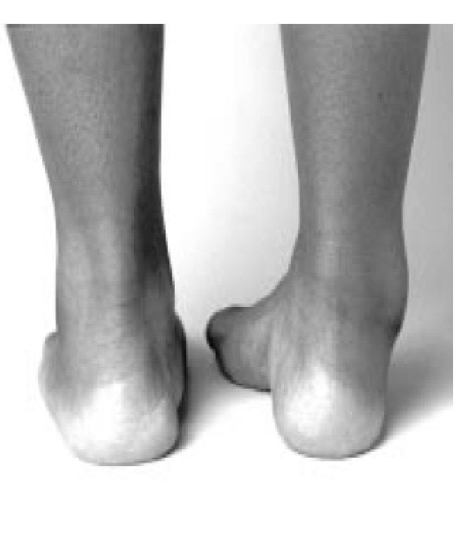
- High medial arch with midfoot cavus
- Hindfoot varus, forefoot adducted and pronated in relation to hindfoot
- Claw toes, peek-a-boo heel sign
Q17What do you ask about in the history of a cavovarus foot?▸
- History of neurological disease, family history (CMT)
- Instability, metatarsalgia, fatigue, shoewear problems
- Callus, ulcers, pain (OA), stress fractures, Muller Weiss
- Functional limitation and expectations
Q18What are the examination findings in cavovarus foot?▸
- LL muscle wasting - inverted champagne bottle appearance
- Forefoot clawing, adduction, pronation; midfoot cavus; hindfoot varus equinus
- Coleman block test for flexibility and forefoot-driven deformity
- Gait: broad based ataxic gait; check lateral ankle instability (ATFL, CFL)
- Full neuro exam (cerebellar), sensation, spine and hands
Q19Explain the Coleman block test.▸
- Decides whether cavovarus is hindfoot or forefoot driven and whether flexible
- Block supports the 5th MT and heel, freeing the 1st MT
- Hindfoot corrects if the deformity is forefoot driven and flexible
- Rationale: hindfoot varus is compensatory to keep the lateral rays on the ground
Q20How is cavovarus foot managed surgically?▸
- Combination of bony procedure with soft tissue procedure, tailored to the individual’s deformities and muscle imbalance
- Flexible: bony + soft tissue procedures for forefoot and midfoot +/- TA lengthening
- Rigid: add midtarsal and hindfoot bony procedures
- Arthritis/progressive disease: fusion
- Soft tissue: PL to PB transfer, Steindler plantar aponeurosis release
- Bone: Dwyer lateral closing wedge calcaneal osteotomy, 1st ray dorsiflexion osteotomy, triple arthrodesis
Q21What additional examination findings are checked in cavovarus foot?▸
- Sole: callosities and ulcers
- Scars and correctability
- Muscle power: PT, PB, tib ant, PL
- Lateral ankle instability (ATFL and CFL)
- Silverskiold test
Q22What is a cavus foot?▸
- Fixed plantar flexion deformity of the forefoot in relation to the hindfoot
- Results in an abnormally high arch
- 2/3 are neurological
Q23What are the causes of cavus foot?▸
- Neurological: CP/stroke/Friedreich's ataxia
- Neurological: syringomyelia/diastematomyelia/spina bifida
- Neurological: SMA, polio, CMT, DMD
- Non-neurological: post-traumatic (Volkmann contracture, talar neck malunion)
- Non-neurological: residual club foot
Q24What are the X-ray findings in cavovarus foot?▸
- DP: talocalcaneal angle and talo-1st MT angle
- Lateral: see-through sign, calcaneal pitch, Meary angle, calcaneal-1st MT angle (Hibb's angle N>150)
- Lateral: lateral kite angle <25 (parallelism), double talar head sign, bell-shaped cuboid
- Look for degeneration; obtain ankle mortise and Saltzman view
- MRI spine if neurological disease is suspected
Q25When is a neurologist referral made in cavovarus foot and what tests are requested?▸
- Refer if neurological disease is suspected
- ECG NCS
- Sural nerve biopsy, muscle biopsy, CK
Q26What is the aim and conservative management of cavovarus foot?▸
- Systemically delineate and treat the underlying cause
- Locally provide a plantigrade, stable, pain-free, shoeable, preferably mobile foot
- Conservative: physio for stretching and accommodative shoewear to relieve pressure
Q27How is surgical management of cavovarus foot tailored to flexibility?▸
- Flexible: bony + soft tissue procedure for forefoot and midfoot; hindfoot should correct with these +/- TA lengthening
- Rigid: add midtarsal + hindfoot bony procedure
- Arthritis/progressive disease: fusion
- +/- lateral ankle ligamentous reconstruction if ankle instability
Q28What soft tissue procedures are used in cavovarus foot?▸
- Hindfoot varus: PL to PB transfer
- Hindfoot varus: PT to cuneiform transfer
- Hindfoot varus: tib ant to cuneiform transfer
- Midfoot cavus: Steindler plantar aponeurosis release
Q29What bony procedures are used in cavovarus foot?▸
- Hindfoot: Dwyer lateral closing wedge calcaneal osteotomy
- Hindfoot: lateral calcaneal page osteotomy
- Midfoot cavus: 1st ray dorsiflexion osteotomy
- Midfoot cavus: TMTJ (Jahss) or tarsal (Japas) osteotomy
Q30How are claw toes in cavovarus foot managed?▸
- PIPJ flexible: Girdlestone-Taylor FDL to EDL transfer
- PIPJ rigid, MTPJ flexible: DuVries (P1 head + P2 base resection/fusion) + EDL tenotomy
- PIPJ rigid, MTPJ rigid not subluxed: DuVries + EDB tenotomy + MTPJ dorsal capsulotomy + EDL lengthening
- PIPJ rigid, MTPJ rigid and subluxed: Weil osteotomy
Q31What is the Jones procedure for the big toe in cavovarus foot?▸
- EHL to MT neck transfer
- Fuse the IPJ
Q32How is equinus managed in cavovarus foot surgery?▸
- Usually corrects after the other deformities are corrected
- TA lengthening is left until other corrections are done
Q33Explain forefoot-driven versus hindfoot-driven cavovarus.▸
- FF driven: PL >> ATT causes 1st MT plantarflexion; PT >> PB causes hindfoot varus
- 1st MT plantarflexion + forefoot pronation -> medial forefoot strikes ground first
- Subtalar joint inverts to bring the lateral forefoot down, so hindfoot varus is compensatory
- Recruited EHL causes claw toe; MTPJ extension tightens the plantar aponeurosis via the windlass, worsening cavus and varus
- Hindfoot cavus (calcaneocavus): weak triceps surae with relatively strong PTT/FHL plantarflexing the entire forefoot
Q34What is Charcot-Marie-Tooth disease?▸
- Hereditary motor sensory neuropathy
- Peroneal muscle atrophy
- Prevalence 1:2500 - most common inherited neuropathy
- Usually autosomal dominant (can be AR or X-linked)
- Affects motor more than sensory
Q35What is the genetic basis of CMT?▸
- Peripheral myelin protein 22 - defective Schwann cell membrane protein
- Causes defective myelin sheath formation
- Duplication of chromosome 17
Q36What are the three main types of CMT?▸
- Type 1: demyelinating, presents 10-20s, AD, cavus foot
- Type 2: axonopathy, 20s+, flaccid foot, less disabling
- Type 3: X-linked recessive, demyelinating + axonal degeneration
Q37What are the clinical features and diagnosis of CMT?▸
- Equinocavovarus +/- rigid hindfoot
- Weakened TA and peroneal muscles - foot drop during swing phase
- Check the hands
- Diagnosis: NCV/EMG, DNA/chromosome analysis
- No role for nerve biopsy
Q38How is CMT managed non-operatively?▸
- Always non op first
- Stretching
- Orthosis: need a solid AFO
- No posterior leaf AFO due to inversion and eversion weakness
Q39How is CMT managed surgically?▸
- Follow cavovarus principles
- Traditionally fusion as the disease is progressive
- Reconstruction has lower rates of degeneration and reoperation compared with triple arthrodesis (Ward JBJS 2008)
Q40What are the differential diagnoses of CMT?▸
- Spina bifida
- SMA
- Polio
- Friedreich ataxia
Q41What is the genetic basis of Friedreich's ataxia?📷▸
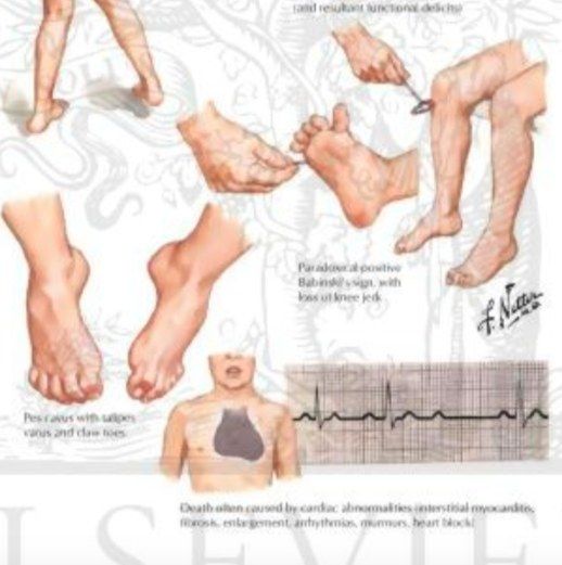
- Autosomal recessive
- Frataxin mutation affecting mitochondria production
Q42How is Friedreich's ataxia diagnosed?▸
- Clinical + family history + genetic testing
- It is the commonest spinocerebellar degenerative disease
Q43How does Friedreich's ataxia present clinically?▸
- Staggering, wide-based gait
- Triad: ataxia, areflexia, upgoing plantar
Q44What are the orthopaedic and systemic associations of Friedreich's ataxia?▸
- Cavovarus foot
- Scoliosis
- Cardiomyopathy - assess before planning surgery
Q45What is the prognosis of Friedreich's ataxia?▸
- Wheelchair-bound around 30 years
- Death around 50 years