Foot and Ankle
Juvenile hallux valgus and physeal bracket
Juvenile hallux valgus management and physeal bracket deformity of the first ray
13 questions
3 source pages
1 images
Images appear with the first question taken from each source page — tap a question to open it.
Q1What is the pathophysiology of a physeal bracket?📷▸
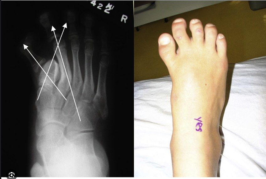
- Incomplete development of primary ossification centres
- Aberrant physeal cartilage extends along the diaphysis and interferes with normal growth
- Leads to a short, wide, triangular or trapezoidal phalanx
- Longitudinally oriented cartilage bracket follows a C-shaped curve
- Autosomal dominant when present in the hand, otherwise sporadic
Q2What conditions are associated with a physeal bracket?▸
- Rubinstein-Taybi syndrome
- Fibrodysplasia ossificans progressiva
- Hallux varus in the feet
- Clinodactyly and polydactyly in the hands
- Apert's syndrome
Q3What is the radiographic staging?▸
- Stage 1: delta-shaped diaphysis with radiolucent cartilaginous epiphysis
- Stage 2: secondary ossification centres at proximal and distal ends
- Stage 3: complete ossification of the bracket epiphysis along one side of the diaphysis
- Stage 4: closure of the physis
- MRI may be needed
Q4How is a physeal bracket managed?▸
- Splinting to loosen soft tissues prior to surgery
- Osteotomies (opening or closing wedge) in older children after closure of the bracket epiphysis
- Physiolysis + interposition (PMMA or fat): earlier intervention allows more correction of angulation and more longitudinal growth
Q5What are the complications of nonoperative and operative treatment?▸
- Nonoperative: structural deformation of the forefoot
- Nonoperative: incomplete longitudinal growth of the affected bone
- Nonoperative: clinodactyly at the interphalangeal joint
- Operative: recurrence of deformity
- Operative: incomplete correction of deformity
Q6How does juvenile hallux valgus differ from adult hallux valgus?▸
- Usually large IMA, large DMAA +/- HVI, congruent MCPJ
- Metatarsus primus varus
- Ligamentous laxity with hypermobile 1st ray
- Physis not yet closed
- High recurrence
Q7What is the management of juvenile hallux valgus?▸
- Address ligamentous laxity: TMT arthrodesis after physis closure for a hypermobile 1st ray
- If physis open: osteotomy at medial cuneiform or wait until closure
- Double osteotomy: medial cuneiform opening wedge + 1st MT osteotomy +/- Akin
- Associated with flexible flatfoot and tight TA
Q8What is the management principle for RA foot deformity?▸
- Relieve symptoms, preserve function, minimize complications
- Goal: stable, pain-free plantigrade foot
- Multidisciplinary: rheumatology, medical, anaesthetics, physio, OT
- Conservative: physio, shoe wear (large toe box, rockerbottom), metatarsal pads or bars, NSAIDs
Q9Why does RA foot surgery favour fusion over reconstruction?▸
- Primary disease is progressive, so reconstruction has a high chance of recurrence
- need to bear in mind the risk of complications as a result of osteopenia, reduced vascularity, and immunosuppression
- Look for other RA complications e.g. C1/2 subluxation, other lower limb arthritis
Q10What are the fusion positions and procedures used in the RA foot?▸
- 1st MTPJ arthrodesis: 20 deg dorsiflexion, 10 deg valgus, neutral rotation
- Lesser toes: principle is to decompress the joint to allow relocation, while maintaining the MT length cascade (Maestro's cascade)
- MTPJ: Fowler (proximal P1 excision) or Stainsby (+ flexor-extensor interposition); IPJ: DuVries
- Midfoot: TN +/- CC fusion; hindfoot: triple fusion
- Ankle: synovectomy; fusion vs arthroplasty (JBJS 2007 systematic review, 70% satisfaction rate in both groups)
Q11What is the pathophysiology of RA foot deformities?▸
- Stage I: synovitis
- Stage II: joint erosion and tendon dysfunction
- Stage III: progressive deformity
- Forefoot: hallux valgus, claw toe, splay foot
- Midfoot: rockerbottom + forefoot abduction; hindfoot: PTT insufficiency and disruption of the talocalcaneal interosseous ligament causing hindfoot valgus
Q12If the IMA is not severe but the HVA is severe, what does that suggest?▸
- MCL rupture (usually acute, sesamoid not subluxed)
- Metatarsus adductus
Q13How does the X-ray localize a flatfoot deformity?▸
- X-ray localization - if at TMTJ, then it is a midfoot driven flatfoot. If at TNJ, is it s hindfoot driven flatfoot