Hip
THR complications - dislocation, gait and HO
Dislocation after cemented THR, Trendelenburg gait and heterotopic ossification
24 questions
3 source pages
1 images
1 fact-check flags
Images appear with the first question taken from each source page — tap a question to open it.
Q1What is the common cause of Trendelenburg gait after THR?▸
- Defective abductor mechanism - think about the lever system
Q2How can a defective abductor mechanism cause Trendelenburg gait after THR?▸
- Instability to generate force: nerve/muscle weakness or inhibition from pain
- Shortened lever arm from altered vertical/horizontal offset - head-neck shortening, LLD with poor length-tension
- Unstable fulcrum (dislocated hip)
Q3How is head offset measured?▸
- From the hip centre to the anatomical axis of the femur
- Relevant when restoring offset in THR
Q4How do you restore abductor function in THR?▸
- Restore the hip centre
- Restore offset
- Restore neck length
Q5What are the problems of increased offset after THR?▸
- Increased lever arm of the neck - increased bending moment at the stem - risk of neck fracture
- If increase with long neck may need larger neck which decreased head neck ratio
- Trochanteric bursitis
Q6What are the problems of decreased offset after THR?▸
- Abductor tension decreased - power decreased (length-tension relationship)
- Decreased abductor lever arm
- Increased joint reaction force
Q7Describe the X-ray findings in this cemented THR with dislocation.📷▸
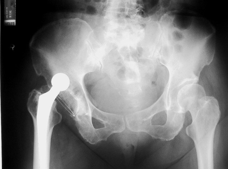
- Cemented THR with dislocation; hard-on-soft bearing
- Acetabular opening angle and anteversion appear within the safety range of Lewinneck, with no signs of loosening by DeLee criteria
- Cemented modular femoral component with small head-neck ratio, no loosening by Harris criteria
- Bony outgrowth at AIIS may cause impingement and dislocation; spine looks degenerated
Q8What is the initial management of a dislocated THR?▸
- Rule out other injuries; AMPLE history; check NV status
- CR in OT under GA to allow muscle relaxation
- Use modified Allis manoeuvre
- Post-reduction check safety range, give abduction pillow, recheck NV status
- CT to check for congruency and any fracture, also implant loosening and alignment (femoral and acetabular version)
Q9What are the causes of dislocation after THR (Knutsor Lancet 2019)?▸
- Preop patient: neuromuscular/neurodegenerative disease (dementia, PD), psychiatric disease (alcoholism, schizophrenia), history of spinal fusion
- Preop disease: revision, DDH, inflammatory arthropathy, fracture
- Intraop technique: approach, soft tissue tension (offsets, releases, capsule repair), impingement (osteophytes, cement, AIIS), component malposition
- Intraop implant: head size, head-neck ratio, neck geometry, neck skirts, constrained cups
- Postop: non-compliance, provocative activities (anterior shoelacing; posterior squat/low chair), infection, wear/loosening, GT nonunion
Q10What is the safe zone for acetabular component position?▸
- Lewinnek 1978: 40 deg abduction + 15 deg anteversion - 6% dislocation inside the zone, 15% outside
- Barrack 2003: anteversion 10-20 deg of both stem and cup minimises impingement and dislocation
- Dorr combined anteversion 2009: 37.6
Q11What is the natural history and incidence of dislocation after THR?▸
- 60% occur <6 weeks, usually due to technical error
- Late dislocation suggests impingement, wear or increased ROM
- Incidence 3% primary, 15% revision (Woo & Morrey JBJS Am 1982)
Q12What are the treatment options for recurrent dislocation after excluding infection, trauma and loosening?▸
- Revision if the hip dislocates in the patient's normal functional range
- Indications: recurrent >=2, late (postop 1 year), malalignment, implant fracture/loosening, Impingement/ PE wear
- Correct the cause: malposition revision, impingement resection, GT distal advancement/increased horizontal offset
- Femoral side: larger head, longer neck, larger offset, GT advancement
- Acetabular side: check anteversion, lipped liner, constrained liner, MDM, resection arthroplasty
Q13What does the stability of a hip replacement depend on?▸
- Component design - head size, neck morphology, head-neck ratio, constrained liner, neck skirt
- Alignment - anteversion 15-30, opening angle 35-45
- Soft tissue tensioning - offset and neck length
- Soft tissue function - abductor complex
Q14What are the resting positions for anterior and posterior dislocation?▸
- Anterior: extension + external rotation
- Posterior: flexion + internal rotation + adduction
Q15How are cup anteversion and stem anteversion defined?▸
- Cup anteversion: angle between the plane of the acetabular cup opening and the sagittal plane
- Stem anteversion: angle between the transcondylar plane including the femoral shaft and the axis of the femoral neck
Q16What is the likely direction of dislocation in this patient and how is it confirmed?▸
- Suspect anterior dislocation by the size of the head, and the rotation and position of the femur
- Confirmed by lateral X-ray
Q17When is a constrained liner indicated and what are its failure modes?▸
- Indication: bone stock okay but soft tissue dysfunction with a well fixed acetabulum
- Covers the femoral head beyond the equator, decreasing dislocation at the expense of decrease ROM
- 4 modes of failure (Dorr J Arthroplasty 2005): fixation failure, biomaterial failure, femoral head dislocation, liner dissociation
- Now superseded by MDM - increases survival and decreases redislocation; theoretical increase in wear due to more bearing surface
Q18What is the principle of a dual mobility (MDM) construct?▸
- Increases the functional arc of motion
- Large head principle (McKee Farrar) vs small head (Charnley)
- 3 primary goals: increase stability, restore nearly physiological ROM, reduce wear
- Anatomical and modular dual mobility - modular risks mal-seating, but can add screws
Q19What is heterotopic ossification and what are its causes?▸
- Ectopic lamellar bone formation in soft tissues
- Traumatic
- Neurogenic - inflammatory neuropeptides including substance P and CGRP are highly upregulated
Q20What are the risk factors for HO?▸
- Patient: hypertrophic OA, history of HO, AS, DISH, Paget's disease
- General: head trauma, spinal cord injury, burn, massive trauma
- Local: approach (Hardinge, anterior > posterior), haemostasis, marrow spilling, immobilisation
- Forceful passive manipulation after a period of immobilisation
Q21What is the pathophysiology of HO?▸
- Unknown trigger with tissue expression of BMPs
- Primitive mesenchymal cells migrate to the injured area, become fibroblasts and secrete collagen/ECM (fibroblastic metaplasia)
- Primitive mesenchymal cells migrate to injured area and transform into fibroblast --> secrete collagen and ECM --> fibroblastic metaplasia --> fibroblast transform into chondrocytes (similar to enchondral ossification) --> some chondrocytes continue to deposit collagen into cartilage matrix while the remaining chondrocytes transform into osteoblasts --> osteoid deposition
- Fibroblasts become chondrocytes (like enchondral ossification); chondrocytes become osteoblasts with osteoid deposition
- Starts within 16 hours after the index operation
Q22What is the Brooker classification of HO, and what grade is shown on this X-ray?▸
- Grade 1: island
- Grade 2: bone spur with >1cm gap
- Grade 3: <1cm gap
- Grade 4: complete osseous ankylosis
- Also mentioned: Alonzo classification
- This X-ray shows HO around a right THR - Brooker III
Q23What laboratory findings are seen in HO and what is their limitation?▸
- ALP, CRP and ESR are elevated
- ALP cannot determine maturity of the heterotopic ossification
Q24How is HO prevented and when is it excised?▸
- Preop: RT 800 Gy within 4-6 hours of surgery (per lecture notes)
- Intraop: minimal subperiosteal stripping, decrease pericapsular trauma
- Postop: RT single dose <72hr; indomethacin 25mg tds x 6 weeks decreases HO by ~10% and decreases severity
- Excise when mature: X-ray trabecular formation/mature rim, normal ALP and ESR, no increased uptake on bone scan
- Indocid MOA: decreases insulin-like growth factor
Fact check
Preoperative radiotherapy 800 Gy within 4-6 hours of surgery prevents heterotopic ossification — unit error / unsafe dose — Single-fraction HO prophylaxis is 700-800 cGy (7-8 Gy), given within 24 hours preoperatively or within 72 hours postoperatively; 800 Gy would be a lethal dose — source