Hip
Periprosthetic fracture and classification zones
Fracture around an arthroplasty, evidence for management and zone classification
30 questions
4 source pages
1 images
Images appear with the first question taken from each source page — tap a question to open it.
Q1How are periprosthetic knee fractures classified?📷▸
- Site: patella > femoral > tibial
- Timing: intra-operative or post-operative
Q2What is the Lewis and Rorabeck classification for femoral periprosthetic fractures?▸
- 1: non-displaced, component intact
- 2: displaced, component intact
- 3: displaced, component loose
- Anterior notching is a debatable risk factor
Q3What is the Su classification for femoral periprosthetic fractures?▸
- I: proximal to the component
- II: at the level of the anterior flange
- III: distal to the anterior flange
Q4What is the Felix classification for tibial periprosthetic fractures?▸
- 1: plateau
- 2: adjacent to tibial stem
- 3: tibial shaft distal to component
- 4: tibial tubercle
- A = implant well fixed; B = implant loose
Q5What is the Keating classification for patellar periprosthetic fractures?▸
- 1: vertical fracture, stable implant, extensor mechanism intact
- 2: horizontal fracture, stable or unstable implant, extensor mechanism disrupted
- 3: intact extensor mechanism but loose implant
- Goldberg: 1 not involving implant, 2 involving implant/quadriceps, 3 inferior pole, 4 fracture dislocation
Q6What is assessed in patient evaluation before treating a knee periprosthetic fracture?▸
- General condition of the patient plus comorbidities
- Function status before fracture
- Prosthesis history: previous OT record, model and size, postop complications, preceding pain before fracture
Q7What pre-operative workup is required?▸
- Bloods
- Nutritional assessment
- +/- joint aspiration if infection is suspected
Q8What is the management principle for knee periprosthetic fractures?▸
- Usually geriatric patients with osteopenic bone
- Stable fixation of fracture and implant in good alignment to allow early weight bearing
- Prevent malunion, which creates abnormal loading and early implant loosening
- Consider fracture site, implant stability and bone stock
Q9What are the general management options?▸
- Conservative (all)
- Long stem fixation spanning the fracture by at least 2 cortical diameters (tibia/femur)
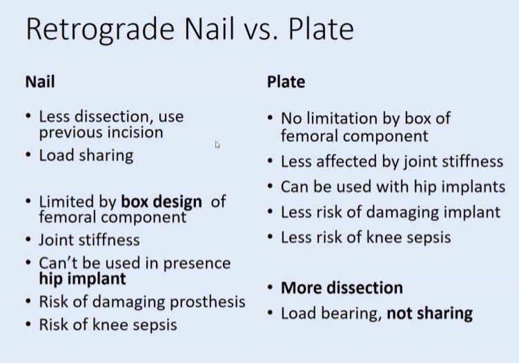
- Nail or plate (femur) - double plate preferred; nail needs at least 2 distal locking screws and may be blocked by notch or patella baja (quote Jones Injury 2016 paper, which describes commonly used knee implants that may or may not allow a nail to pass through)
- Distal femoral replacement (femur)
- ORIF/revision/patellectomy +/- repair (patella)
- Medial approach dissection in the plane of vastus medialis and sartorius
Q10Describe the fracture shown and its classification.▸
- X-ray left proximal femur showing periprosthetic femoral fracture in a previous uncemented THR
- Fracture extends from the middle of the stem to distal to the stem
- Suspected loosening of the stem; compare with previous X-ray
- Vancouver type B2 or B3
Q11What is the Vancouver classification of periprosthetic femoral fractures?▸
- A: trochanteric region
- B: around the stem - 1 stable, 2 unstable, 3 unstable with bone loss
- C: away from the stem
Q12What history, examination and investigations are needed for this fracture?▸
- History and PE: mainly rule out infection and other injuries
- Check previous OT record to know the implant brand and sizes
- Postoperatively also work up for osteoporosis
- Investigations: X-ray lateral, CT for bone stock
Q13What are the aims and surgical principles of definitive management?▸
- Aim: stable, painless, mobile hip allowing early weight bearing
- Restore offset and hip centre; equalise LLD
- Correct positioning of components and soft tissue tension
- Acute setting: seek advice and support from other senior hip surgeons
Q14How are Vancouver type A fractures managed?▸
- Often requires treatment that addresses the osteolysis
- AG fractures <2cm displacement: nonoperative, partial weight bearing, allow fibrous union
- AG fractures >2cm: ORIF with trochanteric claw/cables (loss of abductor function leads to instability)
Q15How is a Vancouver B1 fracture managed?▸
- ORIF using cerclage cables and locking plates
- Subvastus approach; reduction with reduction forceps and colinear clamp; temporary cerclage wires
- LCP: unicortical screws proximally not to violate cement mantle + cerclage wires; distal bicortical screws
- Cortical defect or marked comminution: femoral cortical struct allograft on the anterior aspect of the femur to bypass the defect by at least 2 cortical widths
Q16How are Vancouver B2 and B3 fractures managed?▸
- Revise the stem for B2 and above - ORIF alone in B3 has high reoperation rates: BJJ 2017 Khan showed almost 30% revision rate compared to 15% revision for revision + ORIF
- Femoral component revision with proximal femoral allograft/bone graft or proximal femoral replacement + ORIF
Q17What are the key steps when revising the femoral stem for B2/B3?▸
- Posterior approach - extensile and familiar
- Bypass the fracture with a long, titanium, modular, fluted conical revision stem (e.g. modular restoration stem) - flexible control of leg length, offset and version
- ORIF of the fracture with or without an allograft to maintain the femoral tube
- Decide whether to also revise the acetabulum: compatibility with new stem, premorbid status, fixation and PE wear
Q18How is the acetabulum addressed and what bearing is chosen?▸
- If revising: cementless socket with supplementary screws, with augments available for significant bone defects
- Bearing of choice: ceramic 36mm head on XLPE with a lipped PE liner to minimise posterior dislocation
Q19What other techniques are described for these fractures?▸
- Distally locking, cementless HA-coated long femoral stem (Cannulok; El-Bakoury 2017 100% survivorship at 4 years)
- Exeter group: impaction bone grafting supported by a plate, with a long cemented femoral stem for B2 and B3
- Proximal femoral replacement (Parvizi) with constrained liner as abductors are usually poor; keep as much proximal femoral bone as possible and reapproximate it onto the PFR
Q20What is the new evidence on B2 periprosthetic fractures in cemented stems?▸
- Smitham 2019, Journal of Arthroplasty (Solomon's group, Adelaide)
- B2 fractures in cemented femoral implants can be treated with ORIF without revision of the stem
Q21What is the rationale for ORIF without stem revision in B2 fractures?▸
- Patients too frail to undergo lengthy revision surgery
- CPDT stem can regain stability in the cement mantle after anatomical reduction, provided the cement-bone interface is maintained
- Stem revision can be performed later as a planned elective procedure
Q22What are the criteria for ORIF without stem revision?▸
- Cemented polished double tapered stem
- Cement-bone interface maintained - fracture only disrupts the stem-cement interface
- Stable anatomical reduction achieved at surgery
- Bone stock is adequate
Q23Describe the Knee Society zones.▸
- AP tibia medial to lateral: 1234 base plate, 567 around stem
- Lateral tibia anterior to posterior: 12 base plate, 3 stem
- Femur: 12 anterior flange, 34 posterior condyle, 567 centre
- Patella skyline medial to lateral: 13452
Q24What is the Anderson Orthopaedic Research Institute (AORI) classification of bone defects in TKR?▸
- 1: defect not affecting stability
- 2: defect affecting stability, needs reconstruction with cement/bone/metal
- 3: defect affecting collateral ligaments and patellar tendon, needs constrained implant or major reconstruction
Q25How are tibial bone defects managed after the tibial cut?▸
- Cavitary (contained): cancellous bone grafting
- Segmental (peripheral, typically posteromedial in varus knee)
- <5mm: cement +/- screw
- 5-10mm: metal wedge
- >10mm: metal wedge or structural allograft
Q26What are the problems of using a small femoral component with a thick insert for femoral deficiency?▸
- Elevated joint line - impinges on patella in early knee flexion
- Patella baja with ineffective quadriceps mechanism
- Exposed medial and lateral bone
Q27How do you increase exposure in a medial arthrotomy during TKR?▸
- Rectus snip: proximal extension laterally across quadriceps tendon
- Preserves VL, no brace needed - faster rehab, less injury to lateral genicular artery, but less exposure
- VY plasty (need to cut vastus lateralis also need brace)
Q28What are the two technical problems with a segmental tibial defect?▸
- Smaller tray away from the defect leads to worse load transfer
- Lower tibial cut is limited by Gerdy's tubercle
Q29How is femoral bone deficiency managed in TKR?▸
- Cement
- Metal augment
- Allograft/bone block
Q30What is the alternative to a small femoral component with a thick insert for femoral deficiency?▸
- More wedge + normal femoral component + thin insert