Hip
Revision THA - workup, reconstruction and cages
History, radiographs, failed fixation, revision reconstruction and reinforcement cages
88 questions
7 source pages
1 images
Images appear with the first question taken from each source page — tap a question to open it.
Q1What is a Ganz reinforcement ring?📷▸
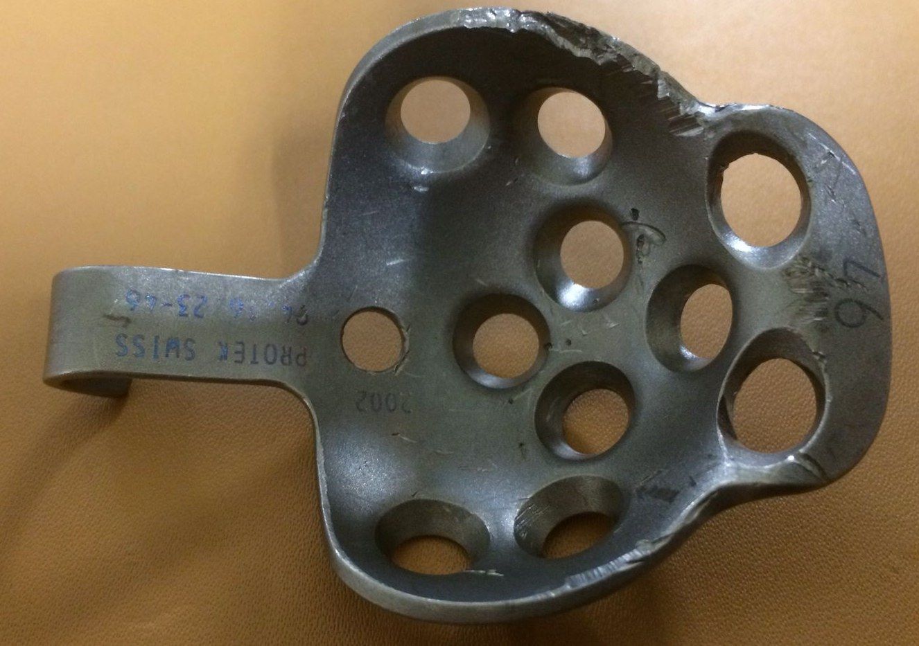
- A reinforcement ring with a hook
- Has screw holes for fixation
Q2How is a Ganz reinforcement ring fixed?▸
- Bone graft on the defect
- Fix the ring with screws
- Then cement PE onto the ring
Q3Why does fixation of the Ganz ring depend only on screws?▸
- There is no ingrowth or ongrowth onto the ring
- Ring fixation depends only on the screws
Q4Describe the X-ray findings and what you would compare with.▸
- Previous cemented THR with femoral lucency over Gruen zones 2, 3, 5, 6, 7, 8, 9, 11, 14 (8-14 anterior to posterior)
- Multiple cortical lytic lesions with endosteal scalloping; no periosteal new bone; no soft tissue calcification
- Compare previous X-ray for stem loosening: subsidence, implant migration, cement breakage, radiolucent line at bone-cement interface
Q5What are the risk factors for infection after THR?▸
- Patient: elderly, immunocompromised - DM, CRF, steroids, alcoholism, recent infection
- Disease: psoriasis, RA, prior hip operation, prior hip infection
- Surgical: prolonged operation; post-operative complications UTI, skin necrosis, haematoma
Q6What should you look for in history and examination?▸
- Immunocompromised conditions, cause of THR, early wound infection, repeated antibiotics, pain-free interval, preceding bacteraemia
- Expect non-mechanical pain
- PE: erythema, sinus
- Classify using Tsukayama (positive intraop c/st, early postop, acute haematogenous, late chronic)
Q7What is the Tsukayama classification of total joint infection?▸
- 1: positive intra-operative cultures
- 2: early acute, <1 month post-op
- 3: acute haematogenous (late acute)
- 4: chronic
Q8What blood tests are used to investigate THR infection?▸
- Leukocytosis, CRP >10mg/L, ESR >30; CRP sensitivity and specificity ~90%
- IL-6 (precursor of CRP) is more accurate but may not be available
- Berbari JBJS Am 2010: accuracy IL-6 > CRP > ESR > WBC
- ESR 75%/70%, CRP 88%/74%, IL-6 97%/91% (sensitivity/specificity)
Q9How useful is hip aspiration and how is it performed?▸
- Spangehl: sensitivity 0.86, specificity 0.94, PPV 0.67, NPV 0.98 (level 1); suggest in high clinical suspicion with abnormal ESR/CRP
- Anterior approach: 1 inch lateral to femoral pulse, 1 inch below inguinal ligament; lateral approach at GT under XR guidance; US or XR guided
- Off antibiotics for at least 1 week; no intra-articular LA (bacteriostatic)
- Send immediately for GS/C+ST, PCR and total cell count (>3000 cells/ml, >80% polymorph); different instruments from 3 different sites, 2/3 samples positive
Q10What are the MSIS criteria and common pathogens for PJI?▸
- MSIS: any one major or 3/5 minor
- Acute <6 weeks: ESR not reliable, CRP >100, WBC >10000 cells/mL, neutrophil >90%
- Chronic: ESR >30, CRP >10, WBC >3000 cells/mL, neutrophil >80%
- Staph aureus and Staph epidermidis account for 2/3; also E. coli, enterococcus
Q11What are the principles of management of a chronic infected THR?▸
- MDT approach with MSK radiologist, microbiologist, histopathologist, plastic surgeon and experienced revision arthroplasty surgeon
- 2-stage revision: removal of implant, debridement and antibiotic-loaded cement spacer
- Systemic antibiotics 6-12 weeks with monitoring; repeat hip aspiration at 6 weeks; CT to reassess bone stock
- Second stage if infection free and serology negative x 2 weeks; must use cemented components (no ingrowth); frozen section negative if <5 polymorphs per 400x HPF
- One stage only if host not immunocompromised, low-virulence organism, no sinus tract, no OM/loosening
- 6 vs 12 weeks antibiotics non-inferior (NEJM 2021 Bernard RCT)
Q12How does the timing of infection determine management?▸
- <4 weeks: acute infection with less biofilm - radical debridement, exchange of modular bearing (thorough debridement), IV antibiotics
- >4 weeks: chronic infection, biofilm present - implant removal and antibiotic-loaded cement spacer
Q13How do the KLIC and CRIME 80 scores guide DAIR?▸
- KLIC for Tsukayama type 2 early acute: kidney 2, liver 1.5, indication (fracture/revision) 1.5, cemented 2, CRP>115 2.5; score >4 = 55% failure rate
- CRIME 80 for type 3 acute haematogenous: COPD 2, CRP>150 1, RA 3, indication fracture 3, male 1, exchange mobile component -1, age >80 2; score >3 = 60% failure
Q14What is the antibiotic-loaded cement recipe used for spacers?▸
- Options are gentamicin, vancomycin, tobramycin
- 1 pack cement (40g) with 1 bottle antibiotic (4g vancomycin); ~10% (~1.x g gentamicin)
- No vacuum, no drain; local concentration falls to 1/2 after 10 days
Q15What are the reported success rates of one-stage and two-stage revision?▸
- UCLH experience: TKR 1 stage 100%, 2 stage 93.2%
- THR 1 stage 94.5%, 2 stage 91.8%
- Ongoing ccc
Q16What history and examination findings are important in a painful THR?▸
- History: age and premorbid state, nature of pain, infection symptoms, current function (HHS), details of last operation, rule out spine, neuro, PVD as causes of pain
- PE: scar, deformity, sinus, ROM
- ROM clues: stiff - HO/impingement/contracture; end range pain - impingement; all range pain - infection; IR/ER pain - femoral stem loose
- Stinchfield test (resisted flexion ~SLR) suggests acetabular loosening; assess abductors
Q17What investigations are needed?▸
- CT
- Scannogram + scoli series
Q18What are the aims of revision THR?▸
- Restore offset and LLD
- Replenish bone stock
- Secure and accurate placement of components
Q19What pre-operative preparation is required?▸
- Counsel patient regarding prognosis of revision THR; check previous implant and approach
- Prepare instruments for implant removal: AO large fragment, ex-plant, straight/curved osteotome, high speed burrs, cement splitters, US device, flexible light source, allograft, GTR plate and cables if planning ETO
- Inform pathology lab for intraop frozen section; prepare cement spacer in case frozen section is positive
- Prepare reconstruction options: long extended coated stem, jumbo cup/metal augment/cupcage/reinforcement rings, allograft
Q20What are the key intra-operative steps for femoral implant removal?▸
- Intraop 1g tranexamic acid to reduce blood loss; old incision, protect sciatic nerve
- Remove bone/cement at Gruen zone 1; try hammering implant out with anchorage at collar/head/hole
- Remove cement mantle with osteotome, reamer, burr, K wires, cement splitter (split radially then longitudinally)
- +/- trochanteric osteotomy/trochanteric page/ETO; if cement mantle is good, use tap in and tap out (cement on cement)
Q21What are the principles of an extended trochanteric osteotomy (ETO)?▸
- Preserve abductor and vastus lateralis (blood supply to the flap); divide the lateralis insertion off the vastus tubercle and elevate vastus lateralis off the linea aspera
- Less than 1/3 of the femoral circumference; length determined by the length of the cement mantle
- Multiple drill holes then break with osteotome; fix with cables
Q22What is a bone window and why may implant removal be difficult?▸
- Bone window by burr - create a trough over the posterior femur, remove the fenestrated part and dissect the bone block
- Difficulty in removal of an AMA is due to bone growth into the fenestration
Q23How is a broken femoral stem removed?▸
- From above: trephine reamer
- From below: cortical window
- From the side: ETO
Q24What ultrasonic and other methods are used to extract cement?▸
- Fish-bone like ultrasonic tip - melts the cement, sinks into it, hardens when USG is off, then hammered out
- Dish-like tip - just melts and lets cement leak through holes
- Drill and tap; segmental cement extraction (long screw with multiple nuts, pour in new cement, hammer out each nut sequentially - cement is weak in tension)
- Powered cement reamer - easy fracture especially over the lateral or anterior side
Q25How is the acetabular implant removed, including a ceramic liner?▸
- Curved osteotome
- Explant
- Ceramic liner: disengage the smooth taperlock by a sharp tap into the liner, then use a suction cup with scallops that contact the peripheral rim of the metal shell to remove the liner
Q26How is reconstruction planned for the femur and acetabulum?▸
- Prefer cementless components if previous cement was used - bone surface become sclerotic, making cement interdigitation difficult
- Femur: long cementless stem with diaphyseal fixation and adequate distal scratch fit
- Acetabulum: identify anatomy with landmarks, navigation, intraoperative imaging, robot
- Rim defect: metal augment, strut graft; medial wall defect: morcelised bone graft, anti-protrusion cage
- Pelvic discontinuity: cup cage construct; rim stable: line-to-line fit with screw fixation; standby dual mobility cup
- Ideally 36mm head
Q27What are the principles and complications of femoral impaction bone grafting?▸
- Retrograde canal filling with impacted particulate graft
- Segmental defect -> contained defect -> allows subsidence + cement creep -> even load distribution -> bone remodelling
- Technique: mesh + wiring creates a neomedullary canal for a cemented femoral stem; advantage: preserves bone stock
- Complications: fracture during impaction, varus/valgus stem, incomplete cement mantle, subsidence (bone graft resorption)
Q28Describe the X-ray findings of this Charnley cemented total hip replacement.▸
- Stem with small head, round back, collared, tapered on AP; cemented PE cup; fixation of trochanteric osteotomy
- Femoral cement-bone interface radiolucency with absent cement at Gruen zones 5 and 6
- Stem in varus with lateral cortical reaction (Harris: probable loosening)
- >2mm radiolucency at DeLee and Charnley zones 1+2 (70% loosening)
- Bone defect both sides: femur Paprosky type 2 with intact diaphyseal bone stock
- Acetabulum likely AAOS type 3 - mixed segmental and cavitary defect
Q29How do you diagnose periprosthetic joint infection in this revision case?▸
- Hip aspiration in 2 separate occasions for C/ST, cell count, alpha defensin
- MSIS major criteria: sinus tract, +ve C/ST in 2 separate tissue/fluid
- MSIS minor criteria: CRP/ESR, synovial WBC count, synovial neutrophil %, single +ve C/ST, >5 neutrophils in HPF
- Chronic: ESR >30, CRP >10mg/L, synovial WBC >3000 cell/uL, PMN >80%
- Acute: ESR not reliable, CRP >100mg/L, synovial WBC >10,000 cell/uL, PMN >90%
Q30If infection is excluded, what causes aseptic loosening and what is your pre-operative assessment?▸
- Mild eccentric wear suggesting osteolysis
- Request initial post-op X-ray: poor cementation technique (<2mm cement mantle) and poor implant positioning (varus stem, increased cup open angle) cause abnormal stress
- Bloods for inflammatory markers and previous X-ray for comparison
- Pre-op CT for bone stock, column and rim integrity; CT angiogram for proximity of great vessels to the cup
- Trace old records for implant size, brand and removal instruments
- If fit for OT, offer operation irrespective of symptoms to prevent further bone stock loss
Q31What are the goals, principles and key surgical considerations of revision hip surgery?▸
- Goal: painless, stable, mobile hip free of infection
- Principles: stable cup fixation with 70% host bone contact, replenish bone stock, restore hip centre, LLD and offset
- Use previous approach, dissect from virgin plane, protect sciatic nerve and gluteus medius
- Frozen section: >5 neutrophils/HPF means treat as infection
- Long stem with extended coating for distal fixation; prepare allograft/ trabecular metal/ jumbo cup/ ring; accept high hip centre in the elderly
- Prepare dual mobility cup and C/XLPE or M/XLPE articulation; slow post-op rehab due to higher dislocation rate
Q32How do you manage acetabular bone defects in revision surgery?▸
- Cementless cup prerequisite: 70% cup coverage, 2/3 rim fit, intact columns
- Paprosky: 1 minimal lysis; 2 <3cm superior migration; 3 >3cm superior migration with column not intact
- Contained defect: avoid defect (high hip centre/ medialise), bone (autograft/allograft, morselized or structural, IBG), metal (wedge, jumbo/oblong cup, cage/ring), cement
- Uncontained defect: convert to contained with mesh, then treat as contained with bone graft +/- augment ring
- Impaction bone grafting: close segmental defects with flexible metal wire mesh, impact 8-10mm morselized trabecular graft, then cemented cup
- Cages offload the graft by bridging ilium or ischium and a liner is cemented in; anti-protrusio cage for pelvic discontinuity
- Jumbo cup >62mm women, >66mm men: simple with more host bone contact, but does not restore bone stock, may raise hip centre and has 10% dislocation rate
Q33What are the reasons for a broken cement mantle and how is an extended trochanteric osteotomy (ETO) performed?▸
- Cement failure when stresses exceed the fatigue endurance limit of the stem-cement interface and the cement itself
- Mantle must be homogeneous and even (2-5mm) with proximal medial cancellous bone <2mm
- Stem must be smooth-contoured, not malaligned, with the tip filling >half of the distal medullary canal
- ETO: drill holes at least 12cm from GT tip, segment is 1/3 of femoral circumference, hinged anterolaterally on periosteum/muscle
- Apply cables distal to the osteotomy before reaming; fix with cerclage wires or cables
- Indications: well-fixed stem, difficult cement removal, varus remodelling of proximal femur, need for enhanced acetabular exposure
Q34What is the aim of assessment when a total hip replacement is loose?▸
- Rule out infection
- Assess symptoms
Q35What history and examination findings are relevant in a loose total hip replacement?▸
- Pain and constitutional symptoms
- Surgical details: timing of OT, initial post-op infection
- Recent infection, wound condition and any sinus
Q36What investigations are required before revision hip surgery?▸
- Obtain previous X-ray for comparison
- Blood test for inflammatory markers
- Hip aspiration on 2 different occasions for C/ST, cell count and alpha defensin
- Pre-op CT for bone stock, column and rim integrity
- CT angiogram for proximity of great vessels to the acetabular cup
- Trace old records for implant size, brand and removal instruments
Q37What are the MSIS 2011 major criteria for periprosthetic joint infection?▸
- Sinus tract communicating with the joint
- +ve C/ST in 2 separate tissue/fluid samples
Q38What are the MSIS 2011 minor criteria for periprosthetic joint infection?▸
- CRP/ESR
- Synovial WBC count and synovial neutrophil %
- 1 C/ST +ve
- 5 neutrophils in HPF
Q39What are the chronic periprosthetic infection thresholds in the MSIS score?▸
- ESR >30
- CRP >10mg/L
- Synovial fluid WBC >3000 cell/uL
- PMN >80%
Q40What are the acute periprosthetic infection thresholds in the MSIS score?▸
- ESR not reliable
- CRP >100mg/L
- Synovial fluid WBC >10,000 cell/uL
- PMN >90%
Q41What initial post-op X-ray findings suggest the aseptic loosening is technique-related?▸
- Poor cementation technique with <2mm cement mantle
- Poor positioning: varus stem position or increased open angle of the cup
- These cause abnormal stress onto the implant
Q42What surgical approach and instruments should be prepared for revision hip surgery?▸
- Use the previous approach
- Prepare fibreoptic light source, XR, IM guide, osteotomes, ultrasonic cement removal device, cables, plates
Q43How are the sciatic nerve and gluteus medius protected during revision dissection?▸
- Protect with hip extension and knee flexion
- Stick to bone during dissection
- Dissect from the virgin plane and extend the previous incision
Q44What is the role of frozen section during revision hip surgery?▸
- Obtain intra-op samples before proceeding
- If >5 neutrophils under high power view -> treat as infection
Q45When can a cementless acetabular cup be used in revision?▸
- Achieve 70% coverage of the cup
- 2/3 rim fit
- Intact columns
Q46What is the AAOS classification of acetabular defects?▸
- Types 1-5: segmental, cavitary, mixed, discontinuity, ankylosed
Q47Describe the Paprosky classification of acetabular defects.▸
- Type 1: minimal lysis
- Type 2: <3cm superior migration, column intact, rim distorted, >50% contact surface; cementless if >2/3 rim fit, otherwise augment ring
- Type 3: >3cm superior migration, column not intact
- X-ray features assessed: superior migration, tear drop, ilioischial line, ischial lysis
Q48How is a contained acetabular defect managed in revision hip surgery?▸
- Avoid the defect: high hip centre or medialise
- Bone: autograft/allograft, structural or morselized, impaction bone grafting
- Metal: metal wedge, jumbo (60mm)/oblong cup, revision ring (Muller/Ganz) if host contact <70%, anti-protrusio cage for discontinuity, mesh for uncontained defect
- Cement
Q49How is an uncontained acetabular defect managed?▸
- Convert to a contained defect using mesh
- Then treat as contained, usually with bone graft + augment ring
- Use cemented fixation as there is no bone ingrowth from bone graft
Q50Which revision stem is chosen for a contained femoral defect?▸
- Cemented stem is poorly fixed due to poor interdigitation with lost cancellous bone and sclerosis
- Prefer extensively coated long stem in good bone stock
- Severe bone loss with a large canal cavity is difficult - needs an extra large implant with risk of fracture and stress shielding (extra large implant for initial stability --> easy fracture during OT, significant stress shielding)
- Alternative: impaction bone grafting + cemented stem
Q51How are segmental or cortical femoral defects managed in revision hip surgery?▸
- Small segmental/cortical defect: cortical onlay strut graft (onlay struft graft)
- Impaction bone grafting + mesh
- Gross proximal bone defect: tumour prosthesis
Q52Describe the technique of impaction bone grafting in the acetabulum.▸
- Close acetabular segmental defects with flexible metal wire mesh
- Fill the contained defect with a layer of morselized 8-10mm trabecular graft, impacted tightly with hammer and impactors
- Cement the cup
Q53What is the role of cages in managing acetabular bone defects?▸
- Reconstruct the defect with allograft or trabecular metal
- Offload the graft by bridging the defect, anchoring to the ilium or ischium
- Liner cemented into the cage
Q54What are the advantages of a jumbo cup?▸
- Defined as >62mm in women and >66mm in men
- Relatively simple
- Increased host bone-cup contact area maximising ingrowth
- Minimises migration by force dissipation over a large area
- Decreases the need for bone graft
Q55What are the disadvantages of a jumbo cup?▸
- Bone stock is not restored
- Limited applicability in oblong defect - may ream away the posterior column or wall
- High dislocation rate due to acetabular size:femoral head ratio (10%) - may use dual mobility
- May raise the hip centre of rotation
Q56How is a Paprosky type IIA (cavitary) acetabular defect reconstructed?▸
- Hemispherical cup
Q57How is a Paprosky type IIB (segmental defect <30% rim) acetabular defect reconstructed?▸
- Hemispherical cup or jumbo cup
Q58How is a Paprosky type IIC (protrusion) acetabular defect reconstructed?▸
- Medial cancellous allograft
- Hemispherical cup
- Impaction bone grafting
Q59How is a Paprosky type IIIA (spherical remodelling, up and out) acetabular defect reconstructed?▸
- Large hemispherical cup
Q60How is a Paprosky type IIIB (oblong remodelling) acetabular defect reconstructed?▸
- Structural autograft/allograft with hemispherical cup
- Trabecular metal shell with superior augment
- High hip centre with hemispherical cup
Q61How is a Paprosky IIIB acetabular defect without discontinuity managed?▸
- Non-biological: cancellous allograft with cage
- Structural allograft (posterior column with cage)
- Biological: trabecular metal with augments
- Triflange custom implant
Q62How is an acute pelvic discontinuity (Paprosky IIIB) managed?▸
- Compression
- Plate with cage and allograft
- Internal plate with trabecular metal
Q63How is a chronic pelvic discontinuity (Paprosky IIIB) managed?▸
- Distraction
- Acetabular transplant
- Trabecular metal with augments
- Triflange implant
Q64Describe Paprosky femoral defect type I.▸
- Minimal metaphyseal bone loss
Q65Describe Paprosky femoral defect type II.▸
- Extension of metaphyseal bone loss with an intact diaphysis
Q66Describe Paprosky femoral defect type IIIA.▸
- Extensive metadiaphyseal bone loss
- At least 4cm of intact cortical bone in the diaphysis
Q67Describe Paprosky femoral defect type IIIB.▸
- Extensive metadiaphyseal bone loss
- less than 4cm intact cortical bone in the diaphysis
Q68Describe Paprosky femoral defect type IV.▸
- Extensive metadiaphyseal bone loss
- Non-supportive diaphysis
Q69What cementless stem options are available for femoral revision?▸
- Primary cementless stem
- Diaphyseal fixation: extensively porous coated cylindrical stems
- Wagner conical stem
- Fluted tapered modular femoral stem
Q70What cemented stem options are available for femoral revision?▸
- Primary cemented stem
- Long cemented stem
- Impaction bone grafting (IBG)
- Cement-in-cement revision
Q71What other femoral reconstruction options exist for revision?▸
- Allograft prosthetic composite
- Tumour prosthesis
Q72What mechanical and biological risk factors predispose to failed hip fracture fixation?▸
- Mechanical: fracture location and pattern, reduction quality, fixation method and quality
- Biological patient factors: osteoporosis, smoking, compliance with rehab, reinjury
- Fracture factors and infection
Q73What do you assess on history and examination of failed hip fracture fixation?▸
- Pain, functional demands, fitness for surgery, patient expectations
- Local: scars, sinus, sciatic nerve
- Systemic: LLD, limb deformity, muscle power, walking status
- XR: reason for failure, complications (AVN, arthritis, broken implant)
- Scannogram, bloods +/- joint aspiration; CT for bone stock, GT healing, screw cut-out
Q74What are the management options for failed fracture fixation in hip fracture?▸
- Joint salvage: revision fixation (unreliable fixation due to osteoporosis, defeats goal of early weight bearing)
- Joint sacrifice: arthroplasty vs Girdlestone vs arthrodesis
- Hemiarthroplasty vs THR
Q75Outline the intraoperative steps of revision surgery for failed hip fracture fixation.▸
- Old incision; watch the sciatic nerve; release contracted tissue (glut max, anterior capsule, iliopsoas)
- Remove implant after dislocation
- Take frozen section samples to rule out infection before proceeding
- Acetabulum: press-fit line to line, augment with screws, standby cemented cup; anatomical hip centre, medialise cup
- Femur: cemented if fracture healed; cementless diaphyseal fixation bypassing fracture if not healed +/- calcar replacement/GT reattachment
- 6. bearing articulation
Q76What are the targets for stability and rehabilitation after revision for failed fracture fixation?▸
- Aim combined anteversion of 35 degrees
- Standby dual mobility cup
- +/- GT osteotomy to retension abductors
- Rehab: hip spica, HO prophylaxis
- Registry: no difference in 15-year cumulative survival between hybrid, cemented and cementless
Q77What pre-operative optimisation is required before revision for failed hip fracture fixation?▸
- Preop anaesthetic and medical optimisation - revision OT is a long operation
- Ulcer prophylaxis
- DVT prophylaxis
Q78What are the general intra-operative considerations in revision for failed hip fracture fixation?▸
- Osteopenic bone
- Careful manipulation of the limb
Q79To what endpoint is the acetabulum reamed and why is the cup medialised in revision?▸
- Ream to the point of exposing bleeding subchondral bone
- Medialisation of the cup decreases joint reaction force (JRF)
Q80What are the indications for a reinforcement ring or cage in acetabular revision?▸
- Rim defect preventing stable cementless cup fixation at the desirable position
- Pelvic discontinuity
Q81What are the advantages of reinforcement rings and cages?▸
- Immediate post-op: span/ bypass defect (span/bypass the defect), support bone graft so it can heal
- Late post-op: incorporated bone graft confers long-term mechanical stability
Q82What does initial stability of a reinforcement ring or cage rely on?▸
- Host bone factors: superior and inferior support
- Superolateral defect - strut bone graft or augment
- Fixation: supplementary screws
- Decreases the seesaw effect
Q83What are the types of reinforcement rings and cages?▸
- Containing: convert uncontained to contained defect (acetabular rim/medial wall mesh cage)
- Lateralising: transfer load to roof and rim while decompressing bone graft (Muller ring)
- Bridging: span major defect (Ganz ring, Burch-Schneider antiprotrusio cage)
- All are used with a cemented PE cup
Q84Describe the Muller ring.▸
- Provides anterior/medial wall support
- Advantage: simple and easy
- Disadvantage: cavitary defect only
Q85Describe the Ganz ring.▸
- = Muller ring + inferior acetabular hook
- Restores an appropriate centre of rotation (COR)
- Decrease superior migration
Q86Describe the Burch-Schneider antiprotrusio cage.▸
- Hemispherical shell with proximal flange + distal nose
- Bridges the acetabular defect by anchorage at the ilium and ischium
Q87What are the indications for a Burch-Schneider cage?▸
- Combined segmental + cavitary defect
- Severe medial wall or posterior column defect
- Pelvic discontinuity
Q88What are the disadvantages of a Burch-Schneider cage?▸
- Large field needed
- Lateralised hip centre
- Technically demanding