Hip
Protrusio acetabuli and inflammatory hip disease
Protrusio on plain film and inflammatory arthropathies affecting the hip
29 questions
2 source pages
1 images
1 fact-check flags
Images appear with the first question taken from each source page — tap a question to open it.
Q1Describe the X-ray findings in ankylosing spondylitis with hip arthropathy.📷▸
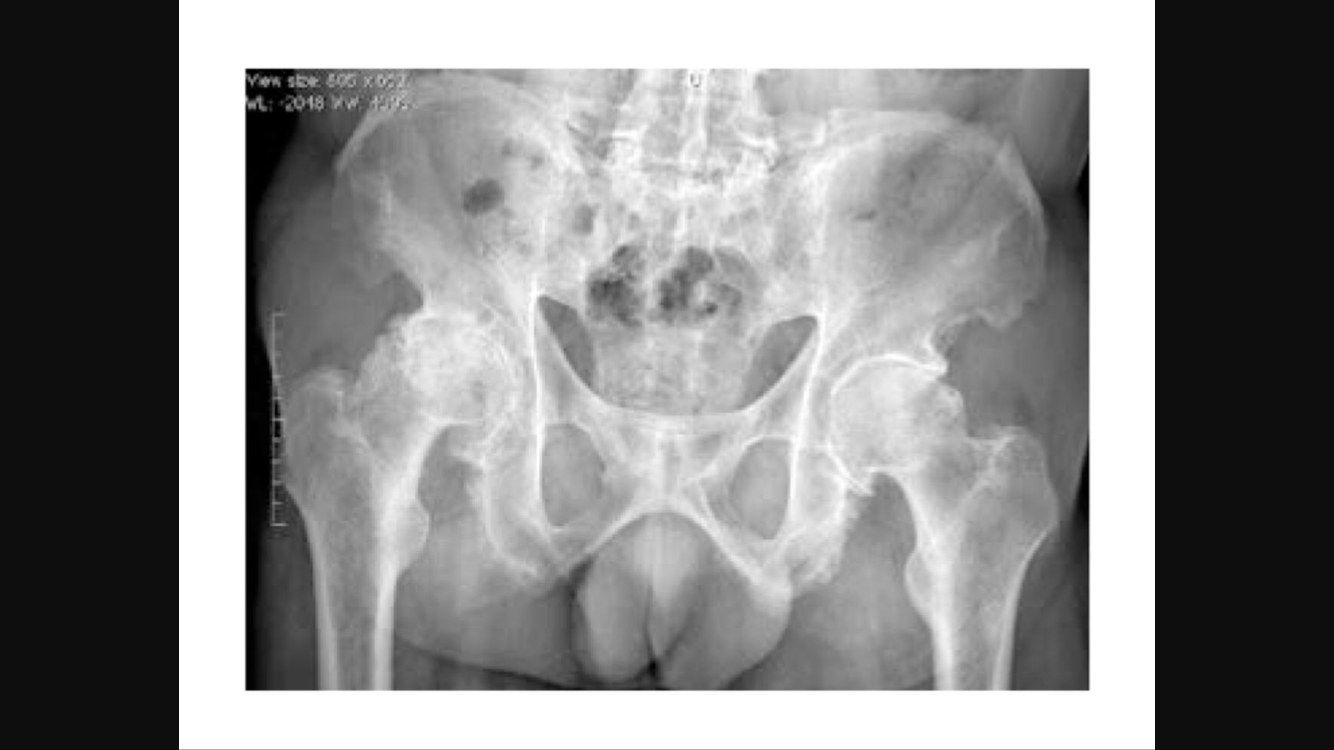
- Decreased joint space over bilateral hip joints, R > L, with OA changes
- Pelvis extended, like an outlet view
- SIJ fused
- Diagnosis: AS with hip arthropathy
Q2How do you diagnose ankylosing spondylitis?▸
- Modified New York criteria 1984: 1 clinical + 1 radiological criterion
- Back pain > 3 months improving with exercise, not relieved by rest
- Limited chest expansion; limited lumbar spine ROM in coronal and sagittal planes
- SIJ involvement: bilateral grade 2-4 or unilateral grade 3-4
- SIJ XR grades: 0 normal, 1 suspicious, 2 erosion/sclerosis, 3 narrowed joint space, 4 fused
Q3How common is hip involvement in AS and why is the pelvis tilted?▸
- 30% of AS have ankylosis of both hips; 90% have bilateral involvement
- Loss of lumbar lordosis and fixed thoracic kyphosis -> pelvis hyperextended (outlet view)
- Fixed hip contracture -> pelvis flexed (inlet view)
Q4Describe the distraction provocation test for the SIJ.▸
- Patient supine
- Examiner applies posterolateral directed pressure to bilateral ASIS
- Positive = reproduction of pain
Q5Describe the compression provocation test for the SIJ.▸
- Patient side-lying
- Examiner compresses the pelvis, pressure over the iliac crest directed at the opposite iliac crest
- Positive = reproduction of symptoms
Q6Describe the thigh thrust test for the SIJ.▸
- Patient supine, hip placed in 90 deg flexion and adduction
- Examiner applies posteriorly directed force through the femur at varying abduction/adduction angles
- Positive = reproduction of buttock pain
Q7Describe the sacral thrust test for the SIJ.▸
- Patient prone
- Examiner delivers an anteriorly directed thrust over the sacrum
- Positive = reproduction of pain
Q8Describe Gaenslen's test.▸
- Patient supine with the symptom leg hyperextended below the bed, asymptomatic leg flexed
- Press on the hyperextended leg
- Positive = reproduction of pain
Q9What are the surgical difficulties in THR for AS and how can you tackle them?▸
- Pre-op: stop biological agent, assess lung and heart function, ulcer prophylaxis; C-spine XR r/o C1/2 instability, may need fibre-optic intubation
- Ortho: decide hip or spine first - usually hip first; expect more bleeding, inform anaesthetist +/- cell saver
- Intra-op: careful positioning (trunk vertical axis parallel to table), posterior approach, sliding trochanteric osteotomy, protect sciatic nerve
- Acetabulum: identify true floor using TAL (Beverland); graft protrusio; set version for tilted pelvis - if PT<20, decrease cup 5 deg for every 10 degrees beyond 20 deg PT; aim combined anteversion 35 deg
- Post-op: HO risk 10%, anterior dislocation, pneumonia, DVT prophylaxis
Q10What is the sequence and rationale for surgery in an RA patient?▸
- 1. Life saving (C1/2 instability)
- 2. Function (ruptured tendon, acute carpal tunnel, most painful joints)
- 3. Sure win procedure
- 4. Lower limb: foot > hip > knee - Foot – eliminate ulcers as source of infection; THR improves flexion range for TKR (typically delay TKR for 6 month due to risk of dislocation)
- 5. Upper limb: proximal to distal
Q11What do you assess in a patient with AS?▸
- Treatment and control of AS
- Associated conditions - Ortho: spine; Others: chest expansion, eye, heart
- Function: walking, sitting, gaze
Q12What are the ASAS classification criteria for spondyloarthritis?▸
- > 3 months back pain + age < 45
- SI joint on imaging (MRI+ or definite XR by modified NY criteria) + >= 1 SpA feature, OR HLA-B27 + >= 2 SpA features
- Sensitivity 82.9%, specificity 84.4%
- SpA features: inflammatory back pain, arthritis, enthesitis, uveitis, psoriasis, dactylitis, IBD, good NSAID response, family history, HLA-B27, elevated CRP
Q13What are the medical and anaesthetic pre-operative considerations in THR for AS?▸
- Medical: stop biological agent; assess chest (lung function test) and heart (echocardiogram); ulcer prophylaxis
- Anaesthetist: C-spine X-ray to rule out C1/2 instability
- Limited C-spine range may need fibre-optic assisted intubation; assess TMJ condition
- Expect more bleeding - inform anaesthetist +/- cell saver
Q14What positioning and draping issues arise in THR for AS?▸
- Cautious during positioning, especially over the neck
- Trunk vertical axis (ear to greater trochanter) parallel to the OT table with eye level perpendicular -> functional axis used as reference for anteversion
- Good padding of all pressure points
- Draping difficult in an adducted position with immobility of the other side (may need to break table)
Q15How do you manage exposure and soft tissues in THR for AS?▸
- Consider posterior approach (better exposure, lower risk of anterior dislocation)
- Difficult exposure: sliding trochanteric osteotomy to increase exposure (needs extensive soft tissue release; hip dislocation + soft bone = fracture risk; consider double cut osteotomy)
- Sequential soft tissue release for scarring; protect the sciatic nerve
Q16How do you identify the true acetabulum in a fused AS hip?▸
- Use TAL (Beverland) as landmark
- Drill through the acetabulum and measure
- Intra-op image intensifier
- Protrusio may need bone graft/cage
Q17What femoral and post-operative problems occur in THR for AS?▸
- Femoral preparation: risk of iatrogenic fracture; osteopenic bone may need a cemented implant
- Post-op: high risk of heterotrophic ossification (heterotopic ossification) - previous hip surgery, complete ankylosis - 10%
- Risk of anterior dislocation - hip precautions; contralateral fused hip causes compensatory flexed position -> stiffness and dislocation
- Pneumonia with limited chest expansion; DVT prophylaxis
Q18Hip or spine first in AS? What is the rationale?▸
- Controversial; lecturer usually does hip first - regains hip mobility to compensate for a stiff spine, may regain horizontal gaze, avoids high-risk spine surgery
- Downsides of hip first: THR dislocation; kyphotic spine + fall -> spine fracture; catastrophic neurological complications during THR rehab
- If spinal imbalance is severe such that THR will not restore horizontal gaze, restore spinopelvic harmony first
- Individualized decision involving multidisciplinary input
Q19How do spinal flexibility and balance guide hip vs spine surgery and cup position (Phan, JBJS 2015)?▸
- Depends on spinal flexibility and overall spinal balance
- Stiff spine, normal balance: increase acetabular anteversion (avoid anterior impingement/posterior dislocation when sitting - the pelvis will not retrovert)
- Stiff spine, unbalanced (retroverted pelvis): decrease anteversion (avoid posterior impingement/anterior dislocation when standing) OR do spine osteotomy first
- Flexible, unbalanced spine: same as stiff unbalanced, but lean more towards hip first (more likely to regain horizontal gaze after THR)
- Flexible, normal balance: unlikely in AS
Q20What are the risk factors for heterotopic ossification?▸
- Patient: age > 60, male, previous HO, diseases (AS, DISH, hypertrophic OA, Paget's disease)
- Injury: head injury, burns
- Surgery: SP > Hardinge, extensive dissection, revision; posterior approach = lowest risk
- Post-op: prolonged immobilisation
Q21What are the radiographic features of protrusio acetabuli?▸
- Femoral head medial to the ilioischial line
- Violation of the ilioischial line (> 3mm in men, > 6mm in women)
- Decreased CEA (centre-edge angle)
- LLD (leg length discrepancy)
Q22How is protrusio acetabuli classified and how does it differ from coxa profunda?▸
- Hirst classification by degree of medial migration (distance between acetabulum and ilioischial line)
- Lecture ranges: 5-10, 10-15, > 15mm
- Not equal to coxa profunda: lateral border of the acetabular teardrop in line with or medial to the ilioischial line
Q23What are the causes of protrusio acetabuli?▸
- Uncommon; rule out other pathologies
- Primary: Otto's disease (arthrokatadysis)
- Congenital: Marfan, OI
- Metabolic: osteomalacia, rickets, osteoporosis
- Inflammatory: RA, psoriasis, AS; Infection: TB
- Others: migration of hemiarthroplasty, Paget's disease
Q24What are the aims and operative plan for THR in protrusio acetabuli?▸
- Aims: stable, painless, mobile joint; restore vertical and horizontal offset and restore LLD
- Choose hybrid THR with ceramic-on-PE articulation
- Posterior approach, protect sciatic nerve; difficult dislocation - extensive capsular incision, may need in-situ osteotomy
- Acetabulum: bone graft the floor and lateralise the hip centre; use TAL as reference; cementless cup with line-to-line fit; cemented standby
- Femur: low level neck cut with enhanced offset to restore abductor tension; Dorr cc ratio > 75% -> cemented
Q25How do you manage the acetabular bone defect in protrusio acetabuli?▸
- Essentially a IIc defect
- Contained: morselized autograft from the femoral head
- Uncontained: mesh + impaction bone graft technique, aim for peripheral fit
- Large reamer first (hourglass deformity), do not deepen the acetabulum further
Q26What history and examination findings are relevant in protrusio acetabuli?▸
- Hx: age, occupation, premorbid walking, PMHx
- Hx: pain and functional limitations
- Local PE: soft tissue, scar, sinus, ROM, FFC
- Systemic PE: LLD, gait, nerve exam, scoliosis, pelvic obliquity, knee FFC, ankle equinus
Q27What investigations are needed in protrusio acetabuli?▸
- Bloods
- CT
- Scannogram
- Mx: conservative or operative
Q28What general technical principles apply to THR in protrusio acetabuli?▸
- Soft bone - careful reaming and broaching
- Bone graft the floor and lateralise the hip centre; use TAL as reference
- Cementless cup line-to-line; cemented standby
- Restore abductor tension: low level neck cut with enhanced offset femoral component
Q29How is reduction and rehabilitation managed after THR for protrusio acetabuli?▸
- Release soft tissue (gluteus maximus, anterior capsule, iliopsoas) before reduction
- Rehab: hip precautions
- Rehab: FWB (full weight bearing)
Fact check
HIRST classification grades protrusio acetabuli by medial migration of 5-10, 10-15 and >15mm — incorrect ranges — Hirst grading is gender-specific by acetabulum-ilioischial line distance: men 3-8/8-13/>13mm (grade III with fragmentation); women 6-11/12-17/>17mm. The 5-10/10-15/>15 grouping resembles other classifications — source