Hip
Pelvic radiography, DDH classification and osteotomy
Reading pelvic radiographs, low DDH, Crowe classification and osteotomy decision
67 questions
8 source pages
4 images
1 fact-check flags
Images appear with the first question taken from each source page — tap a question to open it.
Q1Describe the X-ray findings in a young adult with a dysplastic hip.▸
- Undercoverage of the femoral head and underdeveloped acetabulum
- Acetabular angle ~60 (N 33-38), CEA ~0 (N 25-40), Tonnis angle ~20 (N <10)
- Acetabular index = Hilgenreiner line to acetabular edge
- Superior migration of femur, Shenton's line broken
- Tear drop
- No OA change; also comment on the femur
Q2What causes the pain in a dysplastic hip?▸
- Cartilage wear due to small weight-bearing surface and abnormal loading
- Labral injury
Q3What further investigations are needed in a dysplastic hip?▸
- False profile X-ray (true lateral of hip) for anterior coverage (anterior centre edge angle normal 25-50)
- X-ray with hip in abduction + IR to check congruency (can consider osteotomy)
- MRI for cartilage condition and labral tear, r/o AVN (rule out AVN)
Q4What is the aim of management in an early dysplastic hip with no OA in a young patient?▸
- Relief of symptoms and prevent progression to early osteoarthritis
- Treatment options aim at pain relief +/- improving coverage to reduce contact stress
Q5What are the treatment options for early dysplastic hip with no OA in a young patient?▸
- Conservative - disadvantage: later joint preservation surgery not possible
- Hip arthroscopy: indication labral/chondral pathology; controversial for persistent pain, ?instability after labral debridement
- Periacetabular osteotomy (Ganz): increased coverage and congruency, decreased pain, hyaline cartilage
- Salvage (Chiari, shelf): fibrocartilage, 84% at 17 years
- VDRO if PAO cannot provide sufficient coverage
Q6What are the prerequisites and prognostic factors for PAO?▸
- Prerequisites: congruent joint, no OA, good range, young
- Ganz osteotomy: anterior rotation, lateral rotation, medialisation, intact posterior column
- Poor prognostic factor: >30 y.o
- Bone cyst is not a contraindication in PAO
- Outcomes 60% survivorship in 20yrs
Q7Describe the AP pelvis X-ray findings.📷▸
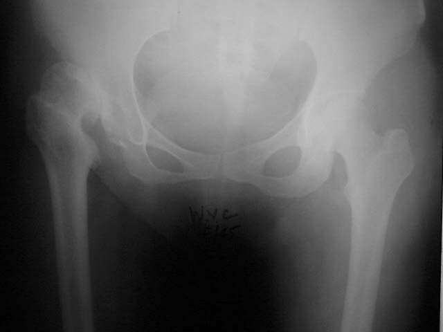
- Right dysplastic hip with superior and lateral dislocation (broken Shenton's line)
- Femoral head loss of sphericity with coxa plana and coxa breva, increased ER (prominent LT), suspected increased NSA, hypoplastic femoral shaft
- False acetabulum in contact with the native acetabulum; anterior superior rim bone loss; teardrop poorly formed
- Tonnis angle >10, acetabular angle of Sharp >45, CEA >45
- Contralateral hip normal; significant LLD; no evidence of previous PAO or VDRO
Q8What is the classification of this hip and why?▸
- Hartofilakidis Type B (low dislocation)
- Lower lip of the false acetabulum is in contact with the upper lip of the true acetabulum
Q9What history and examination findings are relevant in a dysplastic hip?▸
- Hx: age, occupation, premorbid walking, PMHx, risk factors including childhood DDH; pain and limitations
- Local PE: skin, scar, sinus, ROM, FFC, abductor power
- Systemic: gait, nerve, LLD --> scoliosis, pelvic obliquity, contralateral knee flexion contracture, ipsilateral foot equinus
Q10What investigations and templating technique are used?▸
- Bloods to rule out infection +/- joint aspiration
- CT for bone stock, morphology, dimensions, acetabular orientation and femoral anteversion
- Scannogram; +/- MRI/EMG for abductors
- Template: use Ranawat triangle technique to template for anatomical hip centre, measure the superolateral bone defect
- Isocheles triangle, h=1/5 of pelvic height. Hip Center= midpoint of the diagonal line (isosceles triangle, h = 1/5 pelvic height, hip centre = midpoint of the diagonal line)
Q11What are the aims and counselling points for THR in a dysplastic hip?▸
- Offer conservative management, if failed advise THR
- Counsel: overall survival worse (10 yr 87%, 20 yr 55%, Hip Int 2017)
- Aim: restore offset and anatomical hip centre, equalise LLD, correct alignment, good soft tissue tension
- Anatomical centre benefits: better biomechanics (less JRF, abductor function), more bone stock, restore LLD, less impingement
Q12What anatomical abnormalities and natural history are expected in a dysplastic hip?▸
- Soft tissue: elongated redundant capsule, psoas hypertrophy, contracted transversely orientated abductors, shortened rectus/adductor/hamstring
- Nerve: femoral and sciatic nerve shortened
- Bone: shallow, excessively anteverted acetabulum with superanterior bone defect
- Femur: ER, excessive anteversion, increased neck shaft angle, small posteriorly displaced GT, canal stenosis
- Natural history: subluxation --> pain at 20-30; dislocation --> well till 50 (adjacent joint problems)
Q13Why does a dysplastic hip develop early OA?▸
- Loss of coverage of the femoral head + lateralised head
- Decreased abductor lever arm and small contact area --> early OA
Q14What implant and bearing choices are suitable for THR in a dysplastic hip?▸
- Cementless THR or hybrid: both have good longevity in joint registries
- Bearing: metal-on-PE or ceramic-on-PE (good wear profile without the risk of CoC)
- Dual mobility if stability is a concern
- Combined anteversion of 35 degrees
Q15Describe the exposure and soft tissue release in THR for a dysplastic hip.▸
- Larger incision, beware the sciatic nerve
- Contracted soft tissue, pulvinar in the acetabulum, thickened ligamentum teres
- Release sequence: short rotators, capsule, reflected head of rectus, gluteus maximus, iliopsoas (beware descending branch of MFC), adductors
- Finding the acetabulum: use TAL and pulvinar, trace the ligamentum teres; standby X-ray
- Consider transtrochanteric/transfemoral approach (not commonly done)
Q16What are the key points of acetabular bone work in a dysplastic hip?▸
- Usually soft bone - be careful when reaming or broaching
- Acetabulum hypoplastic, deficient anterior/superolateral bone stock
- Aim for cementless fixation with 66% rim fit or 80% host bone contact, standby cemented cup
- Address bone defect: biological or metallic options (trabecular metal, bulk autogenous bone graft, reinforcement rings, cup cage)
- Cotyloplasty: deliberate medial wall fracture to increase coverage, then graft + cementless - difficult revision as bone stock not restored
Q17What are the femoral considerations in THR for DDH?▸
- Small head, short anteverted valgus neck, posterior GT attachment, narrow straight tapered canal
- Subtrochanteric osteotomy if LLD >4 cm - step cut for rotational stability; derotation osteotomy/modular implant if anteversion >40 degrees
- Attempting an uncemented stem in a deformed femur risks fracture
- Cementless modular DDH stem if subtroch osteotomy (press-fit distal fixation, cable/wires + graft); cemented if no osteotomy
- GT osteotomy if abductors need to be advanced and retensioned; proximal femoral resection sacrifices bone stock and makes rotation hard to determine
Q18What is the rehabilitation after THR in a dysplastic hip?▸
- Keep hip in extension, abduction and knee in flexion
- Full weight bearing, hip precautions
- Watch for HO
Q19What is the Crowe classification based on?📷▸
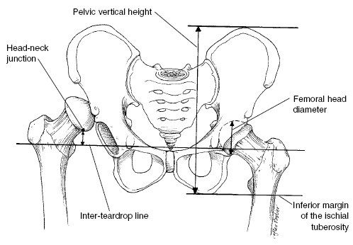
- Radiological classification based on proximal migration of the femoral head compared with pelvic height
- Pelvic vertical height = distance from inferior ischial tuberosity to superior iliac crest
- Measure distance between head-neck junction and inter-teardrop line
Q20What are the Crowe grades by head/neck junction to femoral head diameter?▸
- 1 <50%
- 2 50-75%
- 3 75-100%
- 4 >100%
- 4 = Hartofilakidis B/C
Q21What are the alternative Crowe grades using vertical height?▸
- 1 <10%
- 2 10-15%
- 3 15-20%
- 4 >20%
Q22What are the differential diagnoses for a previously dysplastic or deformed hip?📷▸
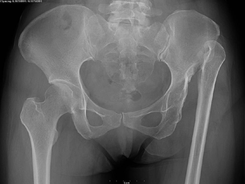
- Infection - septic hip / TB
- Dysplastic hip (treated DDH with childhood surgery)
- Post-traumatic
Q23How do you look for evidence of a septic or TB hip?▸
- History: fever, antibiotics, surgical drainage
- PE: look for sinus
- Ix: inflammatory markers, +/- bone scan, +/- USG for effusion
- If TB hip: 3 months preop + 9 months postop antibiotics
Q24What preoperative planning is needed for THR in DDH?▸
- Consult senior joint surgeon
- PE for LLD, ROM, abductor
- Templating
- CT for bone defect and femur morphology
- MRI / EMG for abductor; prepare implant; ?intra-op culture
Q25What bone and soft tissue considerations exist in high or dislocated hips?▸
- Acetabulum: decide high vs low hip centre
- High hip centre = reconstruction 35 mm higher than inter-teardrop line or 15 mm higher than femoral head centre
- Femur: hypoplastic, narrow and straight, excessive anteversion --> small straight stem / subtrochanteric osteotomy
- Soft tissue: contracture of surrounding soft tissue, sciatic nerve
Q26What is the quoted risk of sciatic nerve injury with limb lengthening?▸
- Kavanagh 1991: <4 cm --> 0% palsy
- >4 cm --> 28% palsy
Q27What does the teardrop indicate?▸
- Tear drop normal if onset of pathology is after 8 years
Q28What is a Brittain ischiofemoral arthrodesis?▸
- Extra-articular arthrodesis of the hip
- Subtrochanteric osteotomy and medial displacement of the femoral shaft with fibular graft to the ischium just below the acetabulum
- Principle: compression force by adductor
- Extracapsular (away from TB infection and progressive femoral head destruction)
- Disadvantages: needs postop immobilisation and difficult revision
Q29What other hip fusion techniques exist?▸
- Extraarticular: iliofemoral fusion
- Intraarticular (Cabenela): dislocate hip, ream away articular cartilage, DHS across joint; preserve abductor, minimal change in anatomy
- Combined: medializing acetabular osteotomy, intra/extraarticular arthrodesis, Davis muscle pedicle graft, cobra plate - stability but stress riser at plate end
Q30What are the indications and contraindications for hip fusion?▸
- Indication: young + recurrent infection
- CI: ipsilateral LL fusion
- CI: contralateral hip fusion
- CI: persistent infection
- CI: poor cardiorespiratory tolerance; poor bone stock
Q31What is the ideal position for hip fusion and its biomechanics?▸
- 25 degrees flexion (balance of sitting/standing)
- 5 degrees adduction/neutral (restore apparent LLD; abduction causes pelvic obliquity and back pain)
- 5 degrees external rotation
- 50% reduction in gait efficiency (increased contralateral pelvic rotation, increased adjacent knee JRF)
- Increased O2 demand and 30% increase in energy consumption
Q32What history and examination findings are relevant before taking down a fused hip?▸
- Hx: age + function, initial indication for fusion, current disability, neurological deficits
- Walking aids + shoe raise
- Posture --> position of fusion; short limb gait + ipsilateral vaulting
- LLD + compensatory deformities; ROM; Thomas test
- Abductor power and adductor jerk test; sinus/scar; gluteal and thigh wasting; Trendelenburg test
Q33What are the options and outcomes for taking down a fused hip?▸
- Options: take down/ corrective osteotomy --> fused in better position; take down + arthroplasty
- Indications: adjacent joint pain (ipsilateral knee, contralateral hip), increasing back/radicular pain
- Also suboptimal fusion position, painful pseudarthrosis
- CI to take down: poor function, residual infection, abductor function (clinical/EMG/MRI)
- Preop: sitting-standing pelvic XR, CT bone stock, MRI/ EMG for abductor, Biopsies or aspiration for potential for reactivation of dormant infection
- Outcome worse than routine THR (74% at 15 yr, Allan Gross JBJS 2021); up to 1/3 revision in 10 yr
Q34What are the difficulties in exposure and identifying the hip centre when taking down a fused hip?▸
- Cannot mobilise the hip - need in-situ dissection, high risk of neurovascular injury
- In-situ double cut osteotomy / trochanteric page osteotomy (cut neck closer to trochanter to save bone stock; landmarks GT, pubofemoral arch)
- Tilt table for visualisation; identify femoral neck orientation and protect with 2 Hoffman
- Hip centre: pulvinar, transverse acetabular ligament, AIIS, greater sciatic notch
- Cup malposition common due to spine/pelvic deformity; XR or robotic guidance; Trimming of residual bone after cup placement; +/- screw augmentation
Q35What soft tissue difficulties and overall results are expected when taking down a fused hip?▸
- Soft tissue: capsule, adductor tenotomy, reflected head of rectus femoris, psoas, HO
- Aim: pain relief, mobile joint, restore hip centre and offset, equalise LLD, balance soft tissue tension, accurate component placement
- High risk surgery (up to 1/3 revision in 10 years)
- Gait will worsen due to inadequate abductor function
- Will improve gradually over months/ years
- Implant choice: hybrid THR
Q36Is abductor weakness an absolute contraindication to take-down, and how is it tackled?▸
- No - but it causes abnormal gait and increases dislocation rate
- Tenodesis of ITB at the greater trochanter (similar to transfer)
- Constrained liner to decrease dislocation
Q37What do you need to know about TB hip when considering take-down?▸
- Traditionally Hardinge and Charnley: THR when no sinus discharge >20 yr, fusion >10 yr, previous medically treated TB
- Kim SJ BJJ 2013 meta-analysis: THR in active TB may be offered before this with extensive debridement + perioperative TB meds
- Pre op 1-3 months Anti TB med
- Post op 9 months anti TB med (in accordance with Infectious Disease Society of America guidelines)
- Sepsis rate post TB ~ 12 %
- Shanmugasundaram classification: stable (normal/atrophic/Perthes) and unstable forms (subluxed/dislocated, protrusio, pestle and mortar)
- Phemister triad: peripheral osseous erosion, joint space narrowing, juxta-articular osteopenia; Tuli stages 1-4
Q38Describe the X-ray findings and the likely stage of AVN.▸
- Collapse of femoral head bilaterally R>L; left Shenton's line broken
- Right hip positive crescent sign
- Positive OA change at the left hip; acetabulum well formed; SIJ normal
- Suggests Ficat IV left hip and Ficat 2B right hip; ask for frog leg lateral if collapse not seen
- Bilateral in 40-80% of cases; Ddx tumour, septic arthritis, BMES (self limiting)
Q39What is the Ficat classification of AVN?▸
- I normal
- II sclerosis or cyst (A focal changes, B crescent sign without flattening)
- III flattening of femoral head without joint space narrowing
- IV flattening of femoral head + OA
Q40What are the causes and risk factors for AVN of the hip?▸
- Traumatic: # NOF or hip dislocation; risk: femoral head fracture 75-100%, NOF I/II <10%, III/IV >80%, basicervical 50%, trochanteric 25%, hip dislocation 5-40% (reduction within 6 hr 2-10%)
- Non-traumatic: idiopathic; intra/extra-vascular and mixed (commonest is mixed)
- Alcohol: 400 ml/week --> RR 9.8-fold (1 unit = 10 ml/8 g alcohol, i.e. 40 units/week, 20 beers/week)
- Steroid: >20 mg/day prednisolone for >30 days or >2 g over 3 months
- Intravascular: protein S/C, SLE, fat embolism, caisson disease, sickle cell anaemia
- Extravascular (marrow adipocyte hypertrophy): Gaucher, radiation
Q41What is the role of MRI and classification in pre-collapse AVN?▸
- Double line sign on T2: hypodense line = edge of necrosis, hyperdense line = hypervascular granulation tissue
- Steinberg (1-6, A <15% / B 15-30% / C >30%) predicts need for head-preserving procedure; <15% observe, >15% offer core decompression
- Kerboul angle: <190 no collapse, 190-240 50% collapse, >240 100% collapse; >200 predicts poor result with preserving procedures
- Shimizu: >1/4 head diameter and >2/3 weight-bearing area --> 74% collapse in <3 years
- Risk of collapse: position, size, presence of bone edema; overall 50-80% progress (symptomatic 70-80%)
- +/- bone scan: increased uptake at the femoral head in AVN, at femoral head and acetabulum in OA
Q42What history and examination findings are needed in AVN of the hip?▸
- Hx: risk factors/causes - drinker? steroid use? SLE? diver? trauma?; current symptoms; functional limitations (HHS)
- PE local: sinus, ROM, FFC, abductor
- Systemic: gait, LLD, document NV status
- Bloods to rule out infection; +/- CT; +/- bone scan
- MRI in pre-collapse stage: size, site, prognostication (Kerboul angle, Shimizu)
Q43What is the aim of AVN management and which parameters guide it (CARE)?▸
- Aim: pain relief, improve function via joint salvage or joint replacement depending on stage
- Care parameters: Collapse of head (Ficat), Age, Reversible cause, Extent of disease (Steinberg ABC, Kerboul, Shimizu)
Q44When do you observe, decompress or graft an AVN hip?▸
- Pre-collapse + small necrotic area + not in weight-bearing zone --> observe
- Stage 1 or 2 + predictor of collapse (large necrotic area in weight-bearing zone by Kerboul, Shimizu, Steinberg) --> core decompression (5-7 yr survival 70-90%)
- Stage 3 + young --> vascularised bone graft (5-7 yr survival 70-80%)
Q45What is the pathophysiology of idiopathic AVN of the hip?▸
- Biological and mechanical
- Intraosseous microcirculation coagulation --> venous thrombosis --> retrograde arterial occlusion --> increased intraosseous pressure --> decreased blood flow to femoral head --> AVN
- Mechanical load on necrotic bone incapable of repair --> mechanical failure and collapse
- Modes of failure: fine cracking of subchondral bone, linear tangential fracture, shearing fracture
Q46What is the pathophysiology of steroid-induced AVN?▸
- Fat cell hypertrophy --> femoral head compartment syndrome
- Hyperlipidaemic state - fat emboli causing endothelial damage
- Direct toxicity - damage to endothelial and smooth muscle cells in the vasculature
- Direct osteocyte death
Q47What is the crescent sign and why does it occur?▸
- Overlying cartilage remains viable from synovial fluid, while subchondral bone is necrotic and collapses
- Ficat IIB = crescent sign without flattening of the femoral head
Q48What is the conservative and joint-preserving management of AVN?▸
- Counsel on natural history: Rate of preservation of the femoral head by stage: stage 1 35%, stage 2 31%, stage 3 13% (Hungerford JBJS 1995)
- Protective weight bearing has no evidence (Fairbank CORR 1996)
- Alendronate may prevent collapse in early disease; LMWH for coagulopathy; statin for steroid-induced (decrease adipogenesis)
- Core decompression (stage I, IIA; preferably reversible cause): 5-7 yr survival 70-90%, # risk 1%; poor results in steroid induced
- Hungerford 1995 JBJS 10-year survival: 90% stage 1, 70% stage 2, 30% stage 3
- Vascularized bone graft for stage 3 young: survival 70-80%; trapdoor for Ficat III/early IV (73% good-excellent at 5 yr)
- Rotational intertrochanteric osteotomy for small lesions, Kerboul <200; fails if >30-50% head involved
Q49Describe the technique of core decompression for AVN.▸
- Indication: pre-collapse lesion, preferably reversible cause (Ficat I, IIA)
- Mechanism: relief of intraosseous hypertension, remove necrotic material, stimulate angiogenesis (mainly pain relieving; effect on natural history unknown)
- Method 1: 8 mm trephine after guide pin, within 5 mm of the articular surface, additional channel by 5 mm trephine
- Method 2: 3.2 mm pin 2-3 times for decompression
- +/- structural support (tantalum rod/strut graft) +/- marrow/BMP to stimulate bone growth
Q50Describe the principles, technique and complications of vascularised bone grafting for AVN.▸
- Indication: pre-collapse or collapsed AVN in young patient (<45 y), preferably reversible cause (Ficat II B/C)
- Principles: decompression, removal of necrotic bone, replacement with corticocancellous graft for structural support, Revascularization
- Graft: fibular (peroneal --> ascending branch of lateral circumflex) or iliac crest (deep circumflex iliac artery); AL approach
- Postop: LMWH then aspirin + NWB walking 6/52
- Complications: subtrochanteric fracture, donor site morbidity (FHL/big toe contracture), harvest fibular graft >6 cm from knee and ankle, superficial peroneal nerve injury
- VBG vs core decompression (CORR 2017 Cao RCT): VBG better functional score (not reaching MCID) and vascularity on SPECT; no difference in progression to THR (10%)
Q51What newer stem cell techniques exist for AVN?▸
- Three-stage procedure: stem cell culture, then cultured stem cells reinjected into a previous core decompression site
- Bone marrow from the pelvis centrifuged in the OR to yield bone marrow concentrate rich in stem cells, transplanted into the necrotic area under X-ray control after core decompression
Q52What are the joint-sacrificing options for AVN?▸
- THR is the mainstay: previous incisions, steroid osteoporosis, acetabular defect, Altered anatomy and bone stock, higher wear/osteolysis, infection, Post op dislocation (alcoholic)
- Aus registry 20y revision rate 12.3%; THR > bipolar (Lee CORR 2004)
- Hip resurfacing: indication AVN of femoral head with no acetabular damage
- Resurfacing CI: poor proximal femoral bone stock (AVN >25% of head), coxa vara/breva, small cup + large neck, osteoporosis, childbearing
- Resurfacing pros: preserve bone stock, less osteolysis, less dislocation; risks: femoral neck fracture 1-2%, metallosis, impingement
- Hip arthrodesis for very young patient
Q53How common is bilateral AVN and which part of the head is usually affected?▸
- Bilateral in 40-80% of cases overall
- 50% of idiopathic cases and 80% of steroid-induced cases
- Usually affects the anterolateral femoral head
Q54Which classification system is used for AVN of the hip, and who described it?▸
- University of Pennsylvania system (Steinberg 1995)
Q55What are the criteria and treatment for each stage of the University of Pennsylvania (Steinberg) system?▸
- 0: Normal MRI → observe
- I: Abnormal MRI, normal X-ray → core decompression
- II: X-ray showing cystic or sclerosis → core decompression, vascularized bone graft
- III: subchondral collapse with crescent sign → vascularized bone graft
- IV: flattening of femoral head → vascularized bone graft, THR
- V: joint narrowing +/- acetabular → THR
- VI: advanced OA → THR
Q56How are the stages further subdivided in this system?▸
- Mild (A), moderate (B) or severe (C) by % of head involvement, surface collapse and location
Q57What is the conversion rate to THR in 5 years (1995 JBJS Urbaniak, <50 years old)?▸
- Stage II – 10%
- Stage III – 20%
- Stage IV – 30%
Q58Which stages are successful in 10 yrs and at what rates (1995 JBJS Hungerford)?▸
- Stage I – 90%
- Stage II – 70%
- Stage III – 30%
- 1% fracture rate
- Poor result with steroid-induced AVN
Q59Describe the X-ray findings in this patient.📷▸
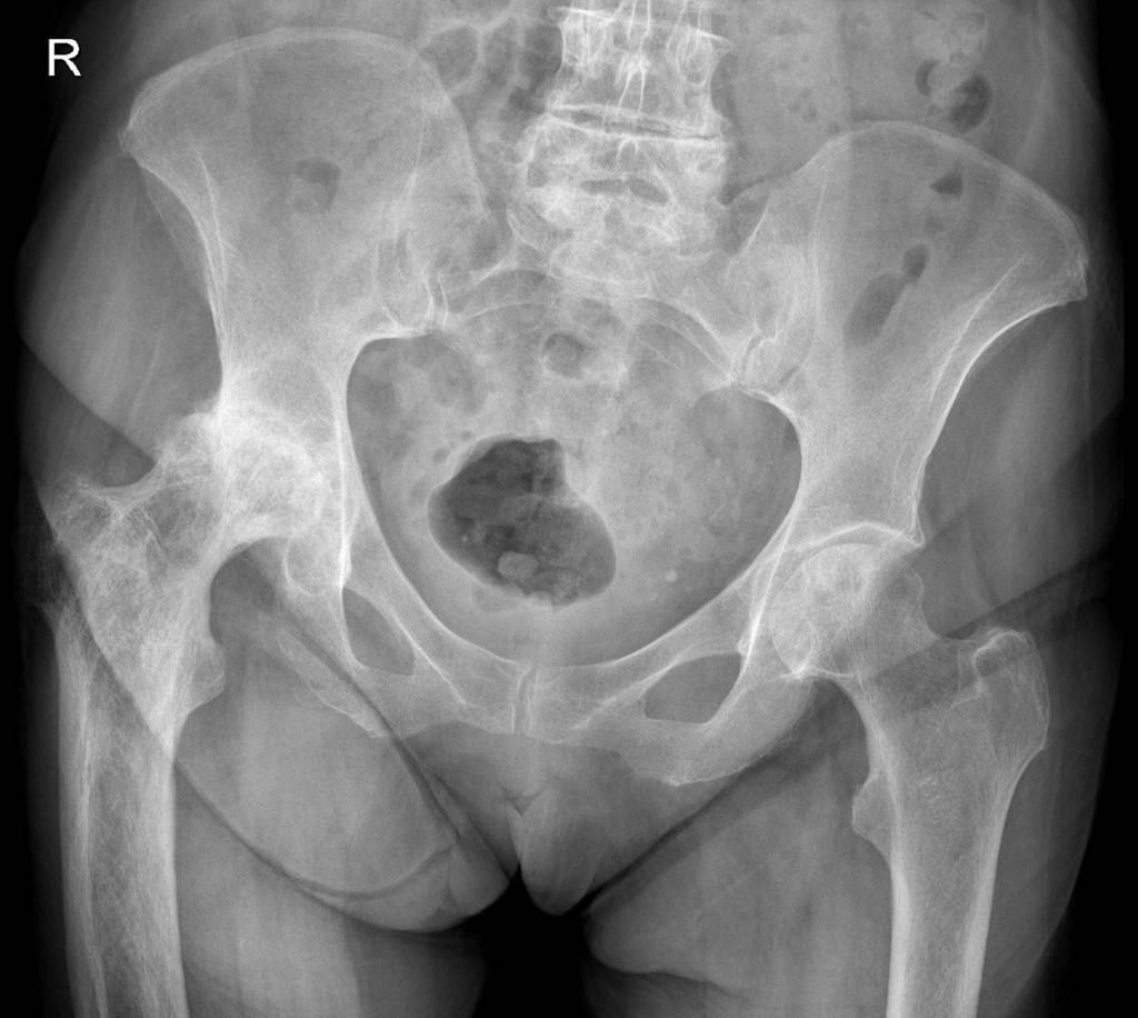
- Coarse trabeculation over the right femur, enlargement of bone and varus deformity
- Thickened iliopectineal and ilioischial lines (Brim sign)
- OA change; subluxation with disruption of Shenton's line
- Diagnosis: Paget's disease
Q60What is the differential diagnosis of these X-ray changes?▸
- Sclerotic bony metastases
- Renal osteodystrophy
- Fibrous dysplasia, multiple myeloma, lymphoma
- Myelofibrosis
Q61What investigations are helpful in Paget's disease?▸
- Raised ALP with normal calcium
- Urine hydroxyproline also raised
- XR of whole lower limb: look for stress fracture
- CT: morphology
Q62What is Paget's disease?▸
- Disorder of high bone turnover
- Abnormal osteoclastic bone resorption with uncoupled osteoblastic bone formation
- Unknown cause: ?paramyxovirus infection in genetically susceptible individuals
- New bone is irregular and woven in nature, less resistant and prone to deformity and fracture
Q63What causes the increased number and activity of Paget's osteoclasts?▸
- Osteoclasts hypersensitive to RANKL and calcitriol
- Marrow stromal cells have increase RANKL expression
- Increased osteoclast precursor recruitment by IL6
- Antiapoptotic oncogene Bcl 2 overexpressed
- Increased proto-oncogene cfos
Q64What are the perioperative considerations for THR in Paget's disease?▸
- Pre-op: ensure the pain generator is the hip (not spinal stenosis, sarcoma or fracture); optimise high output cardiac failure; bisphosphonate (pamidronate) and calcitonin reduce disease activity and bleeding; templating: morphology, ? Need osteotomy
- Intra-op: cell saver, permissive hypotension; bone brittle - risk of iatrogenic fracture
- Acetabulum: protrusio - bone graft, anti-protrusion cage
- Femur: prepare burr for sclerotic and dense bone; Bowing & wide canal: osteotomy/ modular stem; cemented stem has poor interdigitation - trend towards cementless (Parvizi CORR 2002)
- Post-op: HO prophylaxis; Increase risk of osteolysis owing to the increase metabolic turnover of the pathological bone
Q65What history and examination findings should be sought in Paget's disease of the hip?▸
- Clarify symptom: generalised pain / referred pain / Paget's sarcoma / fracture
- Medical treatment; age and functional demand
- PE: ROM, LLD, abduction power
Q66What is the pathophysiology of Paget's disease?▸
- Unknown cause: ?paramyxovirus infection in genetically susceptible individuals; also high arsenic exposure
- Primary abnormality: intense focal resorption by abnormal osteoclasts (abnormal size, activity and quantity) -> large resorption cavities
- Osteoblasts recruited in response but activity is rapid -> disorganised, irregular woven bone
- Woven bone is less resistant and more elastic than lamellar bone -> prone to deformity and fracture
Q67What are the issues with cemented stems in Paget's disease and what is the current trend?▸
- Pathological bone -> poor ossointegration for cementless; bleeding not good for cement interdigitation
- Risk of cement extrusion through an osteotomy site
- Trend towards use of cementless in recent years - previous concerns for the problem with osseous integration are mostly unfounded
- Bone ingrowth biology is similar to the early phases of fracture healing; In Pagets, fracture healing is normal (Parvizi CORR 2002)
Fact check
Sciatic nerve injury after THA: 0% palsy if limb lengthening <4 cm and 28% if >4 cm (Kavanagh 1991) — source attribution unverified; length threshold contested — Modern evidence is mixed: a 508-hip DDH series (Eggli 1999) found no correlation between amount of lengthening and nerve palsy, while other series report risk mainly with lengthening >5 cm; a 2017 systematic review notes the 3-4 cm threshold is suggested but debated — (medium confidence) — source