Knee
Unicompartmental knee arthroplasty
Oxford mobile-bearing unicompartmental knee replacement: indications and outcomes.
13 questions
1 source pages
1 images
1 fact-check flags
Images appear with the first question taken from each source page — tap a question to open it.
Q1What are the X-ray findings in this failed Oxford mobile bearing UKA?📷▸
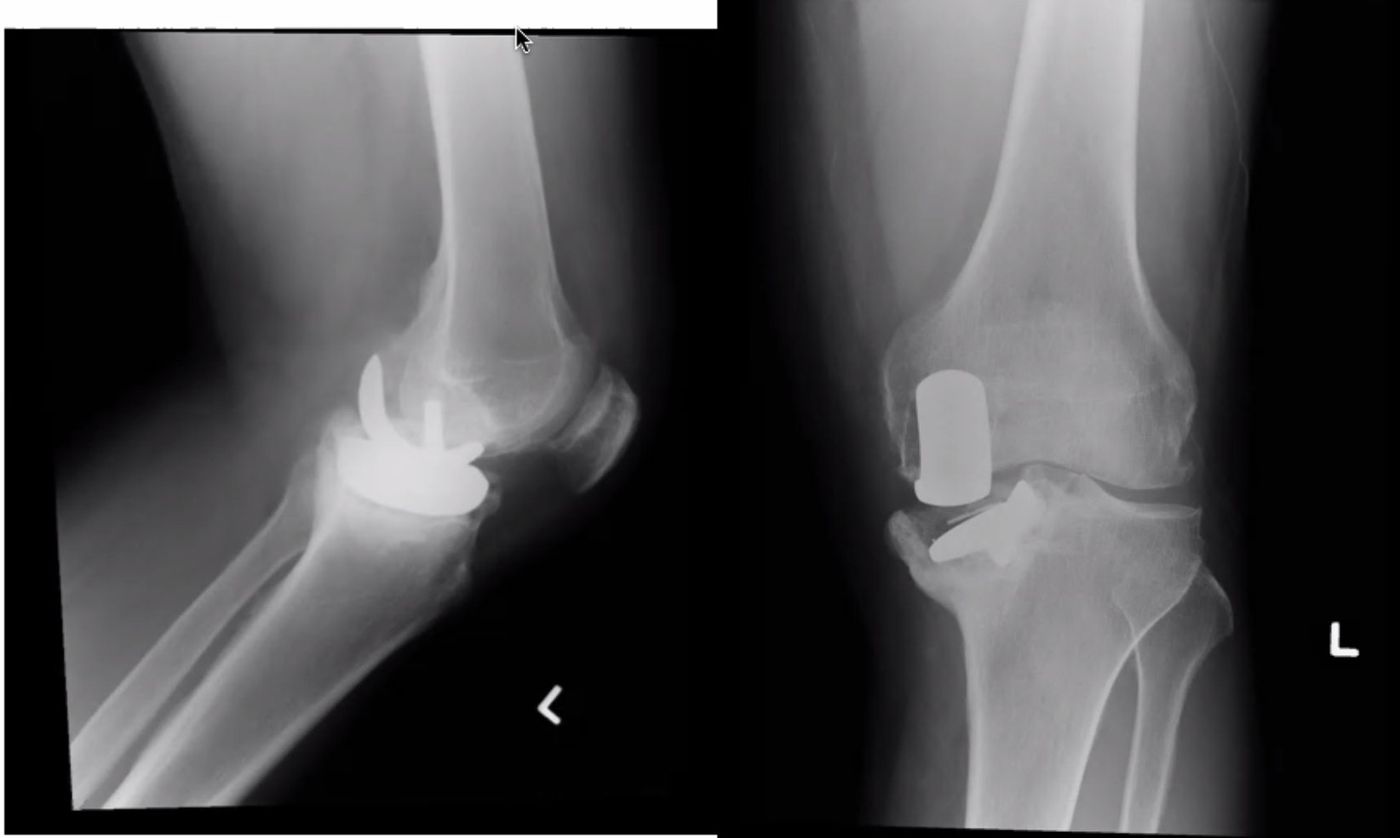
- Loosening of the tibial baseplate with subsidence into varus
- Bearing still on top of the tibial baseplate
- No obvious femoral component loosening
- No lucency or periosteal reaction suggestive of infection
Q2How do you recognise the PE marker on X-ray?▸
- Normal PE marker: line in front, point at back
Q3What history do you take in a failed UKA?▸
- Current symptoms: onset of pain, rest pain, fever or constitutional symptoms
- Functional limitations: OKS
- Details of the previous operation: pre-op indications, intra-op problems and implant sizing, post-op compliance and delayed wound healing
- Other risk factors: obesity, inflammatory joint disease; premorbid state and PMHx
Q4What do you examine in a failed UKA?▸
- Local: sinus, varus/valgus laxity, ROM
- Systemic: is the patient fit for surgery
Q5What investigations are needed for a failed UKA?▸
- WCC, CRP +/- knee aspiration
- Scannogram to check overall alignment
- CT for bone stock
Q6How would you revise a failed UKA?▸
- Pre-op CT to measure bone defect, prepare bone and wedge
- Same incision, raise a full thickness skin flap below the fascial layer; extensile approach (quadriceps snip) if needed
- Remove the implant first (tibia usually easier; femur with ossilating saw (oscillating saw) + flexible osteotomy)
- Take at least 5 intra-op samples at the bone-implant junction for frozen section
Q7What are the anticipated difficulties and solutions when revising a UKA?▸
- Soft tissue: valgus-varus balance - standby constraint; gap balancing, build up bone defect with augment/cones
- Bone: bone defect - use cone or wedge to restore bone stock; implant fixation by zonal fixation
Q8What are the risk factors for bearing dislocation after mobile bearing UKA?▸
- Patient: compliance, infection
- Implant/surgeon: poor tension (MCL, ACL), poor alignment (varus/valgus, rotational), impingement, implant too small/deep flexion
- Too loose spins out; too tight impinges and rotates out
Q9What is the dislocation rate of the Oxford mobile bearing UKA?▸
- 1 in 200 (0.5%) in medial UKA
- 10% in lateral UKA
Q10What is the soft tissue tension principle for bearing stability in UKA?▸
- Too loose -> spin out
- Too tight -> impinge and rotate out
- When there is a discrepancy between 90 degree flexion and deep flexion, use a larger femur implant with a thinner insert
Q11What causes impingement of the mobile bearing?▸
- Cam impingement - the insert hits the medial side of the tibial implant
- Cement, meniscus or osteophytes
- Undersized femur implant due to retained cartilage / osteophytes over the posterior condyle
Q12What happens to the MCL in UKA?▸
- Acute MCL and chronic MCL (overstuff) problems
- The MCL elongates and loosens; the lateral compartment is under stress and develops OA
Q13Why is ACL deficiency a contraindication for UKA?▸
- ACL deficiency causes posteromedial OA from anterior subluxation of the tibia
- In an ACL intact knee the posteromedial cartilage retensions the MCL
- In posteromedial OA the MCL cannot regain tension and becomes contracted
Fact check
Bearing dislocation is 1 in 200 (0.5%) in medial UKA and 10% in lateral UKA — lateral figure outdated/context-specific — 10% applies to the original flat lateral Oxford bearing; the modern domed lateral design has a lower rate of about 1-6% (systematic review 3.7%) — (medium confidence) — source