Knee
TKR in varus and valgus knees
Surgical technique, bone cuts and soft-tissue management for varus and valgus deformity.
25 questions
2 source pages
2 images
Images appear with the first question taken from each source page — tap a question to open it.
Q1What does the pre-op X-ray show in this varus knee?📷▸
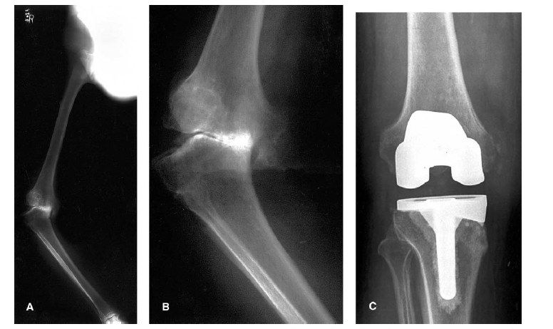
- Long standing and AP XR of the right knee showing a varus knee
- Tibiofemoral angle 45degs
- Medial tibia bone loss
- Widening of lateral joint space
- Tibia appears ER
Q2What are the problems to plan for when performing TKR in a varus knee?▸
- Soft tissue deficiency - plan extensive soft tissue release, constrained implant
- Bony deficiency (contained/uncontained) - cement, cancellous graft, screw on cement, cortical graft, metal augment, tumour prosthesis
- Alignment - pre-op standing scannogram to plan tibial/femoral cuts and guides (intramedullary, extramedullary or navigation)
Q3What is the pre-op preparation for TKR in a varus knee?▸
- Optimize patient fitness for operation
- Prophylactic antibiotic
- Same day admission to a clean ward
- The most important pre-op part is planning (soft tissue, bony, alignment)
Q4What is the sequence of medial release in a varus knee TKR?▸
- Osteophyte
- Medial joint capsule and deep MCL
- PMC (capsule, semimembranosus, posterior oblique ligament)
- Superficial MCL (subperiosteal release / pie crusting)
- Pes anserius (pes anserinus)
- Downsizing tibial side + removing uncapped part; if not balanced use a semi constrained implant (lateral epicondyle advancement / distalize fibular head)
Q5How do you manage a fixed flexion deformity in TKR?▸
- Extension gap tight, flexion gap normal
- Post osteophyte --> post capsule
- May cut more distal femur initially (e.g. 10mm vs normal 8mm)
Q6What is the Anderson classification of bone defects in TKR?▸
- 1: does not affect stability
- 2: affects stability (metaphyseal damage) - cement, bone graft, metal wedge
- 3: deficient metaphysis affecting collateral/patellar tendon attachment - struct allograft, IBG, trabecular metal augment, tumour prosthesis
Q7How are contained and uncontained defects managed by size?▸
- Contained: <5mm cement; large defect cancellous bone graft
- Uncontained: <5mm cement; 5mm-1cm bone graft/screw on cement; >1cm augment/strut graft + screw
- Cannot use cement alone in a large defect (may crack)
Q8How is a tibial bone defect managed during TKR?▸
- Translate the tibial tray away from the defect
- Resect the tibia at a lower level
- Reassess the defect after the tibial cut: <5mm cement, 6-10mm bone graft, >10mm metal wedge
Q9How is a femoral bone defect managed during TKR?▸
- Affecting the chamfer cut - cement
- Affecting the distal femur cut - metal augment
- Massive bone loss - bone block / allograft
Q10What are the pros and cons of bone grafting and the role of a long stem in TKR?▸
- Bone graft replenishes bone stock but has donor morbidity
- Young age tends to preserve bone stock
- Use a long stem tibial component to share stress over the diaphysis (20% stress)
Q11What are the post-op concerns and prophylaxis after TKR?▸
- Chest infection, wound infection, UTI, DVT, knee stiffness
- Post op: Early mobilization (early mobilisation), prophylactic antibiotics, wean off foley as early as possible
- DVT: mechanical +/- medical prophylaxis (enoxaparin, pradaxa) in high risk group
Q12What are the X-ray findings in this valgus knee?📷▸
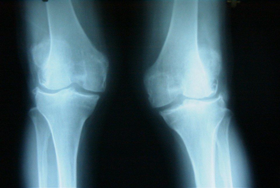
- Bilateral valgus knee, no obvious fracture
- Bone quality good, joint space preserved - no evidence of RA
- Hypoplastic lateral femoral condyle with increased lateral distal femur angle
- Patella lateral subluxation
Q13What are the causes of a valgus knee?▸
- Rheumatoid arthritis
- Post-traumatic
- Iatrogenic (HTO)
- OA with hypoplastic lateral femoral condyle / constitutional valgus
- Haemophilia (Hemophilia), polio, HME
Q14What history do you take in a valgus knee?▸
- Enquire causes (RA, post-traumatic, iatrogenic HTO, OA with hypoplastic lateral femoral condyle/constitutional valgus, haemophilia, polio, HME)
- Disability: KFS
Q15What do you examine in a valgus knee?▸
- Assess patella tracking
- Document peroneal nerve status
- ROM, any FFC
- Collateral laxity
Q16What do you say if a patient with a valgus knee fails conservative management?▸
- "This is a complex primary knee and I would refer to an arthroplasty surgeon"
Q17What is the aim of TKR in a valgus knee?▸
- Stable painless mobile knee
- Restore joint line and mechanical axis
- Restore sagittal and coronal gap balance
- Choose cemented PS knee, standby constraint implant
Q18What are the approach options and pitfalls in valgus knee TKR?▸
- Medial parapatellar: familiar and good exposure, but poor PL exposure, disrupt VMO cause patellar tracking problem, devascularises skin/patella if lateral release needed
- Lateral parapatellar: for a large lateral bone defect needing lateral exposure for augment/cone
- Need a coronal Z plasty
- Prepare distal femur augment for the hypoplastic lateral condyle
Q19What bone problems are encountered in valgus knee TKR?▸
- Hypoplastic lateral femoral condyle / peripheral uncontained defect - use TEA + Whiteside as reference, prepare distal femur augment
- Lateral tibial plateau defect - usually central contained: minimal bone cut, reference off the medial side 5mm
Q20How is coronal imbalance managed in valgus knee TKR?▸
- Contracted lateral soft tissue with attenuation of medial soft tissue
- LFE sliding osteotomy or MCL advancement
- Semiconstraint implant standby
Q21How is sagittal balance managed in valgus knee TKR?▸
- Release osteophyte, PL capsule first
- Then depends on tightness: F: popliteus; E: ITB
- Both: piecrust LCL
Q22How is patella tracking managed in valgus knee TKR?▸
- Lateral retinacular release (1cm from the border)
- Medial imbrication
- ER femoral and tibial implants
Q23What are the risk factors for peroneal nerve palsy after TKR?▸
- Fixed flexion contracture + valgus
- Valgus deformity >12 degrees
- Epidural anaesthesia for pain control
- Previous laminectomy
- Overall 2% esp pre op valgus >12deg; suggest intra-op avoid excessive release; post op recovery room asssessment; prophylactic exploration is not beneficial
Q24How do you manage acute peroneal nerve palsy after TKR?▸
- Remove the compressive dressing
- Check for haematoma, check the drain
- Flex the knee
- Check anaesthetic notes for blocks used
- Check XR: lateral overhang, cement, sawblade cut at fibula
Q25How do you manage late peroneal nerve palsy after TKR?▸
- AFO to prevent foot drop
- Regular follow-up, NCT to document recovery
- +/- exploration if no improvement (controversial)