Basic Science Trauma
Intramedullary nails - types and design
Tibial, Kuntscher, centromedullary, cephalomedullary, Rush pin and Ender nail design and uses
39 questions
6 source pages
2 images
Images appear with the first question taken from each source page — tap a question to open it.
Q1Describe the AO UTN tibial nail.▸
- Slot: anterior
- Herzog bend: 11 degree, proximal 1/3
- Entry: just distal to the tibial articular surface
- Cross-section: cloverleaf; material: stainless steel
- Proximal: 3 slots - middle dynamic; distal: transverse x2, AP x1
Q2What are the special features of the AO UTN tibial nail?▸
- Dovetail proximal - increases torsional rigidity
- Beveled proximal anterior - decreases irritation to the patellar tendon
- Beveled distal posterior - slides smoothly along the dorsal tibial canal, prevents jamming
Q3Describe the Stryker GK tibial nail.▸
- Slot: posterior
- Herzog 15 degrees, proximal 1/5
- Entry: just proximal to the tibial tuberosity (TT)
- Cross-section: cloverleaf; material: stainless steel
- Proximal: transverse x1, AP x1; distal: transverse x2
Q4Compare the AO UTN and Stryker GK tibial nails.▸
- Slot: AO UTN anterior vs Stryker GK posterior
- Herzog bend: AO UTN 11 degrees (proximal 1/3) vs Stryker GK 15 degrees (proximal 1/5)
- Entry: AO UTN just distal to tibial articular surface vs Stryker GK just proximal to TT
- Both are cloverleaf, stainless steel; AO UTN proximal 3 slots (middle dynamic), distal transverse x2 + AP x1; Stryker GK proximal transverse x1 + AP x1, distal transverse x2
Q5What is the framework for describing an intramedullary nail?▸
- Intramedullary nail used for fracture fixation of the femur
- Biological reduction + relative stability and secondary bone healing
- Describe: longitudinal, transverse profile, material, interlocking, reamed/unreamed
- Example given: Stryker GK nail
Q6What are the pros of reaming?▸
- Decrease working length
- Increased width of nail = more rigid
- Provides autogenous bone graft
- Decreases hoop stress generated - less fracture and breakage of implant than an unreamed nail
Q7What are the dangers/cons of reaming?▸
- Disrupted blood supply
- Thermal necrosis
- Pressure -> fat embolism
Q8What constitutes proper reaming technique?▸
- Patient selection
- Equipment: sharp flute with bixcut tip, good clearance by small diameter of flexible shaft keeps pressure low
- Technique: slow advancement, high reaming speed, serial increment, venting
Q9What is the working length in a nail and what factors affect it?▸
- Length of implant spanning the fracture that remains unsupported
- Factors: fracture pattern; construct (nail size, bone contact, locking); type of force
Q10Compare bending and torsional working length.▸
- Bending working length = unsupported area; bending stiffness inversely proportional to square of working length
- Torsional working length = distance between locking bolts; torsional stiffness inversely proportional to working length
Q11What are the three generations of intramedullary nails?▸
- 1st gen: internal splint, minimal rotational stability, close fit (e.g. Kuntscher nail / V nail)
- 2nd gen: locking screw, increased rotational stability (e.g. Russell Taylor nail)
- 3rd gen: anatomical design for a specific bone, increased stability and easier insertion
Q12What are the advantages of interlocking?▸
- Increase torsional rigidity
- Increase bending rigidity
- Control length in comminuted fracture
Q13How can the rigidity of an intramedullary nail construct be increased?▸
- Reaming to allow a wider nail = more rigid
- Interlocking increases torsional and bending rigidity
- Decrease working length (more bone contact)
Q14Describe the general design of the Kuntscher nail.📷▸
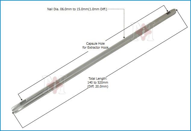
- Stainless steel
- Straight (3-point fixation)
- No interlocking
- Unreamed
Q15What are the effects of the cloverleaf cross-section of the Kuntscher nail?▸
- Increases bending stiffness (higher 2nd moment of inertia)
- Increases rotational control of the fracture
Q16What are the effects of the slot on the Kuntscher nail and where is it placed?▸
- Radial compliance -> better interference fit
- decrease torsional strength
- Place over the tension side
Q17What is the principle of fixation of the Kuntscher nail?▸
- Tight fitting between the elastic nail and stiff bone
- The elastic nail is compressed upon insertion and returns to its original shape, eliminating lateral translation
Q18What are the advantages and disadvantages of the Kuntscher nail?▸
- Advantage: increased callus (elastic)
- Disadvantage - slotted: poor torsional strength
- Disadvantage - telescoping: loose axial coupling between nail and bone if not a tight fit
Q19What is the indication for a Kuntscher nail?▸
- Transverse / short oblique fracture
- Minimal shortening
- Fracture ends interdigitate - rotation instability
Q20Describe the Stryker GK centromedullary nail.📷▸
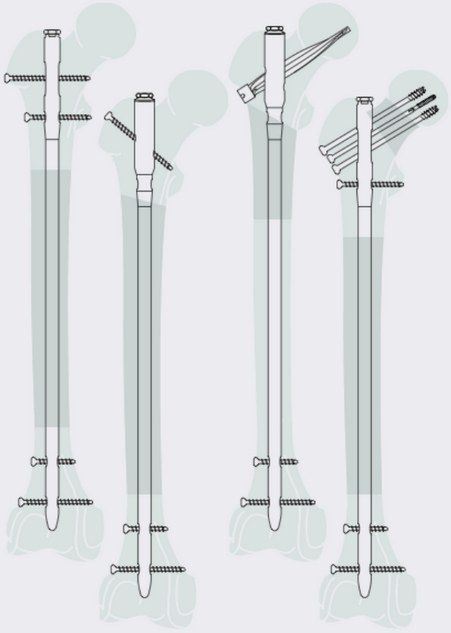
- Straight nail
- GT entry
- Cloverleaf cross-section, posterior slot
- Proximal: diagonal locking; distal: transverse locking
Q21Describe the old AO UFN design.▸
- Slot on the anterior side
- Entry: piriformis entry
- Material: stainless steel
- Anterior curve 1.5m
- Proximal transverse x2; distal transverse x2
- Cross-section: cloverleaf
- Special feature: dovetail proximal
Q22Describe the new AO UFN design.▸
- Unslotted
- Entry: piriformis entry
- Material: titanium alloy
- Anterior curve 1.5/2.0m
- Proximal: transverse x2, 130 degree antegrade, spiral blade; distal: transverse x2
- Cross-section: grooved
- Special: solid, unreamed
Q23What is the purpose of the dovetail proximal feature on the AO UFN?▸
- Prevents spreading of the proximal end when connected to instruments
Q24Compare slotted and non-slotted nails.▸
- Slotted nails allow more flexible insertion (at the site and during insertion)
- Cloverleaf is usually combined with slotting - radial compliance during insertion allows a bigger nail with maximum 3-point endosteal bone contact, decreasing working length -> increasing bending stiffness
- Cons of slotting: poor torsional stiffness (40x), difficult targeting during distal locking
Q25What type of nail is the Gamma nail and what is its indication and material?▸
- Third generation anatomical nail
- Indication: femur fracture
- Material: titanium
Q26Describe the frontal, side and cross-sectional geometry of the Gamma nail.▸
- Frontal view: tapered distal to the lag screw hole (15.5 diameter, allowing a small incision) and also distally
- Frontal view: ML curve 4 degrees
- Side view: anterior curve 3.0mm, radius 1.5m
- Cross-section: unslotted; cannulated
Q27What are the locking features and technique for the Gamma nail?▸
- One lag screw proximally
- 2 distal locking transverse screws (oblong x1 for axial dynamization)
- Technique: GT entry, reamed/unreamed
Q28Describe the design features of the sliding hip screw.▸
- Load transfer with a large thread area
- Reverse buttress thread to reduce cut out
- Asymm depth of groove (asymmetric depth of groove) -> allows lateral page only
Q29What is the role of the set screw in a cephalomedullary nail?▸
- Decreases sliding hip screw rotation
- Decreases hip screw medialization
Q30What type of nail is the Russell Taylor nail and what are its general features?▸
- 2nd generation interlocked nail
- Indication: femur fracture
- Material: stainless steel
- Frontal view: straight nail; side view: anterior curve 2.3m
- Cross-section: unslotted, cannulated, fluted
Q31What are the screw holes and entry technique for the Russell Taylor nail?▸
- 2 proximal sliding hip screw holes
- 4 distal locking screw holes
- Technique: piriformis fossa entry
Q32What are the features of the Enders nail?▸
- Body: flat, C-shaped
- Lead end: blunt and bevelled
- Drive end: flattened eyelet, perpendicular to the curve
Q33What is the indication for an Enders nail?▸
- Initially: TOF fracture
- Later: narrow canal diaphyseal fracture
Q34How is an Enders nail inserted in the femur?▸
- MFC entry point, through the medial femoral cortex
- To within 1.5cm subchondral to the femoral head
- Entry point roof tile
- Insert >3 nails (3-point fixation)
- Tips fan out in AP and lateral
Q35What are the complications of Enders nails?▸
- Fall into bone
- Back out -> impinge on soft tissue
- Joint penetration
Q36What are the features of the Rush pin?▸
- Circular and straight
- Lead end: sharp and bevelled
- Drive end: hooked
Q37What is the indication for a Rush pin?▸
- Narrow canal diaphyseal fracture
Q38How is a Rush pin inserted in the femur?▸
- +/- prebend if 3-point fixation is needed
- Open cortex with a reamer (diameter < nail)
- Entry angle <40 degrees
- Driven into bone by a driver extractor
- Snugly down so the hook grasps the cortex firmly
Q39What are the complications of Rush pins?▸
- Iatrogenic fracture
- Fracture distraction
- Loss of reduction