Soft Tissue
Synovial fluid and articular surfaces
Composition and function of synovial fluid and the resting zone of articular cartilage
13 questions
2 source pages
1 images
Images appear with the first question taken from each source page — tap a question to open it.
Q1What are the functions of synovial fluid?📷▸
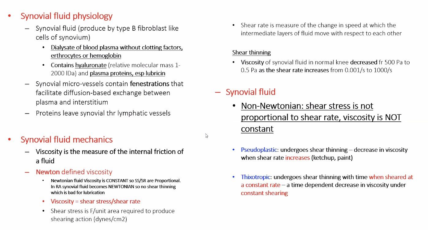
- Lubrication of the joint
- Cartilage nourishment
Q2How is synovial fluid synthesised?▸
- Plasma ultrafiltrate
- Plus secretion from the synovial membrane
Q3What are the two synovial cell types?▸
- Type A - macrophage-derived, involved in phagocytosis
- Type B - fibroblast-like, secrete hyaluronate, fibronectin and collagen
Q4What are the main components of synovial fluid?▸
- Lubricin - provides lubrication
- Hyaluronan - elastic solid during high strain actions
Q5What are the rheological properties of synovial fluid?▸
- Non-Newtonian
- Thixotropic
- Pseudoplastic
Q6Describe the resting zone of the growth plate.▸
- High proportion of ECM to cells, roundish cells
- Cells have high lipid body and vacuole content for later nutritional requirements
- Stem cell characteristics, relatively quiescent with low proliferation
- Low oxygen tension; PTHrP expressing chondrocytes contribute to formation of columnar chondrocytes through the PTHrP –Indian Hedgehog feedback loop)
- Implicated in Gaucher's disease
Q7Describe the proliferative zone.▸
- Chondrocytes are flat and arranged in columns
- Matrix production (type II collagen and proteoglycan) and cell division give longitudinal growth
- High oxygen tension, good blood supply
- Implicated in achondroplasia
Q8What happens in the zone of hypertrophy?▸
- Zone of maturation: Cell division ceases and chondrocytes increase in size 5X; prepare matrix for calcification
- Zone of degeneration: calcium accumulated in mitochondria is released when glycogen is depleted
- Zone of provisional calcification: calcified chondroid matrix becomes scaffold for bone deposition, high ALP
- Implicated in SCFE, rickets, physeal fracture
Q9What forms the primary and secondary spongiosa?▸
- Primary: vascular invagination and woven bone; osteoblasts line up on the calcified cartilage bar (low O2, good blood supply); site of acute osteomyelitis
- Secondary: remodelling - internal (removes cartilage bar and woven bone to lamellar bone), external (funnelisation)
- Implicated in osteopetrosis, osteogenesis imperfecta and scurvy
Q10What are the groove of Ranvier and the perichondral ring of Lacroix?▸
- Groove of Ranvier: contains osteoblasts, fibroblasts and chondrocytes; responsible for appositional growth
- Perichondral ring of Lacroix: dense fibrous tissue anchoring epiphysis to metaphysis
Q11How is the growth plate regulated and what is its blood supply?▸
- Growth factors (IGF, FGF, TGF, PDGF, TNF), vitamins and hormones
- Epiphysis: epiphyseal artery (only to proliferative zone)
- Physis: perichondral artery predominant
- Metaphysis: metaphyseal artery; diaphysis: nutrient artery
Q12What did Urist (Science 1965) discover?▸
- Demineralised bone fragments implanted intramuscularly or subcutaneously induced bone formation
- Led to the discovery of BMP-2 and BMP-7
Q13What are the clinical uses of BMPs?▸
- FDA approved for acute tibial fractures and complex nonunions
- BMP-2 is preferred as it is more effective and less costly