Soft Tissue
Menisci and intervertebral disc
Meniscal anatomy and function, and structure of the intervertebral disc
21 questions
2 source pages
1 images
Images appear with the first question taken from each source page — tap a question to open it.
Q1What are the functions of the meniscus?▸
- Load transmission - increases contact area, decreases peak stress
- Shock absorption via viscoelasticity
- AP stabilisation
- Lubrication and nutrition
- Proprioception (mechanoreceptors in the meniscal horns)
- Mnemonic SSLLA
Q2How much compressive load does the meniscus take up?▸
- 60% of compressive load in extension
- 90% of compressive load at 90 degrees of flexion
Q3What is the composition and macrostructure of the meniscus?▸
- Cells: fibrochondrocytes (middle/inner), fibroblast-like cells (outer half), superficial zone cells
- ECM: water 70%, collagen 22% (outer type I, inner type II), proteoglycans, non-collagen proteins
- Superior superficial layer (random fibres); superior lamellar layer (radial fibres)
- Deep layer - fibres run circumferentially to resist hoop stresses
- Inferior lamellar layer (radial and random); inferior superficial layer (radial)
Q4Describe the lateral meniscus.▸
- O-shaped, covers 80-85% of the lateral tibial plateau, takes 70% of compartment load
- 40-50% contact area reduced after meniscectomy, 200% increase in stress
- Anterior horn just posterior to the ACL insertion, to which it partially blends; posterior horn anterior to the MM posterior horn
- No attachment to the LCL; loose attachment to the capsule
Q5Describe the medial meniscus.▸
- C-shaped, covers 60-65% of the medial plateau, takes 50% of compartment load
- Larger AP diameter than width
- 50-70% contact area reduced after meniscectomy, 100% increase in contact stress
- Anterior root 7 mm anterior to the ACL; posterior root anterior to the PCL
- Attaches to the deep MCL and capsule via the coronary ligament
Q6What are the meniscal ligaments?▸
- Insertional ligament (meniscotibial ligament)
- Intermeniscal ligament
- Meniscofemoral ligament
- Attachment to the deep medial collateral ligament
Q7What is the blood supply of the meniscus?▸
- Blood supply reaches the periphery from perimeniscal plexus (lateral and medial inferior genicular arteries), supplying the peripheral 10-30%
- Central 2/3 nourished by diffusion
- Posterior horn supplied by the middle geniculate artery
- Zones: red-red, red-white, white-white
Q8How does the meniscus bear load?▸
- Compressive axial forces are converted to a radially directed force taken up as circumferential hoop stress
- Radial fibres act as intrasubstance tie-rods resisting longitudinal splitting and excessive compression
- The hoop must be complete - intact circumference and bony attachments
- Tensile modulus: Hoop 110Mpa, Radial 10Mpa
- Barrel analogy: the wedged cross-section extrudes radially, increasing circumference and generating hoop stress
Q9How does the meniscus absorb shock?▸
- Intact menisci dissipate force as biphasic structures
- Reduce 20% of peak force on articular cartilage and bone by increasing TFJ conformity
- The collagen-GAG network resists water movement through the solid phase
- Meniscal tissue is less stiff than articular cartilage because of the lower concentration of proteoglycans; Meniscal tissue is more resistant to the internal movement of water through its tissue than articular cartilage
Q10How does the meniscus provide lubrication and nutrition?▸
- By increasing joint congruity it allows better fluid entrainment and hydrodynamic lubrication
- The sponge phenomenon helps circulate cellular nutrients throughout the joint
Q11How does the meniscus stabilise the knee?▸
- Medial meniscus posterior horn resists anterior tibial translation in an ACL-deficient knee
- Meniscofemoral ligaments are secondary restraints to posterior drawer
- The meniscus construct is a restraint to tibial rotation
Q12What contact area and stress changes follow partial and total meniscectomy?▸
- Partial meniscectomy of the inner third: contact area -10%, peak load +65%
- Total meniscectomy: contact area -75%, peak load +235%
Q13What is the association of SONK with meniscal root tears?▸
- SONK (spontaneous osteonecrosis of the knee) has been suggested to relate to root tears
- Female:male ratio 3:1
Q14What are the functions of the spine and intervertebral disc?📷▸
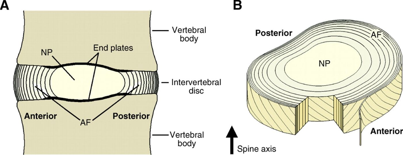
- Spine: carries loads, protects neural elements, supports posture and allows locomotion
- Disc resists compression - nucleus pulposus converts compression into radial force, resisted by annulus fibrosus hoop stress
- Resists bending, shear and torsion (AF); restricts excessive motion
- Shock absorption via viscoelastic NP (time-dependent strain behaviour)
Q15What are the components of the intervertebral disc?▸
- Nucleus pulposus - derived from notochord; mucoprotein gel (water and matrix), chondrocyte -like cells, PG aggregates in a type II collagen network
- Annulus fibrosus - derived from sclerotome; high type I collagen to PG ratio, fibroblast-like cells
- Endplate - semipermeable membrane allowing nutrients and metabolites to diffuse through marrow cavities in the subchondral bone
Q16Describe the structure of the annulus fibrosus.▸
- Anchored to the cartilaginous endplate (inner zone) and attached into osseous tissue through Sharpey fibres (peripheral zone)
- Outer 1/3: concentric oblique fibres at 30 degrees, herringbone pattern, 15-25 lamellae with elastic fibres between
- Inner 2/3: less dense type II collagen matrix lacking lamellar organisation
- Resists hoop stress, distraction and shearing forces in different directions
Q17What is the blood and nerve supply of the disc?▸
- Nutrition mainly by diffusion through the endplate, which is lined by hyaline cartilage
- Only the outer annulus is innervated: sinuvertebral nerve (posterior/posterolateral), grey ramus communicans (lateral), Sympathetic ganglion of the sympathetic trunk (anterolateral disc), sympathetic branches (anterior disc)
- Nerve fibres converge at the DRG
Q18What happens in disc aging?▸
- Decreased vascularity of endplates -> reduced nutritional supply
- Decreased synthesis and increase in proteolytic degradation(different in joint) (e.g. MMP) -> reduced PG concentration
- Loss of water retention and normal biomechanical response to loading (more anisotropic stress state with a more non uniform distribution of stresses)
- Increased type I:II collagen ratio and increased keratin:chondroitin sulphate ratio
- Conversion to fibrocartilage, increased elastic modulus (stiffer), less distinct NP/AF demarcation
- Macroscopic: loss of disc height and dehydration, annular delamination and concentric tears, endplate sclerosis
Q19How do disc aging and degeneration differ?▸
- Both share similar biomechanical alterations and both start with diminished blood/nutritional supply and waste accumulation from reduced endplate porosity
- degen has more distinct initiating factor: mechanical disc overload, genetic factors, immobilisation
- Mechanism: increased load + decreased mechanical strength of the AF
Q20Describe the types of disc herniation.▸
- Bulging - AF intact
- Protrusion - AF partially intact (base > length)
- Extrusion - AF not intact, not bound by AF (base < length)
- Sequestration - fragment separated
Q21Why is discitis more common in children?▸
- Blood vessels cross the endplate and end in the annulus up to late teens
- Explained by Rudert, JBJS 1993