Soft Tissue
Cartilage structure and composition
Collagen types, chondrocyte and fibre differences, perichondrium and cartilage zones
27 questions
3 source pages
2 images
Images appear with the first question taken from each source page — tap a question to open it.
Q1Describe the synthesis of collagen.📷▸
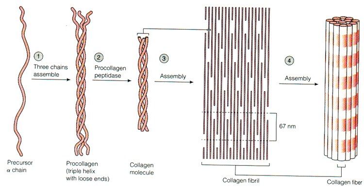
- Amino acids such as glycine-proline-X or glycine-X-hydroxyproline form procollagen polypeptide chains in the endoplasmic reticulum
- Hydroxylation and glycosylation allow three polypeptide chains to form a right-handed triple superhelix (two alpha-1 and one alpha-2 chain)
- Cleaved extracellularly to become tropocollagen, then arranged into quarter-staggered arrays by cleavage of the N and C terminal propeptidases -> collagen microfibrils
- Aggregation of collagen microfibrils by formation of covalent crosslinks by lysyl oxidase -> collagen fibres
Q2What is the collagen chain sequence shown on the source and which structures does it form?▸
- Gly-X-Y-Gly-X-Y-Gly-X-Y
- -> procollagen polypeptide chains (e.g. pro alpha chains) -> procollagen (triple helix) -> collagen microfibril -> collagen fibre
Q3What proportion of whole body proteins is collagen?▸
- 25% of whole body proteins
Q4What are the features of type I collagen?▸
- 90% of total collagen
- Bone, tendon, ligament and skin
- Annular fibrosis
- Associated with OI (osteogenesis imperfecta) and EDS (Ehlers-Danlos syndrome)
Q5Where is type II collagen found?▸
- Hyaline cartilage
- Nucleus pulposus
Q6Where is type III collagen found and what is its role?▸
- Damage and repair collagen
- Skin and blood vessels
- Dupuytren contracture and frozen shoulder
Q7Where is type IV collagen found and with what is it associated?▸
- Basement membrane (BM)
- Nephrotic syndrome
Q8Where are collagen types V, VI, XI and X found?▸
- V, VI, XI - cartilage (articular surface) and MED (multiple epiphyseal dysplasia)
- X - cartilage (deep calcified layer) and early OA
Q9What are the features of articular cartilage?📷▸
- Avascular, aneural, alymphatic, non-immunogenic
- Decreases friction (coefficient 0.002) - 3x more slippery than ice on ice
- Distributes load - resists tensile, shear and compressive forces
- Poor intrinsic healing capacity; nutrition via diffusion from synovial fluid
- Maintains joint congruence and absorbs shock
Q10List the types of cartilage with examples.▸
- Hyaline - nasal, trachea
- Fibrocartilage - ACJ, SCJ, IVD, labrum, meniscus (less water, fewer cells, more matrix)
- Elastic - ligamentum flavum, epiglottis
- Hyaline is the most common and forms articular surfaces, costal cartilages and growth plates
- Cartilage type reflects function: fibrocartilage resists tension, elastic maintains shape (e.g. ear, epiglottis)
Q11What is the composition of articular cartilage?▸
- Cells 5% (chondrocytes) - synthesise matrix
- Water 75%; ECM 20%
- Collagen 10% (type II in hyaline) - resists tensile forces
- Proteoglycan 10% - resists compressive force
- Matrix glycoproteins (fibronectin, chondronectin), degradative enzymes
- Solid matrix = collagen + proteoglycan; fluid phase = water + dissolved ions
Q12What are the biomechanical properties of cartilage and what explains the biphasic nature?▸
- Anisotropic, biphasic, viscoelastic
- Biphasic = water-soaked sponge: solid collagen-proteoglycan phase plus fluid phase (permeability)
- Under high compression, water movement is hindered by frictional drag, reduced porosity and negative PG charges, giving greater stiffness
- Under tension, the crosslinks of the collagen fibres are pulled apart, increasing the water permeability, which leads to decrease in stiffness
- Viscoelastic = sensitive to the speed and duration of loading
- Creep occurs with sustained load; stress relaxation reduces force under constant deformation
Q13Describe the zones of articular cartilage.▸
- Superficial gliding layer 20%: flat chondrocytes (highest number), tangential collagen, highest collagen and water, lowest PG, Water squeezed out to produce squeeze film lubrication
- Middle transition layer 50%: round chondrocytes, oblique fibres at 90 degrees, higher PG
- Deep radial layer 30%: columnar chondrocytes, Collagen fibres are largest diameters, vertically oriented and anchored to subchondral bone, Highest concentration of PG, Lowest H2O content
- Tidemark: smooth, undulating basophilic line for transmission of load between cartilage and bone; cell-free undulating calcification front resisting shear; migrates toward the surface with age
- Calcified zone: collagen X and hydroxyapatite anchor cartilage to subchondral bone with a gradual transition of mechanical properties
Q14What is the lamina splendens?▸
- No cells - a thin layer of collagen fibrils with little proteoglycan
- A cellular layer of flattened chondrocytes 1-3 layers thick
Q15How are load and stiffness transitioned from cartilage to bone?▸
- Calcified zone anchors the layers using collagen X and hydroxyapatite crystals
- Allows gradual transition of mechanical properties between cartilage and bone
- Subchondral and cancellous bone give a gradual transition of Young's modulus to evenly distribute load
- The fibre architecture is described as the arcades of Benninghoff
Q16How does cartilage retain water and what happens in OA?▸
- The negative charge of the proteoglycan creates a repulsion force which is neutralized by the positive ions in the surrounding fluid (i.e. water). The ionic pressure creates swelling pressure which will keep soaking up water until it is resisted by the tension of the collagen fibres
- In OA the collagen network degrades, water content increases, and elasticity/elastic modulus decrease
- Injury beyond the tidemark bleeds and heals with fibrocartilage (type I and II collagen)
- Water content is highest in the superficial zone and decreases with depth; PG concentration shows the opposite gradient
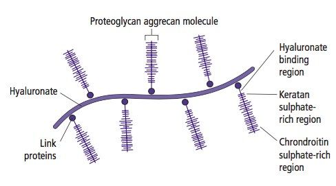
Q17What is the structure of a proteoglycan?▸
- Hyaluronate backbone with many GAG side chains, secreted by chondrocytes
- Link proteins connect onto the protein core: G1, G2 near the N terminus (HA backbone) and G3 near the C terminus
- Keratin sulfate (shorter, closer to HA) and chondroitin sulfate joined by sugar bonds
- The whole structure is called an aggregate
Q18What are the functions of proteoglycans in cartilage?▸
- Fill the interstices between collagen fibrils
- Electrostatic repulsion maintains tension between fibrils, providing compressive strength
- Traps water due to the hydrophilic aggrecan
Q19Describe the microscopic changes of osteoarthritis.▸
- OA (osteoarthritis) is a non-inflammatory joint disease characterized by cartilage loss, new bone formation and capsular fibrosis; Imbalance between repair and degradation
- Abnormal mechanical loading -> chondrocyte damage -> production of proteases, MMP, IL1
- Collagen meshwork breakdown increases permeability
- PG production and degradation both increase but net amount decreases; chains shorten with chondroitin:keratin ratio increased (C4S up, KS down)
- Decreased Young's modulus; stress concentrates on subchondral bone causing subchondral damage
Q20Describe the macroscopic changes of osteoarthritis.▸
- Cartilage: softening, fibrillation, fissures or gross erosions
- Bone: subchondral thickening and osteophytes (metaplasia, endochondral ossification via the Indian hedgehog pathway)
- Synovium: synovial inflammation and joint capsule hypertrophy
Q21What are the changes of cartilage aging (as opposed to OA)?▸
- Decline in the ability of chondrocytes to maintain matrix: density decreases, cell size increases
- chondrocytes become less responsive to the proliferative and anabolic effects of growth factors, with failure of homoeostasis and inability to withstand external mechanical stresses; PG activity decreases
- Marked increase in advanced glycation end products (AGEs) -> more collagen crosslinking -> stiffer, more susceptible to fatigue failure
Q22What are the X-ray findings of osteoarthritis?▸
- Decreased joint space - decreased articular cartilage
- Osteophytes - subchondral bone neovascularisation, proliferation of cartilage and woven bone via endochondral ossification through the Indian hedgehog pathway
- Subchondral cyst - areas of focal bone necrosis
- Subchondral sclerosis
Q23How does articular cartilage respond to injury?▸
- Superficial laceration - does not heal
- Deep laceration - heals with fibrocartilage (type I collagen, no organised zones); haemorrhage -> fibrin clot -> growth factors and fibroblasts
- Contusion - chondrocyte death, matrix damage, fissuring, fibrillation and swelling
Q24What is the function of matrix glycoproteins?▸
- Interact with collagen fibrils and stabilise the matrix framework
- Help chondrocytes bind to matrix macromolecules (type 6 collagen)
- Act as tissue glue binding the various matrix components
Q25What are the matrix regions of cartilage?▸
- Pericellular matrix - plays a role to initiate signal transduction within cartilage with load bearing
- Territorial matrix - protects chondrocytes against mechanical stresses and contributes to resiliency
- Interterritorial matrix - contributes most to the biomechanical properties
- Regions differ in collagen content, fibril diameter/orientation and proteoglycan/non-collagenous protein content
Q26What physiological stress is applied to cartilage?▸
- Cyclic loading (1-5 MPa)
- Moderate frequency (<1 Hz)
- Low rate (<1000 MPa/s)
Q27What structural features differ between zones and types of cartilage?▸
- Type of collagen
- Chondrocyte number
- Size of collagen fibres
- Presence of perichondrium