Hand Soft Tissue
Thumb hypoplasia and radial club hand
Hypoplastic and angulated thumb, radial longitudinal deficiency, radiographic features and deformity
45 questions
6 source pages
1 images
Images appear with the first question taken from each source page — tap a question to open it.
Q1Describe the clinical photo and the likely diagnosis.▸
- Bilateral thumb angulation with the apex radial
- Need an X-ray
- Likely triphalangeal thumb with a delta phalanx (small wedge-shaped ossicle) on the radial side causing angulation
Q2What do you assess and what systemic associations are checked?▸
- Address the patient's complaint and exclude problems: length, instability
- Triphalangism is associated with Holt-Oram, Diamond-Blackfan, Fanconi anaemia, imperforate anus, tibial defects
Q3What is the management of a triphalangeal thumb with delta phalanx?▸
- If small: excise + reconstruct the ligament
- If large: fuse with the proximal or distal phalanx
Q4What is the aetiology of thumb hypoplasia and what examination is needed?▸
- Undergrowth malformation (Swanson). This is due to a undergrowth (according to Swanson); according to OMT (Oberg-Manske-Tonkin), due to a defect in the AER in the limb bud
- Check the rest of the limb for associated longitudinal preaxial deficiency; thenar eminence may be absent
- 60% bilateral; 80% associated with other abnormalities e.g. TAR, Fanconi anemia, VACTERL syndrome
- X-ray to assess bony anatomy and presence of radial deficiency
Q5Describe the LSWAP assessment of the thumb.▸
- Size: nail 70% opposite thumb, 133% index finger girth, nail width 105% of index finger
- Length (reaching IF PIPJ); Alignment sagittal/coronal/axial; Position (abduction, opposition)
- Webspace: I-II IMA 45-55 degrees
- Mobility CMCJ > IPJ > MCPJ; stability UCL > RCL; sensibility, strength, vascularity, growth
Q6Describe Blauth's classification of the hypoplastic thumb.▸
- 1: deficient thumb, extrinsics and intrinsics normal
- 2: deficient intrinsics and collaterals, thumb in adducted position
- 3A: deficient extrinsics, CMCJ stable
- 3B: deficient extrinsics, CMCJ unstable
- 4: floating thumb (pouce flotant); 5: absent thumb/aplasia
Q7What are the aims and principles of management of thumb hypoplasia?▸
- Goal: stable, mobile, sensate, opposable thumb; Lister: position, stability, strength
- 1-3a: deepen webspace +/- soft tissue reconstruction (thenar, extrinsic) +/- stabilise MCPJ (UCL)
- Webspace: Tajima flap, Z-plasty, four-flap plasty; aim 55-60 degrees 1st-2nd MC angle
- Opponensplasty with FDS IV or Huber; release pollex abductus (anomalous FPL-EPL connection 20%)
- 3b-5: ablation +/- reconstruction (toe hand / pollicization)
Q8How is the first MCPJ stabilised in thumb hypoplasia?▸
- Use available local tissue
- Reinforce with FDS IV
- MCPJ chondrodesis (subphyseal arthrodesis) for global instability
Q9What are the four basic surgical principles of pollicization?▸
- Shortening of the index finger ray to a 3-bone system
- Appropriate positioning of the new thumb
- Creation of a wide supple first webspace
- Realignment of the motor system to achieve a good soft tissue balance
Q10Which tendon transfers realign the motor system in pollicization?▸
- EI --> EPL
- EDCII --> APL
- 1st DI --> APB
- 1st PI --> adductor pollicis
Q11What are the principles of pollicization (Buck-Gramcko)?▸
- Severity of radial club hand and thumb hypoplasia does not always correlate; if concomitant problem, wrist first (wrist position needed to balance tendons), thumb later
- Pollicization 1/2-1 year after wrist centralization, before the hand matures at 3 years (Before hand mature @ 3 year)
- Shorten ~1 segment (excise MC shaft, preserve base x insertion of ECRL, preserve head as 'carpal bone')
- Fix in 45 degrees abduction, 100-120 degrees pronation, fix in hyperextend (so will not hyperextend in the end)
- Create webspace; interosseous to abduction (palmar)/adduction (dorsal); shorten extensor, no need to shorten flexor
Q12What are the complications of pollicization?▸
- First webspace contracture
- Stiffness
- Excessive length
- Malrotation
- Lack of opposition
Q13What are the features of radial club hand?▸
- Preaxial longitudinal deficiency with radial deviation of the wrist and radial bowing
- Associated with hypoplastic thumb; comment on the elbow crease
- Failure of formation (Swanson); OMT classification; SHH pathway
- Example: modified Bayne type III
Q14Describe the modified Bayne and Klug classification.▸
- I: deficient distal radial epiphysis
- II: deficient distal and proximal radial epiphysis
- III: partial aplasia
- IV: complete absence (most common)
Q15What history and local examination are required in radial club hand?▸
- Hx: age, developmental Hx, other abnormalities
- Local: presence of radius and radial carpal bones; thumb (size, length, position, webspace, alignment, mobility, stability, sensation, strength, vascularity)
- Elbow range (extension contracture) and bicep function
- Contralateral limb - 50% bilateral
Q16What systemic associations and investigations are needed in radial club hand?▸
- Associations: VACTERL, Holt-Oram syndrome, TAR, Fanconi's anaemia (AR, aplastic anaemia, fatal, needs BMT), trisomy 13/18
- Ix: CBC, renal USG, echocardiogram, spine X-ray
Q17What is the goal of management and when is surgery indicated in radial club hand?▸
- Goal: maximise function; multidisciplinary (paediatrician, physio, ortho)
- Most are non-op: limited life expectancy, well-adapted adults, mild deformity, elbow contracture precludes recon
- Indication for surgery: insufficient carpus support; OT at 6-12 months old
- C/I: lack of elbow flexion or short life expectancy
Q18What are the components of treatment in radial club hand?▸
- Elbow ROM physio/serial casting to preserve ROM; splint taut radial structures from infancy
- Wrist relocation: ulnarization > radialization > centralization (highest recurrence)
- Forearm: radius lengthening + ulnar osteotomy
- Finger pollicization
Q19Describe centralization versus radialization for wrist relocation.▸
- Centralization (at 1 yo): 3rd MC with ulna; radial soft tissue release + tendon transfer; dorsal bilobed flap; single transcarpal K wire
- Release tight radial anlage; tighten ECU, imbricate ulnar wrist capsule, proximalise hypothenar
- Tendon transfer of radial flexor and extensor to ulnar carpus (radial, musculocutaneous nerve and radial artery may be absent)
- Correction limited by the median nerve (pre-op soft tissue distraction lengthening)
- Radialization: 2nd MC with ulna, overcorrect to avoid recurrence, but more pressure on the distal ulnar physis
Q20What are the roles of the multidisciplinary team in radial club hand?▸
- Paediatrician: screen for other organ anomalies
- Physio: maintain wrist and finger motion
- Ortho: deformity correction and preserve forearm growth
- Goal of management is to enhance/maximise the function
Q21What does radial lengthening and ulnar osteotomy involve in radial club hand?▸
- Osteotomy to correct bowing
- +/- lengthening of the radius and ulnar closing wedge osteotomy
- Pin x 3 months (need to be prolonged), then physiotherapy + splintage till adulthood
Q22When is pollicization performed in radial club hand and what are the bone steps?▸
- Hypoplastic thumb addressed around 1 ½ - 2 y.o before hand develop full function
- New thumb tip at PIPJ region (MCPJ becomes CMCJ)
- Reduce 1 phalanx, rotate 140 degrees, cut at MC neck with MCPJ hyperextended
- Plus webspace reconstruction and tendon transfer (abduction/adduction by intrinsic muscles, shortening of extensor tendon)
Q23What is the length of the radius in radial club hand?▸
- Radius is usually 60% of normal length
Q24What nerve, vessel and muscle anomalies occur in radial club hand?▸
- Radial wrist extensors and extrinsic thumb motors usually absent/aberrant
- Radial nerve generally absent below elbow; median nerve and MCN absent
- Radial artery usually absent
- Muscles: absent FPL, FCR, APL, EPL, thenar muscles
Q25A child presents with the hand shown. What findings and syndromic associations should be documented?📷▸
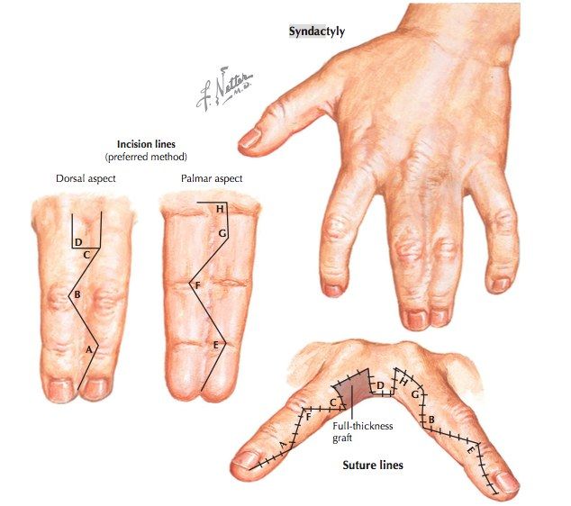
- Immature hand with complete syndactyly of R/F to L/F; nail well formed
- Ulnar deviation of R/F; no brachydactyly
- Screen for associated syndromes: Poland (absent pec major), Apert (craniofacial abnormalities)
- Also consider acrocephalosyndactyly (e.g. Apert) and acrocephalopolysyndactyly (e.g. Carpenter)
Q26How is syndactyly assessed and counselled?▸
- Hx: family Hx, birth Hx; PE: movement, NV status, other limb, Allen test
- X-ray to rule out complex syndactyly
- Single syndactyly unlikely associated (1 in 2000 live births); 30% AD with incomplete penetrance
- Aim: as many independent functional digits as possible, improve hand span and dexterity
- Timing 1-1.5 years; border digits earlier <6 months to prevent angular growth; stage if multiple digits
Q27What are the specific risks of syndactyly release?▸
- Recurrence 8-60% due to web creep
- Need for skin graft
- Injury to NV bundle with necrosis
- Residual angular deformity; nail deformity
Q28Describe the surgical steps of syndactyly release.▸
- Separation with zigzag incision (apex not beyond midline)
- Recreation of commissure with dorsal pantaloon flap or butterfly flap
- Resurface: interdigitating flap radial side, skin graft ulnar side
- Separate bone if complex; nail reconstruction - may consider Buck gramcko flap to recreate the nail fold
- Vessel to one digit; nerve split as proximal as possible; expect 22% skin deficit, FTSG from groin
Q29What is the rule for random flap design in syndactyly release?▸
- Random flap base : height should be 1:1
Q30Classify syndactyly.▸
- Simple: only soft tissue; incomplete (DP spared) or complete (DP involved)
- Complex: side-to-side fusion +/- bone (nail sharing)
- Complicated (accessory digit/ abnormal bone)
Q31Describe Poland syndrome.▸
- Hand + heart (dextrocardia) + chest deformity (also Sprengel)
- Absent pec major sternal head; hypoplasia of breast
- Hand: syndactyly, hypoplasia/absence of metacarpals or phalanges, absent extensors/flexors, carpal coalition, radioulnar synostosis, nail agenesis
- Patho: subclavian artery interruption proximal to internal thoracic artery at 2 weeks
- Surgery: central distraction lengthening; toe-hand transfer for thumb/little finger; release syndactyly
Q32What is the diagnosis and what are the AP X-ray findings?▸
- Diagnosis: Madelung deformity
- Radial shortening and increased radial inclination
- V-shaped carpus
- Carpal ulnar translation
Q33What are the lateral X-ray findings in Madelung deformity?▸
- Volar tilting of the radius
- Dorsal subluxation/dislocation of the ulna
- Volar subluxation of the carpus
Q34What is the differential diagnosis of Madelung deformity?▸
- HME type III
- Malunion
- MED (multiple epiphyseal dysplasia)
- JIA
Q35What are the clinical features of Madelung deformity?▸
- Hx: gymnast? trauma; pain from ulnar impaction; median nerve symptoms (Medial nerve sx?)
- Stiffness: decreased wrist extension, radial deviation and supination
- PE: median nerve, ROM, ulnar impaction signs, tendon attrition
Q36Describe the Vander and Watson classification by aetiology.▸
- 1. Post-traumatic
- 2. Dysplastic (dyschondrosteosis, diaphyseal aclasis aka HME)
- 3. Genetic (e.g. Turner's syndrome, Leri-Weill dyschondrosteosis)
- 4. Idiopathic
Q37How is Madelung deformity managed according to skeletal maturity?▸
- Observe/conservative by avoiding trauma
- Skeletally immature: ulnar epiphysiodesis + radial physeal bar fat interposition (release Vickers ligament, need MRI)
- Skeletally mature: radial corrective osteotomy +/- distal ulnar shortening osteotomy
- DRUJ arthroplasty (very controversial)
Q38Describe the pathophysiology and genetics of Madelung deformity.▸
- AD, female; congenital dyschondrosis of the distal radial physis --> partial growth deficiency --> excessive radial inclination and volar tilt --> ulnar carpal impaction
- Caused by disruption of the ulnar volar physis of the distal radius
- Repetitive trauma/dysplastic arrest - tethered Vickers ligament (thick fibrous band from DR to lunate and TFCC on volar side)
- Leri-Weill dyschondrosteosis: pseudoautosomal dominant, SHOX gene
Q39What are the clinical features of ulnar club hand?▸
- Hand ulnar deviated at the wrist + pronation
- Absent crease at wrist and elbow --> suspect radiohumeral synostosis
- RF and LF syndactyly; +/- loss of ulnar digits
- Hand on flank posture
Q40Describe the Bayne classification of ulnar deficiency.▸
- 0: deficiencies in carpal/hand only
- 1: undersized ulnar with both growth centres present
- 2: partial aplasia
- 3: absent ulna
- 4: radiohumeral synostosis
Q41What are the clinical assessment points in ulnar club hand?▸
- Usually unilateral
- Check elbow ROM
- Assess compensatory wrist and shoulder motion
- Document border syndactyly if present
Q42What are the aims and principles of management of ulnar club hand?▸
- Aim: restore mobility + length; joint stability
- Separate border syndactyly if any at 6 months
Q43How are types 1-3 ulnar club hand managed?▸
- Forearm - ulnar: release ulnar soft tissue, ulnar lengthening; if absent ulna excise fibrous anlage
- Forearm - radius: realignment osteotomy
- Elbow - stable: radial head excision
- Elbow - unstable: radial head excision + one bone forearm
Q44How is type 4 ulnar club hand managed?▸
- Mostly operative as patient is unable to reach the mouth
- Correct pronation and angulation at the radiohumeral synostosis
Q45What is the pathology of ulnar club hand?▸
- Postaxial longitudinal deficiency
- Example: fibular hemimelia