Hand Soft Tissue
Burns, trauma and local flaps
Hand burns and soft tissue coverage, summary of local and regional flaps
29 questions
5 source pages
1 images
Images appear with the first question taken from each source page — tap a question to open it.
Q1What do you look for when describing a burn?📷▸
- Area of burn
- Degree of burn
- Is it circumferential?
Q2What is the emergency concern with a circumferential burn and how is it managed?▸
- Circumferential eschar can cause compartment syndrome
- Requires emergency escharotomy
Q3How is this burn definitively covered and what is the main limitation of the graft?▸
- Debride and achieve coverage with PTSG (pros: large size, easy to take)
- Con: texture
- NO SENSATION in FTSG/PTSG
Q4Define a flap.▸
- A unit of tissue transfer from donor to recipient site
- It maintains its own vascular supply
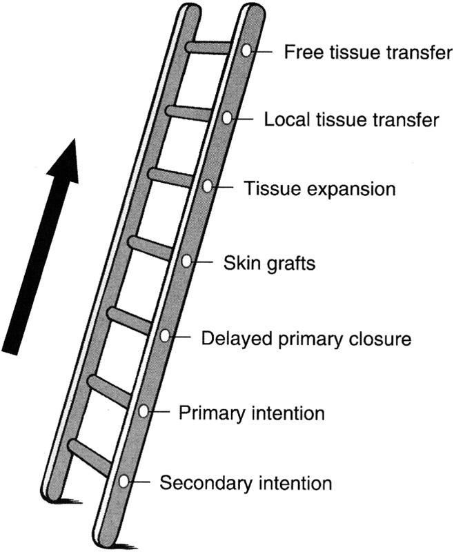
Q5Describe the reconstructive ladder for a hand soft tissue defect and the rationale for each step.▸
- Secondary intention: slow, and tendon may be exposed; may suit traumatic wounds as more scarring means less finger drop
- Primary or delayed primary closure: only if the defect is not too large
- Skin graft: not possible when tendon is exposed
- Flap when critical structures are exposed
Q6What are the indications for a flap?▸
- Tissue loss with exposed bone (no periosteum)
- Exposed tendon (no paratenon)
- Exposed cartilage
- Exposed implant
Q7What systemic and local factors are optimised before flap surgery?▸
- Systemic: smoking (PVD), immunocompromise (delays healing), nutrition and medical optimisation (long GA)
- Local design: point of pivot, axis of rotation, size of plane
- Composition and pedicle: fasciocutaneous or myocutaneous
- Donor site: scar, previous irradiation; recipient site: ensure no infection
Q8What equipment is prepared before flap surgery?▸
- Prepare equipment: Microsurgery instruments and microscope
- Hand-held doppler
- Heparin and vein dilator
Q9How do you classify flaps?▸
- By composition: cutaneous, fasciocutaneous, myocutaneous, osteocutaneous
- By blood supply: axial, random (length:width ratio 1:1)
- By location: local (advancement, transposition, rotation), regional (radial forearm), distant pedicle (groin), distant free (LD)
- Mathes classification applies to fasciocutaneous and myocutaneous flaps
Q10What are the four principles of the perforasome theory?▸
- Each perforasome links to adjacent ones by direct linking vessels (suprafascial/adipose) and indirect subdermal plexus (protective if one is damaged)
- Flap orientation follows direct linking vessels: axial in extremities, perpendicular to trunk midline
- Preferential filling: same source artery first, then adjacent source arteries (e.g. ALT from LFCA then SFA/MCFA)
- Mass vascularity: perforator found adjacent to an articulation is directed away from that same articulation; perforators at a midpoint between two articulations have multidirectional flow (propeller flap)
Q11What are the key steps in flap elevation and insetting?▸
- Elevation: preserve perforators; pedicle skeletonisation NOT required if local flap; tension free
- Insetting: tunnel size = flap base x 2
- Tension-free rotation and careful haemostasis
Q12Why is a radial forearm flap used in this case and what are its pros and cons?▸
- Based on the radial artery, with retrograde flow from the ulnar artery
- Preop Allen test is done
- Advantages: good skin and hairless
- Disadvantage: single vascular supply to hand
Q13What are the key postoperative measures after flap surgery?▸
- Warm room
- Pressure free, no constrictive bandage
- Splint to immobilise
- Give heparin
Q14What is the note attached to this summary of flaps?▸
- Sartorius (notes spell 'Sartoris') is paired with the superficial femoral vessel
- Recall flaps are classified by composition, blood supply and location
Q15Radial forearm flap: composition, nature, blood supply, advantages and disadvantages?▸
- Composition: fasciocutaneous
- Nature: regional, pedicle flap
- Blood supply: radial artery retrograde from ulnar artery communication
- Advantages: good skin and hairless
- Disadvantage: single vascular supply to hand
Q16Groin flap: composition, nature, blood supply, advantages and disadvantages?▸
- Composition: fasciocutaneous
- Nature: distant, pedicle flap
- Blood supply: superficial iliac circumflex
- Advantages: good healing, decrease donor site morbidity, hairless
- Disadvantages: 2 stage, short pedicle, insensate, increase in infection, shoulder stiffness
Q17LD and gracilis flaps: composition, nature, blood supply, advantages and disadvantages?▸
- LD: myocutaneous (printed 'myocutanoeus'), free and pedicle; blood supply thoracodorsal; advantages large muscle for elbow defect, long pedicle; disadvantage donor seroma
- Gracilis: myocutaneous, free and pedicle; blood supply MFCA, nerve anterior branch of obturator nerve; advantage long muscle length for forearm defect; disadvantage unreliable distal skin, need FTSG for coverage
Q18Leg muscle flaps (medial gastroc, lateral gastroc, soleal): composition, nature, blood supply and site?▸
- All are myocutaneous, rotational, pedicle flaps
- Medial gastroc: medial sural artery; site proximal 1/3 tibia
- Lateral gastroc: sural muscular artery from peroneal artery; site very lateral proximal 1/3 tibia
- Soleal: popliteal artery, posterior tibial artery (major), peroneal artery; reverse hemi-soleal (tib perforators) preserves ankle plantarflexion
Q19ALT, fibular vBG and iliac crest vBG flaps: composition, nature and blood supply?▸
- ALT: fasciocutaneous, free and pedicle; LFCA descending branch; large area, fat+
- Fib vBG: osteocutaneous flap, free; peroneal artery pedicles; used for diaphysis
- Iliac crest vBG: bone, free; deep iliac circumflex artery; used for metaphysis
Q20Describe the clinical photo and the likely diagnosis after a history of restraint.▸
- Swollen hand with tense palm and fingertip
- Consistent with compartment syndrome of the hand
- Requires urgent assessment and release
Q21How many compartments are in the hand and how are they released?▸
- 7 interossei released through 2 dorsal incisions
- Thenar and adductor released through 1 incision similar to a Wagner approach
- Hypothenar released through 1 incision at the ulnar palmar-dorsal skin junction
Q22How is the forearm released in compartment syndrome?▸
- Release the carpal tunnel, volar compartment, mobile wad and lacertus fibrosus
- Carpal tunnel incision with a large radially based flap at the distal 1/3 forearm covering median nerve and tendons
- Curve radially to release the mobile wad, then back ulnar when crossing the elbow to release lacertus fibrosus
Q23Describe the clinical photo and give the likely diagnosis with differentials.▸
- Unilateral involvement with a possible surgical scar
- Gross wasting of the right upper limb over hand and forearm
- Elbow flexed, forearm pronated, wrist flexed, thumb adducted, intrinsic minus hand
- Impression: Volkmann's contracture; DDx nerve (polio), spine (syringomyelia), systemic (CMT if bilateral)
Q24What is Volkmann's contracture?▸
- Contracture of the forearm, wrist and hand
- Results from muscle necrosis
Q25What history is important in Volkmann's contracture?▸
- Previous injury and treatment
- Hand function
Q26What is the Tsuge classification of Volkmann's contracture severity?▸
- Mild: finger flexor involvement
- Moderate: finger and wrist flexors
- Severe: finger and wrist flexors plus extensors
Q27What are the treatment principles for Volkmann's contracture?▸
- Depends on hand function and functional demands
- r/o infection if previously performed ORIF
- Physiotherapy for stretching and dynamic splinting
- Tendon lengthening/transfer (for FPL/FDP)
Q28What is syringomyelia and how does it present?▸
- A syrinx (fluid-filled cavity) within the spinal cord that progressively expands and causes neurological deficits
- Secondary to tumour, trauma or previous infection (meningitis)
- Presentation: scoliosis (25-80%), pain and temperature affected first (decussation), anterior horn involvement gives a lower motor neuron syndrome, cranial nerves if it extends proximally
- Charcot joint, often at the shoulder
Q29How is syringomyelia treated?▸
- Observe
- Operate if severe neurological deficit (motor weakness, sensory deficit, cerebellar or cranial nerve signs)
- Operate if the curve is expected to progress (large curve, young onset)
- Decompression then +/- fusion